Search results (125 results)
-
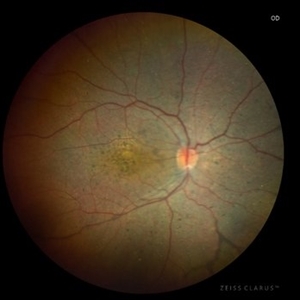 LCA Type 8
Apr 10 2025 by Joshua Friedman
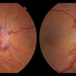
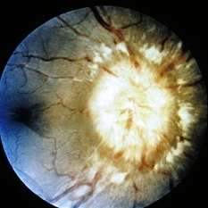
LCA Type 8 due to a pathogenic mutations in CRB1. 5-year-old male with a visual acuity of count fingers at 3 feet. Note the pseudopapilledema, para-arteriolar sparing, and nummular intraretinal pigment migration.
Photographer: Stephen Tsang, MD, PhD
Condition/keywords: Leber Congenital Amaurosis
-
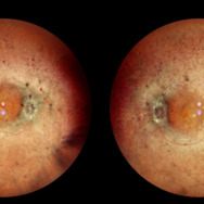 Retinitis Pigmentosa With Papilledema
Sep 29 2024 by Tejaswita Verma
Fundus pictures of RE and LE of a 30 year old male with a 20 day history of diminution of vision in LE more than RE. Vision was 6/9P and 6/24P in RE and LE respectively. History of RP in father. Alleged history of trauma to left side of forehead 4 months back with subgaleal haematoma over left side of frontal region on CT done 4 months back, No significant intracranial abnormality. He was started on oral steroids with tapering and F/U after 2 weeks.
Photographer: DR. TEJASWITA VERMA
Imaging device: MIRANTE
Condition/keywords: papilledema, retinitis pigmentosa
-
 Pseudotumor cerebri
Nov 2 2022 by pedro fernandes souza neto
Fundus photography of an 27-year-old woman with severe papiledema secondary to idiopathic intracranial hypertension. (Photo 1 - before treatment / Photo 2 - After Treatment)
Photographer: Pedro Fernandes, Universidade Federal da Bahia
Condition/keywords: papilledema, pseudotumor cerebri
-
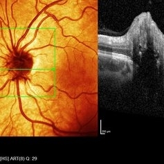
 Right eye SD- OCT-RNFL of optic nerve head drusen showing hypo reflective centre with hyper reflective margins.
Aug 5 2022 by Kavitha Duraipandi, MD DNB FICO FRCS
A 20 year old patient referred to the clinic with blurred disc margins to rule out papilledema.
Photographer: Natalie Fox- Bussell
Condition/keywords: optic nerve drusen, optical coherence tomography (OCT)
-
 Blue autofluroscence of Right eye optic nerve head showing auto fluorescence of the drusen
Aug 5 2022 by Kavitha Duraipandi, MD DNB FICO FRCS
A 20 year old patient referred to the clinic with blurred disc margins to rule out papilledema.
Photographer: Natalie Fox- Bussell
Condition/keywords: Blue autofluroscence, Heidelburg Spectralis
-
 Left eye SD- OCT-RNFL of optic nerve head drusen showing hypo reflective centre with hyper reflective margins.
Aug 5 2022 by Kavitha Duraipandi, MD DNB FICO FRCS
A 20 year old patient referred to the clinic with blurred disc margins to rule out papilledema.
Photographer: Natalie Fox- Bussell
Condition/keywords: optic nerve drusen, optical coherence tomography (OCT)
-
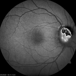 ONH-drusen-OD
Mar 24 2022 by Elite Bor-Shavit, MD
Fundus autofluorescence of a 41-years old patient with combined true papilledema and optic nerve head drusen, treated with Diamox and monitored.
Condition/keywords: optic disc drusen, papilledema
-
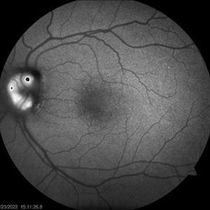 ONH-drusen-OS
Mar 24 2022 by Elite Bor-Shavit, MD
Fundus autofluorescence of a 41-years old patient with combined true papilledema and optic nerve head drusen, treated with Diamox and monitored.
Condition/keywords: optic disc drusen, papilledema
-
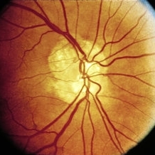
 Papilledema
Apr 23 2021 by Amin Masjedi
Fundus photograph of a 32-year-old female with IIH.
Photographer: Amin Masjedi,MD
Condition/keywords: papilledema
-
 Papilledema
Jan 22 2021 by AGNES KIM
Fundus photograph of 22-year-old woman with papilledema in right eye only. OS ruled out.
Condition/keywords: papilledema
-
 Papiledema
Dec 19 2019 by Zura Glonti
OS. Fundus photograph of 25-years-old man with papilledema, caused by meningioma.
Photographer: Zurab Glonti
Condition/keywords: meningioma, papilledema
-
 Papilledema
Dec 19 2019 by Zura Glonti
OD. Fundus photograph of 25-year-old man with papilledema, caused by meningioma.
Photographer: Zurab Glonti
Condition/keywords: meningioma, papilledema
-
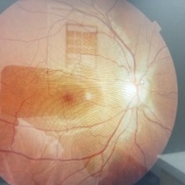
 Papilledema Color
Apr 26 2019 by Carissa Hurdstrom
Papilledema color
Photographer: Carissa Hurdstrom
Imaging device: Topcon 50DX
Condition/keywords: color photo, papilledema, stereo pair
-
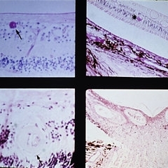
 Slide 11-21
Feb 26 2019 by Lancaster Course in Ophthalmology
Optic neuritis in a patient with measles endophthalmitis ( x 16). Note the swelling of the optic disk identical to that in papilledema, but inflammatory cells are the key to the diagnosis. (Thomas Jefferson University, No. 68-3043.)
Condition/keywords: endophthalmitis, optic neuritis
-
 Slide 11-15
Feb 26 2019 by Lancaster Course in Ophthalmology
Drusen of optic nerve. Clinical appearance. Drusen may be confused with papilledema. (Courtesy of H. G. Scheie, M.D.)
Condition/keywords: drusen, optic nerve
-
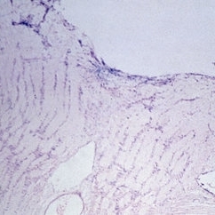
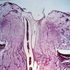
 Slide 11-13
Feb 26 2019 by Lancaster Course in Ophthalmology
Papilledema in a patient with severe hypertension. Edema is less severe than in other forms of choked disk. Low-power view of disk ( x 16).
Condition/keywords: papilledema
-
 Slide 11-12
Feb 26 2019 by Lancaster Course in Ophthalmology
Myelin artifact ( x16). Myelin has been squeezed from nerve into retinal vessels and subretinal space, and may be confused with papilledema. (Scheie Eye Institute, No. 5031.)
Condition/keywords: myelin
-
 Slide 11-11
Feb 26 2019 by Lancaster Course in Ophthalmology
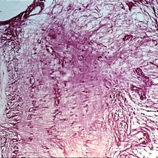
Papilledema. Forward bowing of fine glial fibers in front of the lamina (xllO). (Thomas Jefferson University, No. 71-0897.)
Condition/keywords: papilledema
-
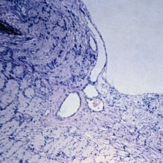
 Slide 11-10
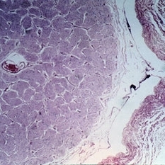
Feb 26 2019 by Lancaster Course in Ophthalmology
Papilledema. Enlargement of the nerve sheath (X 16).
Condition/keywords: papilledema
-
 Slide 11-9
Feb 26 2019 by Lancaster Course in Ophthalmology
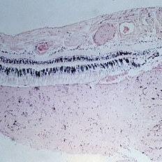
Papilledema. Section through the center of the disk shows swelling of the nerve fibers and lateral displacement of the sensory retina ( x16). (Thomas Jefferson University, No. 71-0897.)
Condition/keywords: papilledema
-
 Slide 11-8
Feb 26 2019 by Lancaster Course in Ophthalmology
Papilledema. Clinical characteristics include blurred margins, elevation, concentric folds of retina, and congested vessels. Hemorrhages and cotton wool exudates are commonly seen.
Condition/keywords: papilledema
-
 Slide 9-18
Feb 26 2019 by Lancaster Course in Ophthalmology
Malignant hypertension with retinal arterioles that are thickened and have fibrinoid necrosis (arrows). Retinal exudates (asterisk) and papilledema are also present. Papilledema is evidenced by fullness of the optic nerve head and peripapillary crowding of the retina (lower right).
Condition/keywords: fibrinoid, malignant hypertension, papilledema, retinal arteriole, retinal exudates
-
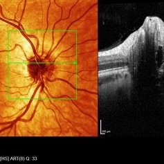
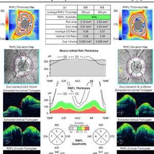 OCT in Patient With IIH Showing Thickened RNFL
Jan 16 2019 by John S. King, MD
18-year-old African American female with increased BMI with a history of headaches, nausea, transient diplopia and vision loss that she notices when getting up from her bed (and goes away after standing upright) for the last two weeks. Went to PCP and was treated for the flu, and after no improvement and visual symptoms known, was sent to ED. MRI did not show any masses and showed empty sella turcia. Vision 20/30 OD and 20/20 OS; no RAPD; IOP 15OU; no anterior segment or vitreous inflammation; discs are elevated with obscuration of the disc margins and some of the smaller vessels; there are no SVPs; there are mild Patton's lines temporally (see Initial Photos). The optic disc cube shows 360 degrees of RNFL thickening (see OCT). Was referred to near-ophthalmologist, Dr. Doyle. She obtained additional work-up, and LP opening pressure was high, and MRV showed bilateral transverse sinus stenosis. Patient showed steady improvement with medical therapy, that included weight loss and oral diamox. On her last visit with Dr. Doyle, vision has remained stable at 20/20-20/25 without an enlarged blindspot; there are SVPs and optic disc edema has resolved (see Post Treatment Photos); she is currently on 1000 mg of diamox and has lost 15 pounds, and no stinting procedure needed.
Imaging device: Cirrus
Condition/keywords: benign idiopatic intracranial hypertension, optic disc edema, papilledema
-
 Mild Patton's Lines in IIH - Initial Photos
Jan 16 2019 by John S. King, MD
18-year-old African American female with increased BMI with a history of headaches, nausea, transient diplopia and vision loss that she notices when getting up from her bed (and goes away after standing upright) for the last two weeks. Went to PCP and was treated for the flu, and after no improvement and visual symptoms known, was sent to ED. MRI did not show any masses and showed empty sella turcia. Vision 20/30 OD and 20/20 OS; no RAPD; IOP 15OU; no anterior segment or vitreous inflammation; discs are elevated with obscuration of the disc margins and some of the smaller vessels; there are no SVPs; there are mild Patton's lines temporally (see Initial Photos). The optic disc cube shows 360 degrees of RNFL thickening (see OCT). Was referred to near-ophthalmologist, Dr. Doyle. She obtained additional work-up, and LP opening pressure was high, and MRV showed bilateral transverse sinus stenosis. Patient showed steady improvement with medical therapy, that included weight loss and oral diamox. On her last visit with Dr. Doyle, vision has remained stable at 20/20-20/25 without an enlarged blindspot; there are SVPs and optic disc edema has resolved (see Post Treatment Photos); she is currently on 1000 mg of diamox and has lost 15 pounds, and no stinting procedure needed.
Photographer: Gretchen Harper
Imaging device: Topcon 50
Condition/keywords: idiopathic intracranial hypertension, optic disc edema, papilledema, Patton's Lines
-
 Mild Patton's Lines in IIH - Initial Photo
Jan 16 2019 by John S. King, MD
18-year-old African American female with increased BMI with a history of headaches, nausea, transient diplopia and vision loss that she notices when getting up from her bed (and goes away after standing upright) for the last two weeks. Went to PCP and was treated for the flu, and after no improvement and visual symptoms known, was sent to ED. MRI did not show any masses and showed empty sella turcia. Vision 20/30 OD and 20/20 OS; no RAPD; IOP 15OU; no anterior segment or vitreous inflammation; discs are elevated with obscuration of the disc margins and some of the smaller vessels; there are no SVPs; there are mild Patton's lines temporally (see Initial Photos). The optic disc cube shows 360 degrees of RNFL thickening (see OCT). Was referred to near-ophthalmologist, Dr. Doyle. She obtained additional work-up, and LP opening pressure was high, and MRV showed bilateral transverse sinus stenosis. Patient showed steady improvement with medical therapy, that included weight loss and oral diamox. On her last visit with Dr. Doyle, vision has remained stable at 20/20-20/25 without an enlarged blindspot; there are SVPs and optic disc edema has resolved (see Post Treatment Photos); she is currently on 1000 mg of diamox and has lost 15 pounds, and no stinting procedure needed.
Photographer: Gretchen Harper
Imaging device: Topcon 50
Condition/keywords: idiopathic intracranial hypertension, optic disc edema, papilledema, Patton's Lines
 Loading…