Search results (24 results)
-
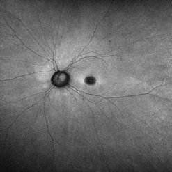
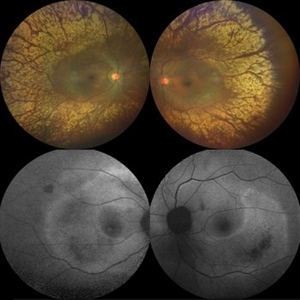
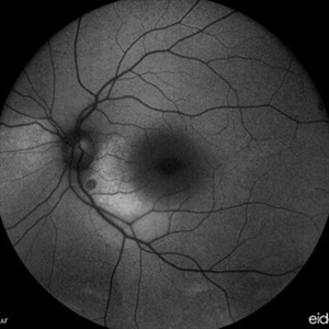
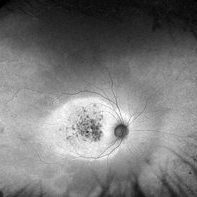
 Choroidal Metastasis
Apr 11 2024 by Corey Grant
Ultra-Widefield fundus photography and fundus autofluorescence images of a 61 year old female with Choroidal Metastasis affecting both eyes. Patient presented with blurred vision and flashes for a few weeks. Patient visual acuity was cc20/100 PH20/60 in the right eye and cc20/200 in the left eye. Patient admits to history of smoking for many years bit no known history of cancer prior to the visit. Physician recommended going to the ER for full body PET CT and stated that the first line of treatment is usually systemic chemo therapy. Patient will be reassessed in one month.
Photographer: Corey Grant
Imaging device: OPTOS CALIFORNIA RGB
Condition/keywords: cancer, choroidal metastasis, fundus autofluorescence (FAF), fundus photography, hyperautofluorescence, hypoautofluorescence, Optos, OPTOS CALIFORNIA RGB, Retina, ULTRA WIDE FIELD
-
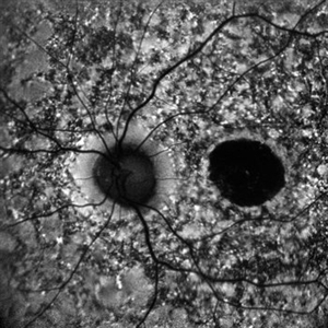
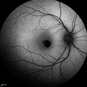
 Dome Shaped Macula
Jan 7 2025 by Jordyn Beckman
Autofluorescence photograph of 36 year old woman with Dome Shaped Macula with hypoautofluorescence and hyperautofluorescence centrally.
Photographer: Jordyn Beckman
Imaging device: California Optos
Condition/keywords: autoflorescence, convex protrusion, dome shaped macula, high myopia, hyperautofluorescent centrally, hypoautofluorescence
-
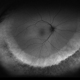
 Pigmentary Retinal Dystrophy
May 5 2020 by Olivia Rainey
Ultra-widefield fundus autofluorescence image of an 44-year-old male with pigmentary retinal dystrophy affecting both eyes. He presented with decreased night vision for 6 months prior to his appointment. He stated that his recovery time from transitioning from dark to light areas is reduced. He stated that his peripheral vision has never been very good for most of his life. He admits to environmental hearing loss. Patient denies family history of blin. His vision was 20/20 in both eyes. His full field ERG, visual fields were not consistent with RP. Genetic testing with ID Your IRD and annual follow up has been recommended.
Photographer: Olivia Rainey, OCT-C, COA
Imaging device: Optos California
Condition/keywords: fundus autofluorescence (FAF), hyperautofluorescence, hypoautofluorescence, inferior retina, left eye, Optos, ultra-wide field imaging
-
 Pigmentary Retinal Dystrophy
May 5 2020 by Olivia Rainey
Ultra-widefield fundus autofluorescence image of an 44-year-old male with pigmentary retinal dystrophy affecting both eyes. He presented with decreased night vision for 6 months prior to his appointment. He stated that his recovery time from transitioning from dark to light areas is reduced. He stated that his peripheral vision has never been very good for most of his life. He admits to environmental hearing loss. Patient denies family history of blin. His vision was 20/20 in both eyes. His full field ERG, visual fields were not consistent with RP. Genetic testing with ID Your IRD and annual follow up has been recommended.
Photographer: Olivia Rainey, OCT-C, COA
Imaging device: Optos California
Condition/keywords: fundus autofluorescence (FAF), hyperautofluorescence, hypoautofluorescence, inferior retina, Optos, pigment, ultra-wide field imaging
-
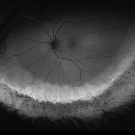
 Retinitis Pigmentosa
May 26 2017 by Olivia Rainey
Ultra-wide-field fundus autofluorescence image of the left eye of an 39-year-old female with Retinitis Pigmentosa. She had slightly atypical appearance due to asymmetry: sectoral atrophy in left eye, compared to 360 degree bone spicule formation in right eye. Ddx: Pigmentary degeneration vs infection vs X-linked RP carrier due to asymmetry. Recommended genetic testing through My Retina Tracker, as well as visual field and ERG testing. Patient's vision was sc20/100 PH 20/70 in the right eye and sc20/80 PH 20/40 in the left eye.
Photographer: Olivia Rainey
Imaging device: Optos
Condition/keywords: autofluorescence imaging, hyperautofluorescence, hypoautofluorescence, left eye, Optos, peripheral bone spicules, retinitis pigmentosa, ultra-wide field imaging
-
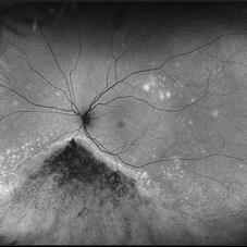
 Retinitis Pigmentosa
May 26 2017 by Olivia Rainey
Ultra-wide-field pseudocolor image of the left eye of an 39-year-old female with Retinitis Pigmentosa. She had slightly atypical appearance due to asymmetry: sectoral atrophy in left eye, compared to 360 degree bone spicule formation in right eye. Ddx: Pigmentary degeneration vs infection vs X-linked RP carrier due to asymmetry. Recommended genetic testing through My Retina Tracker, as well as visual field and ERG testing. Patient's vision was sc20/100 PH 20/70 in the right eye and sc20/80 PH 20/40 in the left eye.
Photographer: Olivia Rainey
Imaging device: Optos California
Condition/keywords: autofluorescence imaging, bone spicule, hyperautofluorescent ring, hypoautofluorescence, Optos, peripheral bone spicules, retinitis pigmentosa, ultra-wide field imaging
-
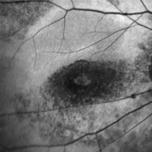
 Stargardt's Disease
Apr 20 2024 by Tejaswita Verma
Fundus autofluorescence image of the right eye of a 39 year old male showing hypoautofluorescence in a case of Stargardt's disease.
Photographer: DR. TEJASWITA VERMA
Imaging device: MIRANTE
Condition/keywords: fundus autofluorescence (FAF), hereditary macular dystrophy, hypoautofluorescence, Stargardt disease
-
 Stargardt's Disease
Apr 20 2024 by Tejaswita Verma
Fundus autofluorescence image of the left eye of a 39 year old male showing hypoautofluorescence in a case of Stargardt's disease.
Photographer: DR. TEJASWITA VERMA
Imaging device: MIRANTE
Condition/keywords: fundus autofluorescence (FAF), hereditary macular dystrophy, hypoautofluorescence, Stargardt disease
-
 All That Glitters is Not Gold
Mar 4 2021 by SHISHIR VERGHESE, MS, FVRS, FAICO (Retina)
Enhanced tapetoretinal reflex in a 70-year-old female patient with retinitis pigmentosa along with corresponding auto-fluorescence showing patchy hypoautofluorescence with surrounding granular hyperautofluorescence
Photographer: SHISHIR VERGHESE, ARAVIND EYE HOSPITAL AND POSTGRADUATE INSTITUTE OF OPHTHALMOLOGY, COIMBATORE
Imaging device: ZEISS, CLARUS
Condition/keywords: retinitis pigmentosa, tapetoretinal reflex
-
 Autosomal Recessive Bestrophinopathy
Apr 7 2022 by Nassim Alejandro Abreu Arbaje, MD
Fundus autofluorescence photo of a 22 year old boy with macular hypoautofluorescence due to the pigmentary changes in the posterior pole and some hyperautofluorescence due to the pooling of lipofuscin
Photographer: Nassim Abreu
Imaging device: Topcon Triton Plus
Condition/keywords: Autosomal recessive bestrophinopathy
-
 Autosomal Recessive Bestrophinopathy
Apr 7 2022 by Nassim Alejandro Abreu Arbaje, MD
Fundus autofluorescence photo of a 22 year old boy with macular hypoautofluorescence due to the pigmentary changes inside the temporal arcades and some hyperautofluorescence due to the pooling of lipofuscin
Photographer: Nassim Abreu
Imaging device: Topcon Triton Plus
Condition/keywords: Autosomal recessive bestrophinopathy
-
 B-FAF in Stargardt's Disease
Jul 4 2024 by Tejaswita Verma
Blue fundus autofluorescence showing hypoautofluorescence picture of a 28 year old male with 6/60 vision in BE in a case of Stargardt's disease.
Photographer: DR. TEJASWITA VERMA
Imaging device: MIRANTE
Condition/keywords: fundus autofluorescence (FAF), hereditary macular dystrophy, Stargardt disease
-
 Both Eyes Fundus Autofluorescence in Case of CNVM with Angioid Streaks
Nov 29 2024 by Anand Temkar
A 45 year old male came with chief complaint of blurring vision in right eyes since past 4 days. His vision is 6/12 in right eye and 6/9 in left eye. His vision was 14 mmHg in right eye and 16 mmHg in left eye. He was diagnosed with Angioid Streaks in both eyes about a year ago, then he developed choroidal neovascularization in his left eye 8 months ago, for which he received AntiVEGF injections x 3. Left eye is a stable eye now. Patient presented with right eye choroidal neovascularization in a case of Angioid Streaks on recent follow up. We have advised him right eye AntiVEGF injections x 3. In this image we can see fundus hypoautofluorescence in right eye due to hemorrhages and angioid streaks and in left eye fundus hypoautofluorescence is noted due to angioid streaks.
Photographer: Dr.Anand Temkar- Retina Foundation, Ahmedabad
Imaging device: Mirante
Condition/keywords: Angioid Streaks, choroidal neovascular membrane (CNVM), fundus autofluorescence (FAF)
-
 Central Serous Chorioretinopathy in Pregnancy. OS-AF
Dec 31 2020 by Cosimo Antonio Calabro
Woman (37-years-old) in pregnancy (third trimester). Central serous chorioretinopathy in both eyes. The AF image shows a diffuse hypoautofluorescence in the macular area; a hyperautofluorescent sickle between macula and papilla; a small hypoautofluorescent spot, in the same area, under the papilla.
Photographer: Cosimo Antonio Calabrò
Condition/keywords: bilateral chronic central serous retinopathy
-
 Congenital Simple Hamartoma of the RPE Autofluorescence
Aug 3 2015 by Bindu Rajesh
Fundus autofluorescence image of a 26-year-old male depicting hypoautofluorescence nasal to the fovea corresponding to the hamartoma visible clinically.
Imaging device: Heidelberg Spectralis
Condition/keywords: congenital, hamartoma, retinal pigment epithelium
-
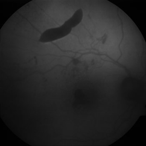 Extrafoveal PED with RPE rip AF
Dec 23 2012 by Alex P. Hunyor, MD
80-year-old female with subfoveal occult CNV and large extrafoveal PED which underwent spontaneous RPE rip. Autofluorescence image shows hypoautofluorescence in crescentic area of absent RPE due to rip, and also RPE atrophy adjacent to fovea. Intervening small areas of hypoautofluorescence are due to subretinal haemorrhage.
Condition/keywords: pigment epithelial detachment (PED), retinal pigment epithelium (RPE) tear
-
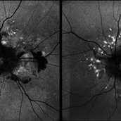
 FA ICG AZOOR
Oct 14 2017 by Navneet Mehrotra, DNB
fundus autofluorescence OD showing peripapillary hypoautofluorescence surrounded by an area of hyperautofluorescence with well demarcated margins suggestive of AZOOR.
Photographer: Ashish jain, Retina Foundation, Ahmedabad
Imaging device: Heidelberg spectralis
Condition/keywords: acute zonal occult outer retinopathy (AZOOR)
-
 FA ICG AZOOR
Oct 14 2017 by Navneet Mehrotra, DNB
Fundus autofluorescence OS showing peripapillary hypoautofluorescence surrounded by an area of hyperautofluorescence with well demarcated margins suggestive of AZOOR.
Photographer: Ashish jain, Retina Foundation, Ahmedabad
Imaging device: Heidelberg spectralis
Condition/keywords: acute zonal occult outer retinopathy (AZOOR)
-
 LCA Type 2
Apr 10 2025 by Joshua Friedman
LCA Type 2 (RPE65) showing characteristic hypoautofluorescence and retinal thinning. 8F with best corrected visual acuity of 20/400 (OD) and 20/150 (OS). Small white intraretinal spots and RPE mottling are visible on color fundus photography. Blue light autofluorescence reveals near-complete loss of signal, while OCT demonstrates widespread outer retinal thinning.
Photographer: Stephen Tsang, MD, PhD
Condition/keywords: Leber Congenital Amaurosis
-
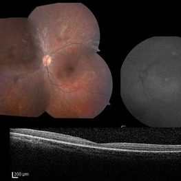
 Multimodal Imaging in CHRPE
Mar 6 2025 by Gerardo - Montante Montelongo, MD
Fundus photograph of an 83-year-old male with a history of Diabetes, smoking, cataract surgery on the right eye in 2022, and open-angle glaucoma. Asymptomatic. Indirect ophthalmoscopy revealed 80% excavation, peripapillary atrophy, and a hyperpigmented perifoveal lesion with 35% atrophy, 10% drusen, and 5.1 mm diameter, corresponding to a CHRPE. At multimodal imaging, FFA shows hypoautofluorescence of the lesion, OCT shows preservation of internal retinal layers, atrophy of external retinal layer, with an RPE disruption, and posterior shadowing. USG shows a flat hyperechoic lesion 5.1 mm in diameter and 1.32 mm in thickness, solid and with high internal reflectance.
Photographer: Gerardo Montante-Montelongo, MD, Mexican Institute of Ophthalmology
Imaging device: Clarus 700
Condition/keywords: congenital hypertrophy of the retinal pigment epithelium (CHRPE), multimodal imaging
-
 Plaquenil-toxicity
Mar 31 2023 by Niloofar Piri, MD
Fundus autofluorescence image of the right eye in a highly myopic Caucasian patient who was screened for plaquenil toxicity elsewhere for 15 years. Medicine was stopped 3 years ago. She presented to us with deteriorationg central vision and scotoma for the past 3 years. FAF demonstrtaes classic symmetric bull's eye pattern of hypoautofluorescence in parafoveal area both eyes with some extension to the arcades, which is the result of severe plaquenil toxicity. Notice the hyperAF area surrounding the bull'e eye which is demonstarting stressed RPE cells filled with Lipofuscin. This area will likely turn to a larger hypoAF later. It is critical to diagnose it early to prevent the ongoing damage and loss of RPE and photoreceptors from the long term deposited medicine in RPE cells.
Photographer: Sean Kelso, Saint Lousi University
Condition/keywords: bull's eye atrophy, bull's eye maculopathy, hydroxychloroquine toxicity, plaquenil toxicity
-
 Stargardt Disease
Apr 8 2024 by T. P . VIGNESH, MBBS,MS
Fundus autofluorescence image of the right eye revealing foveal hypoautofluorescence and multiple hypoautofluorescent specks in the background radiating from posterior pole towards periphery.
Photographer: Bharathi
Imaging device: ZEISS CLARUS
Condition/keywords: fundus autofluorescence (FAF), Stargardt disease
-
 Stargardt Disease
Apr 8 2024 by T. P . VIGNESH, MBBS,MS
Fundus autofluorescence image of the left eye revealing foveal hypoautofluorescence and multiple hypoautofluorescent specks in the background radiating from posterior pole towards periphery .
Photographer: Bharathi
Imaging device: ZEISS CLARUS
Condition/keywords: fundus autofluorescence (FAF), Stargardts Disease
-
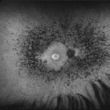
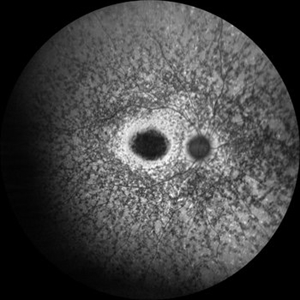
 THE ADDED VALUE OF WIDE-FIELD FUNDUS AUTOFLUORESCENCE IN CONE-ROD DYSTROPHIES
Oct 7 2019 by Mariana Oliveira
This wide-field fundus autofluorescence belongs to a 43 year-old female with CORD and no family history of inherited retinal disease. Her complains were reduced visual acuity and photophobia beginning in early adulthood. Note the central hyperautofluorescence with patchy hypoautofluorescence depicting atrophy of the retinal pigment epithelium in the macular area, an equatorial ring of preserved retinal autofluorescence and peripheral changes portraying rod involvement. The latter would be easily missed without wide-field imaging.
Condition/keywords: cone dystrophy
 Loading…