File number: 37257
Comments
-
 Suber S. Huang, MD, MBA, FASRS (November 8 2019)
This is a superb image. Please re-submit with full clinical details including testing and classify as an inherited retinal dystrophy and cone dystrophy. Thank you!
Sign in to comment.
 Initializing download.
-
 By Mariana Oliveira
Department of Ophthalmology, Centro Hospitalar e Universitário de Coimbra - CHUC, Coimbra, Portugal
Co-author(s): João Pedro Marques - Uploaded on Oct 7, 2019.
- Last modified by Mariana Oliveira on Nov 13, 2019.
- Rating
- Appears in
- Miscellaneous
- Condition/keywords
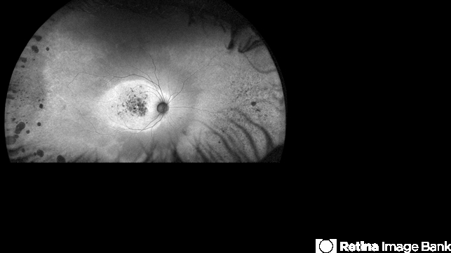
- cone dystrophy
- Description
- This wide-field fundus autofluorescence belongs to a 43 year-old female with CORD and no family history of inherited retinal disease. Her complains were reduced visual acuity and photophobia beginning in early adulthood. Note the central hyperautofluorescence with patchy hypoautofluorescence depicting atrophy of the retinal pigment epithelium in the macular area, an equatorial ring of preserved retinal autofluorescence and peripheral changes portraying rod involvement. The latter would be easily missed without wide-field imaging.


---thumb.jpg/image-square;max$79,0.ImageHandler "Cone Dystrophy")
---thumb.jpg/image-square;max$79,0.ImageHandler "Cone Dystrophy")
---thumb.jpg/image-square;max$79,0.ImageHandler "Cone Dystrophy")
---thumb.jpg/image-square;max$79,0.ImageHandler "Cone Dystrophy")
---thumb.jpg/image-square;max$79,0.ImageHandler "Cone Dystrophy")
---thumb.jpg/image-square;max$79,0.ImageHandler "Cone Dystrophy")
---thumb.jpg/image-square;max$79,0.ImageHandler "Cone Dystrophy")
