Search results (140 results)
-
 Amelanotic Choroidal Melanoma
May 18 2020 by McGill University Health Centre
The enucleation specimen in (A) shows an amelanotic, mushroom-shaped tumor arising from the choroid. Microhemorrhages are present within the tumor and also surround the tumor base (arrow). True retinal detachment is present (arrowhead). The subretinal fluid is mixed: clear (1), hemorrhagic (2), and fibrinoid (3).
Condition/keywords: enucleation, mushroom-shaped
-
 Amelanotic Choroidal Melanoma
May 18 2020 by McGill University Health Centre
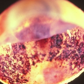
The enucleation image shows a large amelanotic tumor with large areas of hemorrhage and necrosis. Note the several dilated blood vessels and an adjacent retinal detachment with lipofuscin pigment on its surface (arrow).
Condition/keywords: amelanotic melanoma, enucleation, mushroom-shaped
-
 Amelanotic Mushroom-Shaped Choroidal Melanoma
May 18 2020 by McGill University Health Centre
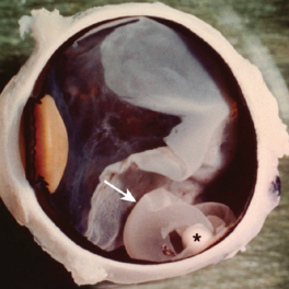
This enucleation specimen demonstrates an amelanotic, mushroom-shaped, slightly hemorrhagic tumor near the optic nerve (arrow). True retinal detachment is present, and the retina is folded (arrowhead). The subretinal fluid is hazy (*).
Condition/keywords: amelanotic melanoma, enucleation, mushroom-shaped
-
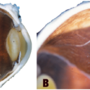
 Amelanotic Mushroom-Shaped Choroidal Melanoma
May 18 2020 by McGill University Health Centre
The enucleation specimen in (B) shows an amelanotic, mushroom-shaped, slightly hemorrhagic tumor near the optic nerve (arrow). The shape is due to infiltration of the retina by a rupture of the Bruch membrane. A retinal detachment artifact is present.
Condition/keywords: enucleation, mushroom-shaped
-
 Carcinoid
May 18 2020 by McGill University Health Centre
These are usually secondary tumor foci, arising most frequently from a primary tumor in the gastrointestinal tract or lung. Melanoma should be considered as part of the differential diagnosis, because neither carcinoid nor melanoma tumors have a necrotic component. This enucleation specimen shows a welldifferentiated neuroendocrine tumor in the choroid. The overlying pigment is caused by proliferation of the retinal pigment epithelium (arrowhead). Focal microhemorrhages are visible on the surface of the retina (arrows).
Condition/keywords: carcinoid, enucleation, focal microhemorrhages
-
 Central Retinal Artery of Optic Nerve
May 18 2020 by McGill University Health Centre
Image showing the central retinal artery (arrow) in the optic nerve.
Condition/keywords: central artery, enucleation, optic nerve
-
 Choroidal Melanoma
May 18 2020 by McGill University Health Centre
Choroidal melanoma is often asymptomatic and diagnosis is incidental. The tumors may grow beneath the retina, or may break through the Bruch membrane and disrupt the retina. Tumors breaking through the Bruch membrane and disrupting the retina have a characteristic “mushroom” shape. This enucleation specimen shows a pigmented dome-shaped choroidal melanoma (arrow). The cataractous lens is dislocated (*) and the retina is folded (•).
Condition/keywords: enucleation
-
 Choroidal Melanoma
Nov 3 2022 by pedro fernandes souza neto
Transillumination of Enucleation specimen of Choroidal Melanoma: anterior chamber is closed. Total secondary retinal detachment with subretinal serous fluid and some subretinal hemorrhages are present.
Photographer: Eduardo Marback, Federal University of Bahia, Brazil
Condition/keywords: enucleation, melanoma
-
 Choroidal Melanoma
Nov 3 2022 by pedro fernandes souza neto
Enucleation specimen of Choroidal Melanoma: anterior chamber is closed. Total secondary retinal detachment with subretinal serous fluid and some subretinal hemorrhages are present.
Photographer: Eduardo Marback, Federal University of Bahia, Brazil
Condition/keywords: enucleation
-
 Ciliary Body Melanoma
May 18 2020 by McGill University Health Centre
Uveal melanoma is the most common primary eye malignancy in adulthood, occurring mainly after age 60. The uveal tract — composed of the iris, ciliary body, and choroid — can be affected by uveal melanoma. Despite advances in treatment of the primary tumor, metastatic disease occurs in almost half of patients, generally affecting the liver and lungs via hematogenous dissemination of the primary tumor. Tumors have different levels of pigmentation, and some are amelanocytic (nonpigmented). The differential diagnosis for amelanotic choroidal melanoma is metastatic disease. Large tumors displace the lens. Of the 3 locations in the uveal tract, tumors of the ciliary body have the worst prognosis. The enucleation specimen in (A) shows a firm, dome-shaped, deeply pigmented tumor arising from the ciliary body (arrow). The lens has been removed, and a diffuse retinal detachment artifact is present.
Condition/keywords: enucleation, melanoma
-
 Ciliary Body Melanoma
May 18 2020 by McGill University Health Centre
Uveal melanoma is the most common primary eye malignancy in adulthood, occurring mainly after age 60. The uveal tract — composed of the iris, ciliary body, and choroid — can be affected by uveal melanoma. Despite advances in treatment of the primary tumor, metastatic disease occurs in almost half of patients, generally affecting the liver and lungs via hematogenous dissemination of the primary tumor. Tumors have different levels of pigmentation, and some are amelanocytic (nonpigmented). The differential diagnosis for amelanotic choroidal melanoma is metastatic disease. Large tumors displace the lens. Of the 3 locations in the uveal tract, tumors of the ciliary body have the worst prognosis The enucleation specimen in (B) shows a large, dome-shaped, mixed melanotic and amelanotic choroidal melanoma. The anterior chamber is closed, and the angle is infiltrated (arrow). Total secondary retinal detachment with subretinal serous fluid and some subretinal hemorrhages are present (arrowhead). The lens is cataractous.
Condition/keywords: enucleation, melanoma
-
 Ciliary Body Melanoma
May 18 2020 by McGill University Health Centre
Uveal melanoma is the most common primary eye malignancy in adulthood, occurring mainly after age 60. The uveal tract — composed of the iris, ciliary body, and choroid — can be affected by uveal melanoma. Despite advances in treatment of the primary tumor, metastatic disease occurs in almost half of patients, generally affecting the liver and lungs via hematogenous dissemination of the primary tumor. Tumors have different levels of pigmentation, and some are amelanocytic (nonpigmented). The differential diagnosis for amelanotic choroidal melanoma is metastatic disease. Large tumors displace the lens. Of the 3 locations in the uveal tract, tumors of the ciliary body have the worst prognosis. This enucleation specimen shows a pigmented, nodular-shaped ciliary body melanoma (arrow) with extensive necrosis (*). A retinal detachment is present with subretinal fluid (arrowhead), and the retina is folded (•).
Condition/keywords: enucleation, melanoma
-
 Common Artifacts in Macroscopic Ocular Globe Evaluation
May 18 2020 by McGill University Health Centre
This sample was retrieved from a patient with a blind, painful eye. Blind, painful eye may be the end stage of several conditions including glaucoma, retinal detachment, and endophthalmitis, among others. Evisceration specimens are generally submitted in fragments. Different intraocular structures are identifiable: retina, cornea and capsular bag, choroidal tissue, and hematic material.
Condition/keywords: enucleation, evisceration, intraocular structures
-
 Cross-Section of Enucleated Specimen
May 18 2020 by McGill University Health Centre
The eye is an organ with 3 layers (from the outermost to the innermost): fibrous layer (sclera and cornea), uveal tract (iris, ciliary body, and choroid), and retina. The lens divides the eye into the aqueous chamber (filled with aqueous humor) and vitreous chamber (filled with vitreous humor). The iris divides the aqueous chamber into anterior and posterior chambers. This image illustrates the aqueous chamber (1), posterior chamber (2), and vitreous chamber (3).
Condition/keywords: aqueous chamber, cross-section, enucleation, posterior chamber intraocular lens (PCIOL), vitreous chamber
-
 Cysticercosis
May 18 2020 by McGill University Health Centre
Ocular cysticercosis is a disease that is caused by the encystment of cysticercus larvae from certain tapeworms in the eye. In this enucleation specimen, a choroidal cyst (arrow) containing a larva (*) is clearly visible. Note the retinal detachment overlying the cyst.
Condition/keywords: cyst, cysticercosis, enucleation
-
 Endophytic Retinoblastoma
May 18 2020 by McGill University Health Centre
Image (A) shows an endophytic grayish tumor located on the retina of this enucleation specimen (arrow). Higher magnification of the same specimen (B) shows small hemorrhagic areas (arrow). The choroidal layer is not compromised.
Condition/keywords: enucleation, retinoblastoma
-
 Enucleated Cataractous Lens Seen Through the Pupil
May 18 2020 by McGill University Health Centre
The anterior view of enucleated eye shows a cataractous lens seen through the pupil.
Condition/keywords: cataract, enucleation
-
 Enucleated Eye Showing Choroidal Melanoma
May 18 2020 by McGill University Health Centre
This enucleation specimen shows an aphakic eye with a large, solid choroidal tumor. The tumor is heavily pigmented; it shows different shades in some areas. The tumor reaches the ciliary body.
Condition/keywords: aphakic eye, choroidal tumor, enucleation
-
 Enucleated Eye with Cataractous Lens
May 18 2020 by McGill University Health Centre
The cornea is transparent and thin. The lens is cataractous. The ora Serrata (arrow) demarcates a transition zone in the uveal tract between the pars plana of the ciliary body and the retina.
Condition/keywords: cataract, enucleation
-
 Enucleated Eye with Macular Edema
May 18 2020 by McGill University Health Centre
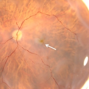
In an ophthalmoscopic-like view of an enucleation specimen shows macular edema. Note the folds surrounding the foveal area (arrow).
Condition/keywords: enucleation, macular edema
-
 Enucleated Eye with Malignant Melamoma
Apr 13 2020 by Sophia El Hamichi, MD
A 84-year-old female with advanced malignant melanoma of the her left eye requiring enucleation.
Photographer: Belinda Rodriguez, Murray Ocular Oncology and Retina, Miami
Condition/keywords: enucleation, gross pathology, malignant melanoma
-
 Enucleated Eye with Retinal Atrophy
May 18 2020 by McGill University Health Centre
This image illustrates marked retinal atrophy with several bone-spicule-shaped pigment deposits in the peripheral retina. The macular area is preserved but has a rim of depigmentation. Note the thin blood vessels and the pallor of the optic nerve.
Condition/keywords: atrophy, enucleation
-
 Enucleated Eye: Phthisis Bulbi
May 18 2020 by McGill University Health Centre
The enucleation specimen shows an atrophic eye (< 16 mm in its largest dimension) with complete disorganization of the posterior structures.
Condition/keywords: enucleation, phthisis bulbi
-
 Enucleated Specimen with Pseudo-Retinal Detachment
May 18 2020 by McGill University Health Centre
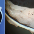
In (A), a pupil–optic view of an enucleation specimen shows pseudo–retinal detachment. Note the absence of fluid beneath the retina (arrow). In (B), high magnification of a pseudo–choroidal detachment shows the bare sclera separate from the choroid and retinal pigmented epithelium.
Condition/keywords: choroidal detachment, enucleation
-
 Enucleation of an Eye with Advanced Choroidal Melanoma with Implant and Donor Sclera Replacement
Jan 11 2021 by Sophia El Hamichi, MD
Surgery of the left eye affected with advanced melanoma: Upper left image: separating the sclera from the conjunctiva and the tenon by performing a peritomy, then separating the rectus muscles that will be later sutured to the donor sclera, to preserve post-op motility. Upper right image: cutting the optic nerve. Middle left image: the globe is enucleated. Middle right image: dissection of the globe showing the melanoma. Tissue is then sent to pathology Lower left image: putting the porous polyethyline implant inside the donor sclera and marking muscles' insertion. Lower right image: reinsertion of the rectus muscles on the donor sclera, then covering with tenon and conjunctiva.
Photographer: Belinda Rodriguez, Murray Ocular Oncology and Retina, Miami
Condition/keywords: donor sclera, enucleation, implant, melanoma
 Loading…