Search results (140 results)
-
Venous Pulsations in Large Choroidal Melanoma
Oct 9 2025 by Virginia Gebhart
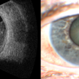
81 year old female diagnosed with large, pigmented collar button tumor. Given size of lesion enucleation was recommended. CT scans show no evidence of metastatic disease. Pt is doing well s/p enucleation.
Condition/keywords: bscan ultrasound, choroidal melanoma
-
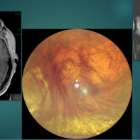
 Choroidal Melanoma
Oct 3 2025 by Virginia Gebhart
63 year old male with new choroidal melanoma. Pt states vision has been poor for 2 years, worsening in the last year. Bscan ultrasound shows lesion extends into the macula up to the optic nerve. Recommended enucleation due to size of lesion (7.7 x 15.6 x 15.2) and poor prognosis of visual recovery. Surgery will be scheduled pending CT scan results.
Photographer: Virginia Gebhart, Retina Consultants of Carolina
Imaging device: Optos California
Condition/keywords: choroidal melanoma, exudative detachment
-
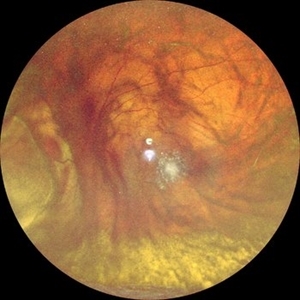
 Choroidal Melanoma
Oct 1 2025 by Virginia Gebhart
60 year old male referred by optometrist for retinal detachment. Pt had been having symptoms of flashing lights and shadow in vision for approximately 1 month. Exam and diagnostics consistent with choroidal melanoma with exudative detachment inferior. Due to size of lesion (10.8 x 14.8 x 13.2) enucleation was recommended. Pt will be scheduled for surgery pending CT scan results.
Photographer: Virginia Gebhart, Retina Consultants of Carolina
Imaging device: Optos California
Condition/keywords: choroidal melanoma, exudative detachment, melanoma
-
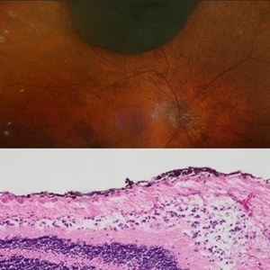
 Massive Choroidal Melanoma
Jun 18 2025 by Corey R Lacher, MD
A 57-year-old patient presented with no light perception vision in her right eye. B-scan ultrasonography revealed evidence of a large choroidal melanoma. External photography demonstrated detached retina visible just posterior to the lens. The patient subsequently underwent enucleation, and histopathologic examination confirmed the diagnosis of choroidal melanoma. The tumor measured 24 mm anteroposteriorly, 24 mm horizontally, and 25 mm vertically.
Photographer: Beth Malpica
Condition/keywords: choroidal melanoma
-
 Choroidal Melanoma
Mar 27 2025 by Virginia Gebhart
91 year old female with resolved choroidal melanoma. Pathology report confirms dispersed melanoma in the vitreous and on the entire retina surface. Pt is going well 1 month s/p enucleation, once healed she will be referred to an ocularist. No evidence of metastatic disease.
Condition/keywords: biopsy, choroidal melanoma
-
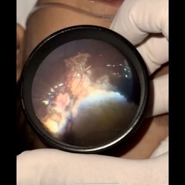
 Choroidal Melanoma Masquerading as PEHCR
Mar 3 2025 by Tejaswita Verma
A 65 year old diabetic male presented with large nasal retinal mass giving the appearance of organized dehaemoglobinized subretinal hemorrhage with breakthrough vitreous hemorrhage , with 6/6P vision. Enucleation specimen showed histopathology confirmed choroidal melanoma.
Photographer: DR. TEJASWITA VERMA
Imaging device: MIRANTE
Condition/keywords: vitreous hemorrhage
-
 Ciliary Body Melanoma
Feb 12 2025 by Virginia Gebhart
91 year old female with large collar button tumor emanating from the ciliary body with resolving vitreous hemorrhage. Melanoma cells in the AV as well as studded on the entire retina surface. Pt scheduled for enucleation. CT scans of chest and abdomen showed no evidence of metastatic disease.
Photographer: Virginia Gebhart, Retina Consultants of Carolina
Imaging device: Optos California
Condition/keywords: asteroid hyalosis, ciliary body mass, ciliary body melanoma, vitreous hemorrhage
-
 Choroidal Melanoma
Feb 6 2025 by Virginia Gebhart
81 year old female with large pigmented collar button ciliochoroidal mass extending into the mid-vitreous cavity. Clinical exam and ultrasound finding consistent with melanoma. Due to size of tumor, pt scheduled for enucleation. CT scan of abdomen showed no evidence of metastatic disease.
Photographer: Virginia Gebhart, Retina Consultants of Carolina
Imaging device: Optos California
Condition/keywords: ciliochoroidal melanoma, collar button, melanoma
-
 Iris Melanoma
Jan 28 2025 by Korey Starkey
Slit-lamp image of 90-year-old patient with iris melanoma and new hemorrhage affecting the right eye. Patient re-presented after nearly 1 year, now seeking treatment. Given iris location of tumor, multiple clock hours of iris involved, and increase in size of the known malignant transformation; safest approach was enucleation.
Photographer: Korey Starkey
Imaging device: Slit lamp camera
Condition/keywords: anterior chamber, hemorrhage, iris melanoma, slit lamp photo
-
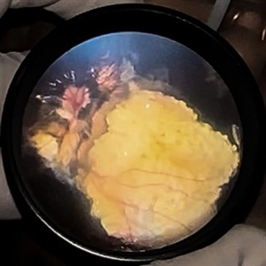
 Choroidal Melanoma Masquerading as Subretinal Hemorrhage With Breakthrough VH
Jan 23 2025 by Tejaswita Verma
A 65 year old diabetic male presented with large nasal retinal mass giving the appearance of organized dehaemoglobinized subretinal hemorrhage with breakthrough vitreous hemorrhage , with 6/6P vision. Enucleation specimen showed histopathology confirmed choroidal melanoma.
Photographer: DR. TEJASWITA VERMA
Imaging device: MIRANTE
-
 Ciliary Body Melanoma
Nov 2 2024 by Virginia Gebhart
53 year old male with a large mass behind the lens as well as prominent scleral vessels. Clinical exam and ultrasound findings consistent with melanoma. Pt will be scheduled for enucleation pending CT scan results. Edit: Sadly patient has canceled all appointments and has requested no further contact
Photographer: Virginia Gebhart, Retina Consultants of Carolina
Imaging device: Optos California
Condition/keywords: ciliary body mass, ciliary body melanoma, ciliary body tumor
-
 Calcified Retinoblastoma After Intra-arterial Chemotherapy
Apr 6 2024 by Hector Gabriel Moreno Solano, MD, MHA
Fundus photography of a 5 yea-old Mexican child with bilateral retinoblastoma following unilateral enucleation and 4 cycles of intra-arterial chemotherapy in her only remaining eye. The image shows a successfully treated tumor with a completely calcificied regression.
Photographer: Héctor Gabriel Moreno-Solano, MD, MHA
Imaging device: SmartPhone (IPhone 11 pro Max)
Condition/keywords: pediatric retina, pediatric tumor, retinoblastoma
-
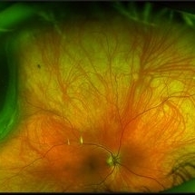 Choroidal Melanoma
Mar 26 2024 by Xitlali Caterina
Ultra-widefield fundus photograph of a 40-year-old woman with Choroidal Melanoma in right eye. Patient present with 20/50+2 vision in the right eye. Patient reported having frequent headaches located frontal area of their head and sometimes radiated to the right side as well. Patient also noted eye pain in both eyes that has remained constant for many years, as well as light sensitivity. The physician stated that since this is a medium-sized tumor, the treatment options include I-125 brachytherapy or enucleation. He recommended I-125 brachytherapy.
Photographer: Xitlali Caterina
Imaging device: Optos California RGB
Condition/keywords: fundus photography, Optos, OPTOS CALIFORNIA, superior retina, ultra-wide field imaging, ultra-widefield image
-
 Calcified Retinoblastoma after intra-arterial chemotherapy
Jan 19 2024 by Hector Gabriel Moreno Solano, MD, MHA
Fundus photography of a 5- Year-old Mexican child with bilateral retinoblastoma following unilateral enucleation and 4 cycles of intra-arterial chemotherapy in her only remaining eye. The image shows a succesfully treated tumor with a completely calcificied regression.
Photographer: Hector Solano, Hospital General de Zona #20 IMSS, Puebla
Imaging device: SmartPhone (IPhone 11 ProMax)
Condition/keywords: pediatic retina, pediatric tumor, retinoblastoma
-
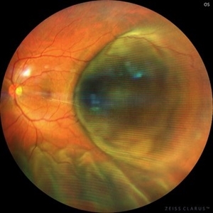 Choroidal Melanoma with exudative retinal detachment
Jul 19 2023 by Mariam Cernichiaro-Espinosa, MD
40-year old male with choroidal melanoma and secondary exudative retinal detachment. His visual acuity was 20/200. It was treated with enucleation.
Photographer: Mariam Cernichiaro-Espinosa, Asociación para Evitar la Ceguera en México, I.A.P. Mexico City, Mexico.
Imaging device: Zeiss Clarus
Condition/keywords: exudative retinal detachment
-
 Choroidal Melanoma
May 24 2023 by pedro fernandes souza neto
Transillumination of Enucleation specimen of Choroidal Melanoma
Photographer: Isabela Valladares Cesar Evangelista, Centro Oftalmológico de Minas Gerais
Condition/keywords: Choroidal melanoma
-
 Choroidal Melanoma
Nov 3 2022 by pedro fernandes souza neto
Enucleation specimen of Choroidal Melanoma: anterior chamber is closed. Total secondary retinal detachment with subretinal serous fluid and some subretinal hemorrhages are present.
Photographer: Eduardo Marback, Federal University of Bahia, Brazil
Condition/keywords: enucleation
-
 Choroidal Melanoma
Nov 3 2022 by pedro fernandes souza neto
Transillumination of Enucleation specimen of Choroidal Melanoma: anterior chamber is closed. Total secondary retinal detachment with subretinal serous fluid and some subretinal hemorrhages are present.
Photographer: Eduardo Marback, Federal University of Bahia, Brazil
Condition/keywords: enucleation, melanoma
-
 Ciliary Body Melanoma
Jul 4 2021 by Gerardo Rivera Arroyo
Clinical image taken in a slit lamp with a gonioscopy of a 39-year-old female patient with ciliary body melanoma before enucleation and pathological study.
Condition/keywords: ciliary body melanoma, gonioscopy
-
 Ciliary body melanoma
Jul 4 2021 by Gerardo Rivera Arroyo
Clinical image taken in a slit lamp with a gonioscopy of a 39-year-old female patient with ciliary body melanoma before enucleation and pathological study.
Condition/keywords: ciliary body melanoma, gonioscopy
-
 Enucleation of an Eye with Advanced Choroidal Melanoma with Implant and Donor Sclera Replacement
Jan 11 2021 by Sophia El Hamichi, MD
Surgery of the left eye affected with advanced melanoma: Upper left image: separating the sclera from the conjunctiva and the tenon by performing a peritomy, then separating the rectus muscles that will be later sutured to the donor sclera, to preserve post-op motility. Upper right image: cutting the optic nerve. Middle left image: the globe is enucleated. Middle right image: dissection of the globe showing the melanoma. Tissue is then sent to pathology Lower left image: putting the porous polyethyline implant inside the donor sclera and marking muscles' insertion. Lower right image: reinsertion of the rectus muscles on the donor sclera, then covering with tenon and conjunctiva.
Photographer: Belinda Rodriguez, Murray Ocular Oncology and Retina, Miami
Condition/keywords: donor sclera, enucleation, implant, melanoma
-
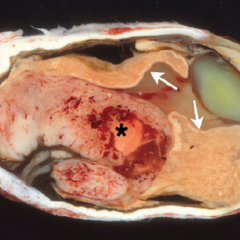 Mushroom-shaped Choroidal Melanoma
May 18 2020 by McGill University Health Centre
The enucleation specimen in (A) shows a diffuse melanoma infiltrating the choroid and ciliary body. In the center, a large area of necrosis and hemorrhage is present (*) and the retina is infiltrated (arrows). Note hypopyon in the anterior chamber and the cataractous lens.
Condition/keywords: enucleation, mushroom-shaped
-
 Amelanotic Mushroom-Shaped Choroidal Melanoma
May 18 2020 by McGill University Health Centre
The enucleation specimen in (B) shows an amelanotic, mushroom-shaped, slightly hemorrhagic tumor near the optic nerve (arrow). The shape is due to infiltration of the retina by a rupture of the Bruch membrane. A retinal detachment artifact is present.
Condition/keywords: enucleation, mushroom-shaped
-
 Amelanotic Mushroom-Shaped Choroidal Melanoma
May 18 2020 by McGill University Health Centre
This enucleation specimen demonstrates an amelanotic, mushroom-shaped, slightly hemorrhagic tumor near the optic nerve (arrow). True retinal detachment is present, and the retina is folded (arrowhead). The subretinal fluid is hazy (*).
Condition/keywords: amelanotic melanoma, enucleation, mushroom-shaped
-
 Amelanotic Choroidal Melanoma
May 18 2020 by McGill University Health Centre
The enucleation image shows a large amelanotic tumor with large areas of hemorrhage and necrosis. Note the several dilated blood vessels and an adjacent retinal detachment with lipofuscin pigment on its surface (arrow).
Condition/keywords: amelanotic melanoma, enucleation, mushroom-shaped
 Loading…