 Initializing download.
-
 By McGill University Health Centre
The MUHC-McGill University
Co-author(s): Sabrina Bergeron, P. Zoroquiain, E. Esposito, S. Corredor Casas, P. Logan, A. N. Odashiro, Miguel N. Burnier, Paulina García de Alba Graue, McGill University Health Center-McGill University Ocular Pathology & Translational Research Laboratory - Uploaded on May 18, 2020.
- Last modified by Caroline Bozell on May 19, 2020.
- Rating
- Appears in
- Enucleated Eye
- Condition/keywords
- atrophy, enucleation
- Description
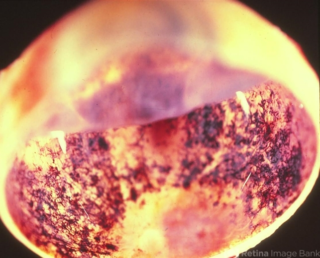
- This image illustrates marked retinal atrophy with several bone-spicule-shaped pigment deposits in the peripheral retina. The macular area is preserved but has a rim of depigmentation. Note the thin blood vessels and the pallor of the optic nerve.


