Search results (140 results)
-
 Retinoblastoma
Jul 4 2012 by John T. Thompson, MD
Retinoblastoma filling enucleated eye
Condition/keywords: enucleation, pediatric tumor, retinoblastoma
-
 Choroidal Melanoma
Jan 30 2019 by Karen Panzegrau
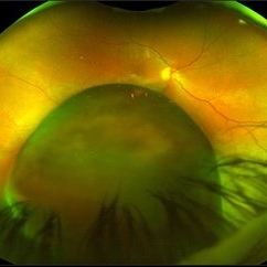
Ultra-wide field optos image of a 27-year-old male patient who presented with loss of vision for about 6-8 weeks. Previous choroidal nevus seen. Recommended annual monitoring. No exam for since 10/2014. Brachytherapy vs enucleation was discussed. Brachytherapy was decided as treatment. Full metastatic work up is being performed.
Photographer: Karen Panzegrau
Imaging device: Optos
Condition/keywords: choroidal nevus, exudative retinal detachment, malignant neoplasm of eye, Optos, ultra-wide field imaging
-
 Knife Stabbing Enucleation
-
---thumb.jpg/image-square;max$300,300.ImageHandler) Subretinal Neovascular Membrane With RPE
Subretinal Neovascular Membrane With RPE
Oct 11 2013 by Maurice F. Rabb
This healthy 81 year old Caucasian female has a family history of macular degeneration. She has no history of hypertension, and has not been on ASA or anticoagulants. She had a subretinal neovascular membrane with an RPE rip of the left eye, which advanced to disciform macular degeneration with eventual massive hemorrhage and total exudative retinal detachment. She developed neovascular glaucoma OS due to total retinal detachment and had enucleation of the left eye.
Condition/keywords: subretinal neovascular membrane
-
 Retinoblastoma OD FA 6-2-2015-12-15-42 PM Proof
Jun 4 2015 by Kathy Karsten, COT
15-year-old male born with retinoblastoma. S/P enucleation OS at 3 months of age. S/P chemo/radiation lesions OD
Photographer: Kathy Karsten, COT
Imaging device: Topcon TRC 50-DX
Condition/keywords: retinoblastoma
-
 Wide Field Fundus Montage of Intraocular Mass with Retinal Detachment
Aug 28 2019 by Gayathri Mohan
50 year old female came with diminution of vision in the LE. Wide field fundus photograph showing an intraocular mass temporally along with an exudative retinal detachment inferiorly. Ultrasonography showed an intraocular mass with collar button appearance suggestive of a Choroidal melanoma. She underwent enucleation and histopathology confirmed a spindle cell choroidal melanoma
Photographer: Dr. Gayathri Mohan, Retina Foundation
Imaging device: Nidek Mirante SLO
Condition/keywords: choroidal mass, collar button
-
---thumb.jpg/image-square;max$300,300.ImageHandler) Subretinal Neovascular Membrane With RPE
Subretinal Neovascular Membrane With RPE
Oct 11 2013 by Maurice F. Rabb
This healthy 81 year old Caucasian female has a family history of macular degeneration. She has no history of hypertension, and has not been on ASA or anticoagulants. She had a subretinal neovascular membrane with an RPE rip of the left eye, which advanced to disciform macular degeneration with eventual massive hemorrhage and total exudative retinal detachment. She developed neovascular glaucoma OS due to total retinal detachment and had enucleation of the left eye.
Condition/keywords: subretinal neovascular membrane
-
 Enucleated Eye with Malignant Melamoma
Apr 13 2020 by Sophia El Hamichi, MD
A 84-year-old female with advanced malignant melanoma of the her left eye requiring enucleation.
Photographer: Belinda Rodriguez, Murray Ocular Oncology and Retina, Miami
Condition/keywords: enucleation, gross pathology, malignant melanoma
-
 Large Choroidal Melanoma
Mar 29 2019 by Olivia Rainey
Ultra-wide field pseudocolor image of a 59-year-old male with a large choroidal melanoma affecting his right eye. Patient presented with a superior visual field defect 3 months prior to examination, but reports that the vision has been "bad" in the right eye since he was 3 years old. The risks, benefits and alternatives for treating the melanoma were thoroughly discussed and the patient has elected to proceed with enucleation, which we will schedule for the near future. Patient reports a brother with "blood cancer" and a father with throat cancer.
Photographer: Olivia Rainey
Imaging device: Optos
Condition/keywords: Optos, ultra-wide field imaging
-
 Enucleated Eye: Phthisis Bulbi
May 18 2020 by McGill University Health Centre
The enucleation specimen shows an atrophic eye (< 16 mm in its largest dimension) with complete disorganization of the posterior structures.
Condition/keywords: enucleation, phthisis bulbi
-
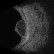
 B-Scan Showing Intraocular Mass
Aug 28 2019 by Gayathri Mohan
50 year old female came with diminution of vision in the LE. Ultrasonography showed an intraocular mass with collar button appearance suggestive of a Choroidal melanoma. She underwent enucleation and histopathology confirmed a spindle cell choroidal melanoma
Photographer: Dr. Gayathri Mohan - Retina Foundation
Imaging device: Nidek ,Mirante
Condition/keywords: collar button, melanoma
-
 Posterior View of Enucleated Eye with Extraocular Extension of an Intraocular Tumor
May 18 2020 by McGill University Health Centre
Posterior view of enucleated eye showing the extraocular extension of an intraocular tumor and extensive areas of hemorrhage, including a subdural hemorrhage.
Condition/keywords: enucleation, extraocular extension, intraocular tumor
-
 Large, Irregularly Shaped Choroidal Melanoma - B Scan (Transverse)
Feb 13 2020 by Michael Seider, MD
Large, irregularly shaped choroidal melanoma with overlying subretinal fluid and inferior exudative retinal detachment in the right eye of a 93-year-old woman. Note the extensive overlying orange pigment (lipofuscin) which is hyper-autofluorescent. B-Scan ultrasonography confirms low tumor internal reflectivity, adjacent retinal detachment and multi-lobulated shape. Especially because of the poor baseline vision and the severe vision loss expected from radiotherapy (because of the larger tumor size and proximity to the optic nerve), this older woman elected primary enucleation.
-
 Exophytic Retinoblastoma
May 18 2020 by McGill University Health Centre
This type of tumor grows from the retina toward the choroid. In this enucleation specimen, the retina is completely detached, and the tumor is growing inside the subretinal proteinaceous fluid. Note the distance between the tumor and the optic nerve head.
Condition/keywords: enucleation, retinoblastoma
-
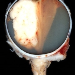
 Choroidal Melanoma
Nov 3 2022 by pedro fernandes souza neto
Transillumination of Enucleation specimen of Choroidal Melanoma: anterior chamber is closed. Total secondary retinal detachment with subretinal serous fluid and some subretinal hemorrhages are present.
Photographer: Eduardo Marback, Federal University of Bahia, Brazil
Condition/keywords: enucleation, melanoma
-
 Enucleated Eye Showing Choroidal Melanoma
May 18 2020 by McGill University Health Centre
This enucleation specimen shows an aphakic eye with a large, solid choroidal tumor. The tumor is heavily pigmented; it shows different shades in some areas. The tumor reaches the ciliary body.
Condition/keywords: aphakic eye, choroidal tumor, enucleation
-
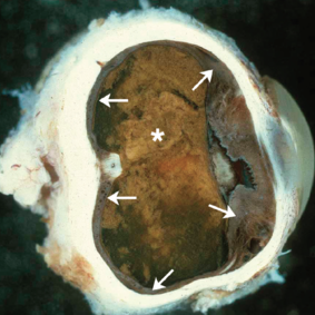 Sympathetic Ophthalmia
May 18 2020 by McGill University Health Centre
Sympathetic ophthalmia is characterized by bilateral diffuse granulomatous uveitis that occurs 2 weeks to many years after traumatic penetration or perforation of the eye. It threatens the sight of the uninjured (sympathizing) eye. In this enucleation specimen, thickening of the uveal tract is evident (arrows). Complete proteinaceous retinal detachment (*) is also present, along with posterior synechia (adhesion of the iris to the anterior capsule of the lens).
Condition/keywords: enucleation, sympathetic ophthalmia
-
 Very Large Choroidal Melanoma in Monocular Patient - BScan
Feb 13 2020 by Michael Seider, MD
Very large choroidal melanoma in the left eye of a 51-year-old man with long-standing poor vision in the right eye from a childhood injury (with traumatic macular hole and chorioretinal scarring). Note the large superior choroidal tumor with overlying subretinal hemorrhage and extensive inferior exudative retinal detachment. B-Scan ultrasound shows the collar-stud shape of the lesion and the overlying subretinal hemorrhage which is hyper-reflective compared to the vitreous and slightly hypo-reflective compared to the tumor. The patient has optic disk drusen in both eyes. He elected enucleation.
-
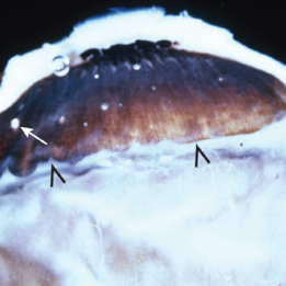
 Magnification of the Eye Wall from an Enucleated Eye
May 18 2020 by McGill University Health Centre
Magnification of the eye wall showing the neurosensory retina (1), the retinal pigment epithelium (arrow), a thin layer overlying the choroid (2), and the sclera (3).
Condition/keywords: choroid, enucleation, neurosensory retina, retinal pigment epithelium, sclera
-
 Slide 3-16
Feb 20 2019 by Lancaster Course in Ophthalmology
Enucleated eye with Toxocara endophthalmitis, showing severe posterior inflammation with retinal detachment and disorganization.
Condition/keywords: endophthalmitis, enucleation, toxocariasis
-
 Central Retinal Artery of Optic Nerve
May 18 2020 by McGill University Health Centre
Image showing the central retinal artery (arrow) in the optic nerve.
Condition/keywords: central artery, enucleation, optic nerve
-
 Amelanotic Mushroom-Shaped Choroidal Melanoma
May 18 2020 by McGill University Health Centre
This enucleation specimen demonstrates an amelanotic, mushroom-shaped, slightly hemorrhagic tumor near the optic nerve (arrow). True retinal detachment is present, and the retina is folded (arrowhead). The subretinal fluid is hazy (*).
Condition/keywords: amelanotic melanoma, enucleation, mushroom-shaped
-
 Intermediate Uveitis
May 18 2020 by McGill University Health Centre
This enucleation specimen shows: “snowballs” or localized inflammatory foci (arrow); and a “snow bank” or inflammation at the ora serrata, the anterior-most limit of the retina. These are caused by a reaction to the subjacent uveitis (arrowhead).
Condition/keywords: intermediate uveitis
 Loading…