Search results (40 results)
-
 Endolaser in Status-Post Vitrectomy
Aug 28 2023 by Aditya S Kelkar, MS, FRCS, FASRS,FRCOphth
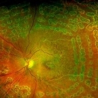
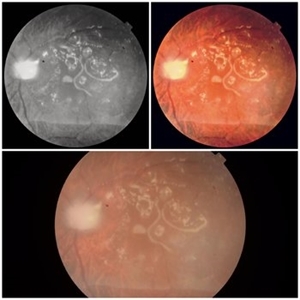
Endolaser in Status-Post Vitrectomy.
Photographer: Optom Komal Jangam, National Institute of Ophthalmology, Pune, India.
Imaging device: OPTOS DAYTONA
Condition/keywords: endolaser, pars plana vitrectomy (PPV), vitrectomy
-
 Retinal Detachment Following Scleral Buckling, Retinectomy, Laser, and Oil
Jan 31 2022 by Ahmad B. Tarabishy, MD
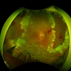
Ultra wide-field fundus photograph of a 55-year-old gentleman who is 4 days after surgery with scleral buckling, pars plana vitrectomy, perfluoron tamponade, membrane peeling, direct fluid-PFO-oil exchange, nasal and temporal retinectomies, and endolaser photocoagulation. Visual acuity was 20/150 under oil.
Photographer: Megan McLandsborough, Lakeland Eye Clinic
Imaging device: Optos California UWF Camera
Condition/keywords: endolaser, Membrane Peel, PPV, proliferative retinopathy, proliferative vitreoretinopathy (PVR), Retinal Detachment, retinal detachment with retinal defect, scleral buckle, submacular perfluorocarbon liquid (PFO)
-
Surgery for optic nerve pit
Oct 24 2022 by Manish Nagpal, MD, FRCS (UK), FASRS
This video showcases the steps of surgery for optic nerve pit associated with sensory fluid under fovea, fovea sparing ilm peel is carried out followed by endolaser to the temporal edges of the disc margins followed by air fluid exchange and gas.
Photographer: Manish Nagpal
Condition/keywords: endolaser, ONP, optic nerve pit, video, vitrectomy
-
Surgery for retinal detachment with multiple tears
Oct 24 2022 by Manish Nagpal, MD, FRCS (UK), FASRS
This video shows the steps of repairing a retinal detachment with multiple tears. vitreous is removed followed by air fluid exchange and then endo drainage to flatten the retina and followed by endolaser.
Photographer: Manish Nagpal
Condition/keywords: endodrainage, endolaser, RD, tears, video, vitrectomy
-
Vitrectomy for bullous retinal detachment with superior tear
Jan 2 2023 by Manish Nagpal, MD, FRCS (UK), FASRS
Vitrectomy for bullous Retinal detachment with superior tear| In this case vitrectomy is being done for a retinal detachment with superior tear. Once the vitreous is removed, air fluid exchange is carried out. Perfluorocarbon heavy liquid is injected to flatten the posterior pole and push the fluid to the periphery for endo drainage. This is followed by endo drainage from the superior break. Once the retina flattened endolaser was carried out.
Condition/keywords: air fluid exchange, bullous retinal detachment, endo drainage, endolaser, holes, Prophylaxis, RD, reattachment of retinal detachment, tear, video, vitrectomy
-
Vitrectomy for Myopic Retinal Detachment with multiple tears
Jan 2 2023 by Manish Nagpal, MD, FRCS (UK), FASRS
Vitrectomy for Myopic Retinal detachment with multiple tears and lattice degenerations | Vitrectomy is carried out and triamcinolone staining is used to stain the hyaloid attachment. The hyaloid attachment is extremely adherent. With high vacuum the cutter engages the stained hyaloid and gradually peels it off the mobile retina. After this Perfluorocarbon heavy liquid is injected to flatten the posterior pole and push the fluid to the periphery till the edge of the tear. This is followed by endo drainage from the tear. Once the retina flattened endolaser was carried out.
Condition/keywords: air fluid exchange, endo drainage, endolaser, holes, lattice degeneration, myopia, myopic retinal retachment, RD, reattachment of retinal detachment, tear, video, vitrectomy
-
Vitrectomy for Myopic Retinal Detachment with multiple tears status post prophylaxis
Jan 3 2023 by Manish Nagpal, MD, FRCS (UK), FASRS
Vitrectomy for Myopic Retinal detachment with multiple tears and lattice degenerations status post prophylaxis laser done | Vitrectomy is carried out. Once the vitreous is removed the retina is freely mobile. After this Perfluorocarbon heavy liquid is injected to flatten the posterior pole and push the fluid to the periphery till the edges of the tear. This is followed by endo drainage from infero nasal tear. Scars of laser marks are seen around it. Once the retina flattened endolaser is carried out.
Condition/keywords: air fluid exchange, endo drainage, endolaser, holes, lattice degeneration, myopia, myopic retinal detachment, prophylaxis, RD, reattachment of retinal detachment, tear, video, vitrectomy
-
Vitrectomy for PDR for TRD and Subhyaloid Haemorrhage
Jan 4 2023 by Manish Nagpal, MD, FRCS (UK), FASRS
Vitrectomy for PDR and TRD and subhyaloid haemorrhage using Cutter based dissection along with the use of a forceps. The subhyaloid haemorrhage is cleared first using aspiration of the cutter after making a opening in the hyaloid. A 25 gauge bevelled cutter is used to dissect all the epiretinal proliferations and tractional components. The ports of these cutters can reach very close to the retinal surface and cut flush without causing any iatrogenic damage to the retinal surface. Forceps is also used to gently peel off a adherent proliferation. Bleeders are stopped raising pressure and applying diathermy. Once the retina is flattened endolaser is done 360 degree to achieve long term regression.
Condition/keywords: diabetic retinopathy, diathermy, endolaser, forceps, peeling, TRD, video, vitrectomy
-
Vitrectomy for PDR in TRD with Vit haemorrhage
Jan 5 2023 by Manish Nagpal, MD, FRCS (UK), FASRS
Vitrectomy for PDR and TRD and Vitreous haemorrhage using Cutter based dissection. The Vitreous haemorrhage is cleared first. A 25 gauge bevelled cutter is used to dissect all the epiretinal proliferations and tractional components. The ports of these cutters can reach very close to the retinal surface and cut flush without causing any iatrogenic damage to the retinal surface. Forceps is also used to gently peel off a adherent proliferation. Bleeders are stopped raising pressure and applying diathermy. Air fluid exchange is done and viscous subretinal fluid drained from a hole adjacent to diathermy superiorly. Once the retina is flattened endolaser is done 360 degree to achieve long term regression.
Condition/keywords: diabetic retinopathy, diathermy, endolaser, forceps, PDR, peeling, TRD, video, vitrectomy
-
Vitrectomy for retinal detachment with superior holes
Jan 2 2023 by Manish Nagpal, MD, FRCS (UK), FASRS
Vitrectomy for Retinal detachment with multiple holes superiorly | In this case vitrectomy is being done for a retinal detachment with superior holes. Once the vitreous is removed, air fluid exchange is carried out. This is followed by endo drainage from the superior break. Once the retina flattened endolaser was carried out.
Condition/keywords: air fluid exchange, bullous retinal detachment, endo drainage, endolaser, holes, Prophylaxis, RD, reattachment of retinal detachment, video, vitrectomy
-
Vitrectomy for Subhyaloid blood over macula in Diabetic retinopathy
Nov 29 2022 by Manish Nagpal, MD, FRCS (UK), FASRS
Subhyaloid blood over macula in diabetic retinopathy| This is a case of non resolving subhyaloid haemorrhage over macula in a case of diabetic retinopathy.. Vitrectomy is carried out and then using the cutter a opening is made in the hyaloid to give a oulet to the blood. Typically the blood in the subhyaloid plane does not clot and easily aspirates out|. After this endolaser PRP is carried out to achieve good regression of the retinopathy. Air fluid exchange is carried out.
Photographer: Manish Nagpal
Condition/keywords: diabetic macular oedema, diabetic retinopathy, endolaser, PRP, SHH, subhyaloid blood, subhyaloid haemorrhage, video, vitrectomy
-
Vitrectomy TRD in Proliferative diabetic retinopathy
Jan 2 2023 by Manish Nagpal, MD, FRCS (UK), FASRS
Vitrectomy for PDR and TRD using Cutter based dissection| This is a case of subhyaloid hemorrhage and Tractional retinal detachment in a diabetic patient. The subhyaloid hemorrhage is aspirated using the cutter . 25 gauge bevelled cutter is used to dissect all the epiretinal proliferations and tractional components. The ports of these cutters can reach very close to the retinal surface and cut flush without causing any iatrogenic damage to the retinal surface. Forceps are used to peel adherent membranes Bleeders are stopped raising pressure and applying diathermy. Once the retina is flattened endolaser is done 360 degree to achieve long term regression.
Condition/keywords: cutter, diabetic retinopathy, endolaser, forceps, PDR, peeling, proliferative diabetic retinopathy (PDR), PRP, tractional retinal detachment, TRD, video, vitrectomy
-
 24 Hours Post Scleral Wound Closure+ Scleral Buckle+25 g Vitrectomy+Silicon Oil
Jan 23 2015 by Carlos Quezada-Ruiz, MD, FASRS
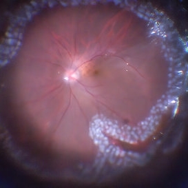
24 hours post op fundus photograph of a 43-year-old man who had perforating injury to the right eye with a small piece of plastic while he was hammering. OD LP, subconjunctival hemorrhage, clear cornea, hyphema, irido and ciclodyalisis as well as a luxated lens with traumatic cataract and a dense vitreous hemorrhage. B-US showed rhegmatogenous retinal detachment with a tear and a big inferior hemorrhagic choroidal detachment. 360 peritomy revealed 2-entry scleral wounds were found in zone II (M V and M VI) and closure was performed. 25 G PPV was performed with the infusion canal placed in the AC through the limbus. Lensectomy and removal of a dense recent vitreous hemorrhage revealed a white detached retina with an exit wound through the temporal inferior segment of the optic nerve with a nasal GRT and sub retinal hemorrhage as well as temporal inferior choroidal, PVD was induced and PFOs helped stabilizing the retina while vitrectomy and sub-retinal hemorrhage was removed through the GRT. Fluid air exchange was made and 360 endolaser over the buckle indentation was done and silicon oil was used as endotamponade. This picture was taken 24 hrs after the surgery.
Photographer: Lilibeth Rodriguez, Instituto de la Visión. Torreon, Mexico.
Condition/keywords: central retinal artery occlusion (CRAO), giant retinal tear, trauma
-
 360 Endolaser Barrage
Feb 2 2022 by Manish Nagpal, MD, FRCS (UK), FASRS
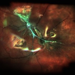
Intraoperative photo of a 360 laser barrage done for a case of retinal detachment with a large superior tear.
Photographer: Manish Nagpal, Retina Foundation, Ahmedabad, India
Imaging device: Sony PMW -10 MD surgical camera
Condition/keywords: laser, laser photocoagulation, tear
-
 COATS Disease Optical Coherence Tomography Posttreatment
Sep 2 2022 by FLOR ANGELICA JACOME GUTIERREZ
OCT of a 14 yo male with coats disease stage 2a and epiretinal membrane after 2 months of pars plana vitrectomy with ERM peeling, endolaser, intravitreal aflibercept and SF6 18% with VA 20/30. 3 months previously indirect laser, intravítreal aflibercept and subtenon triamcinolone where offered.
Photographer: Dr. Guillermo Salcedo Villanueva
Imaging device: Optovue
Condition/keywords: Coats' disease
-
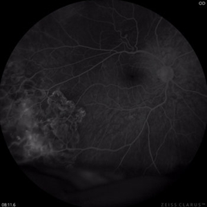 Coats Disease FAG Posttreatment
Sep 2 2022 by FLOR ANGELICA JACOME GUTIERREZ
FAG of a 14 yo male with coats disease stage 2a and epiretinal membrane after 2 months of pars plana vitrectomy with ERM peeling, endolaser, intravitreal aflibercept and SF6 18% with VA 20/30. 3 months previously indirect laser, intravítreal aflibercept and subtenon triamcinolone where offered.
Photographer: Dr. Guillermo Salcedo Villanueva
Imaging device: Zeiss CLARUS 700 (FA)
Condition/keywords: Coats' disease
-
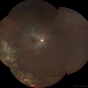 Coats disease Posttreatment
Sep 2 2022 by FLOR ANGELICA JACOME GUTIERREZ
Fundus image of a 14 yo male with coats disease stage 2a and epiretinal membrane after 2 months of pars plana vitrectomy with ERM peeling, endolaser, intravitreal aflibercept and SF6 18% with VA 20/30. 3 months previously indirect laser, intravitreal aflibercept and subtenon triamcinolone where offered.
Photographer: Dr. Guillermo Salcedo Villanueva
Imaging device: Zeiss Clarus 700
Condition/keywords: Coats' disease
-
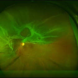 Combined RD in PDR patient
Sep 8 2017 by Eitae Kim, MD
52-year-old woman with diabetes visited visual disturbance. Fundus exam shows superior tractional-rhegmatogenous retinal detachment with proliferative vitreoretinopathy. I performed PPV, membranectomy, endolaser and silicone oil injection.
Photographer: Eitae Kim, BOIM retina center, Pureun eye hospital
Condition/keywords: ultra-wide field imaging
-
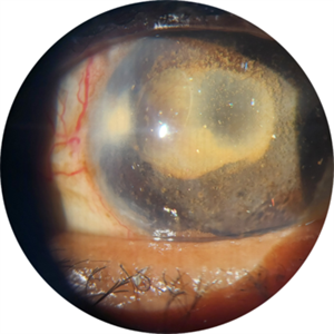 Crystallized silicone oil particles in the anterior chamber
Oct 26 2023 by Anmol Naik, MS, DNB, FMRF, FICO, MNAMS
Anterior segment image of a 67-year-old Indian woman who had proliferative diabetic retinopathy with traction retinal detachment with neovascular glaucoma. Patient underwent vitrectomy with membrane peeling with endolaser followed by silicone oil injection 1 year back. Patient was lost to follow up and presented a year later with this picture. She had crystallized silicone oil particles in the anterior chamber rendering a polychromatic lustre like appearance; a unique and rare finding.
Photographer: Anmol Naik
Condition/keywords: Polychromatic lustre in Anterior Chamber
-
 Emulsified Silicone Oil in Macular Hole
Jun 7 2024 by Vaidehi Jethwa
Fundus photograph of 72 year old man was having a/h/o Left Eye trauma by a cow horn, 8 years Ago, and developed Left Eye total Retinal detachment and was operated for Left Eye vitrectomy with FAX, SOI, Endolaser on 11 /04/2015 and was advised Left Eye silicone Oil removal.
Photographer: Dr. Vaidehi Jethwa, M and J institute of Ophthalmology, Ahmedabad, Gujarat.
Imaging device: Zeiss Visucam lite
Condition/keywords: macular hole, silicon oil emulsification in vitreous cavity
-
 Encircling Buckle Effect
Jul 7 2015 by Hamid Ahmadieh, MD
Late FA image of the right eye of a 30-year-old man who underwent pars plana vitrectomy , endolaser photocoagulation and an encircling band placement a couple of years before following a penetrating trauma at the vitreous base area at the 7 o'clock meridian.
Photographer: Nayereh Hadipour, Negah Eye Center,Tehran, Iran
Imaging device: Specteralis
Condition/keywords: pars plana vitrectomy (PPV)
-
 Exudative Macular Detachment After Intensive Laser Photocoagulation
Mar 12 2016 by Sjakon G Tahija, MD
Fundus photograph of 44-year-old man with exudative detachment of the macula after vitrectomy and ILM peeling for proliferative diabetic retinopathy combined with intensive endolaser photocagulation.
Photographer: Avris Siahaan, Klinik Mata Nusantara
Condition/keywords: exudative detachment, pan-retinal photocoagulation (PRP)
-
 Fibrosis and Traction Following Traction Retinal Detachment Repair
Oct 13 2020 by Sophia El Hamichi, MD
A 29-year-old female with a history of diabetes mellitus type 1, presented with proliferative diabetic retinopathy OU and tractional retinal detachment OD. The patient underwent retinal detachment repair with pars plana vitrectomy, endolaser and silicone oil placement. After one month of her surgery, the patient presented with retinal fibrosis and tractions depicted in the image.
Photographer: Belinda Rodriguez, Murray Ocular Oncology and Retina, Miami
Condition/keywords: pars plana vitrectomy (PPV), post-op, proliferative diabetic retinopathy (PDR), proliferative vitreoretinopathy (PVR), tractional retinal detachment
-
 Fibrosis and Traction Following Traction Retinal Detachment Repair
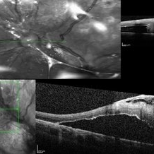
Oct 13 2020 by Sophia El Hamichi, MD
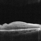
A 29-year-old female with a history of diabetes mellitus type 1, presented with proliferative diabetic retinopathy OU and tractional retinal detachment OD. The patient underwent retinal detachment repair with pars plana vitrectomy, endolaser and silicone oil placement. After one month of her surgery, the patient presented with retinal fibrosis and traction. The image on the top shows the OCT of the fibrosis post op, that was not present in the pre op (OCT image on the bottom).
Photographer: Belinda Rodriguez, Murray Ocular Oncology and Retina, Miami
Condition/keywords: optical coherence tomography (OCT), pars plana vitrectomy (PPV), proliferative diabetic retinopathy (PDR), proliferative vitreoretinopathy (PVR), tractional retinal detachment
-
Iatrogenic Macular Hole and Subretinal Migration of PFCL
Feb 7 2023 by Aditya S Kelkar, MS, FRCS, FASRS,FRCOphth
The video demonstrates a surgical scenario where the fovea gives away by the force imparted by the jet of an injecting PFCL (Perfluorocarbon heavy Liquid) and the PFCL migrates subfoveally. Intraoperative OCT confirms the presence of a macular hole. The situation is managed by ILM peeling and mobilizing subfoveal PFCL peripherally by injecting another bubble of PFCL over the posterior pole. A peripheral drainage retinotomy is then created to aspirate the subretinal PFCL followed by fluid-air exchange, PFCL-air exchange, and endolaser around the retinotomy. Post-operative OCT at 3 weeks’ follow-up shows a sealed macular hole.
Condition/keywords: Iatrogenic macular hole, Intraoperative complications, Subretinal PFCL
 Loading…