Search results (870 results)
-
 Active CNVM
Jul 12 2023 by Harsh Vardhan Singh, MS
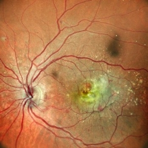
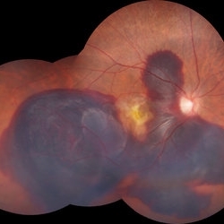
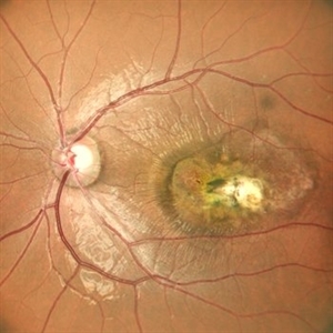
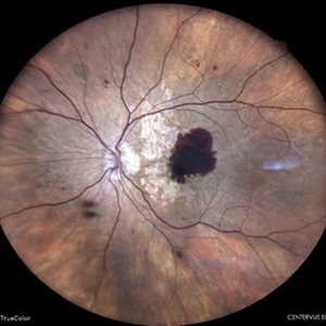
55-year male with left eye sub-retinal hemorrhage due to Active CNVM, Colour fundus photograph of left eye subretinal hemorrhage due to Active CNVM
Photographer: Harsh Vardhan Singh
Condition/keywords: choroidal neovascular membrane (CNVM), CNVM, subretinal hemorrhage
-
 Active CNVM
Jul 12 2023 by Harsh Vardhan Singh, MS
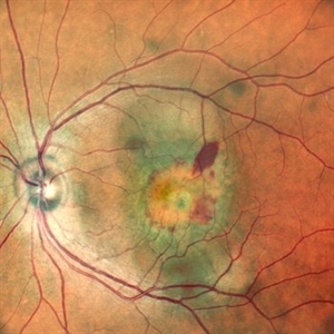
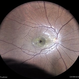
55-year male with left eye sub-retinal hemorrhage due to Active CNVM, Colour fundus photograph of left eye subretinal hemorrhage due to Active CNVM; Red-free image of left eye sub-retinal hemorrhage due to Active CNVM
Photographer: Harsh Vardhan Singh
Condition/keywords: choroidal neovascular membrane (CNVM), CNVM, subretinal hemorrhage
-
 Choroidal Neovascular Membrane
Oct 14 2022 by Vaidehi Sathaye
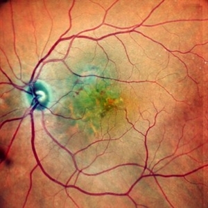
Fundus photograph of LE of a 68 year male patient with a choroidal neovascular membrane
Photographer: Dr. Vaidehi Sathaye
Imaging device: Mirante
Condition/keywords: CNVM
-
 CNVM
CNVM
Dec 14 2021 by VIRAL SHAH
23 year-old female complained of dim vision in right eye for 1 week. She is not myopic.
Photographer: VIRAL SHAH, NETRALOK RETINA CLINIC, AHMEDABAD
Condition/keywords: choroidal neovascular membrane (CNVM)
-
 CNVM
CNVM
Jul 16 2021 by ASRS Staff
OCTA of a 50-year-old male with CNVM.
Imaging device: Nidek Mirante
Condition/keywords: choroidal neovascular membrane (CNVM)
-
 CNVM
CNVM
Aug 26 2021 by ASRS Staff
Fudus Photograph of a 50-year-old male with CNVM.
Imaging device: Nidek Mirante
Condition/keywords: choroidal neovascular membrane (CNVM)
-
 CNVM
CNVM
Mar 17 2015 by Jason Griffith
Photograph of a 45 year old male with CNVM.
Photographer: Jason Griffith, Tennessee Retina, Nashville, TN
Imaging device: Topcon TRC-50EX
Condition/keywords: choroidal neovascular membrane (CNVM)
-
 CNVM: Post-CSCR
Apr 4 2024 by T. P . VIGNESH, MBBS,MS
Fundus photograph of an 45-year-old man with CNVM post CSCR .
Photographer: Bharathi S
Imaging device: Zeiss Clarus
Condition/keywords: chronic central serous chorioretinopathy (CSCR), CNVM
-
 Hereditary Macular Dystrophy with CNVM
Apr 4 2025 by Tejaswita Verma
Fundus photo of a 46 y/o male with 6/36 vision, Stargardt's disease with CNVM
Photographer: DR. TEJASWITA VERMA
Imaging device: MIRANTE
Condition/keywords: CNVM, hereditary macular dystrophy, HMD
-
 Idiopathic Choroidal Neovascularization
Mar 2 2023 by Corey Grant
Optical coherence tomography and ultra-wide field fundus photograph of a 51 year old male with idiopathic choroidal neovascularization affecting his right eye. The patient had no symptoms at the time of the appointment and his vision was Dcc20/20-2 OU. The physcian stated that there wasn't active exudation on the exam or ocular imaging and based on the clinical findings, he has recommended we defer any treatments.
Photographer: Corey Grant
Imaging device: Heidelberg Spectralis, OPTOS California
Condition/keywords: choroidal neovascularization (CNV), CNVM, fundus photograph, OCT, optical coherence tomography (OCT), Optos, Right Eye, ultra-wide field imaging
-
 Macular Mount Everest
Aug 8 2025 by Anand Temkar
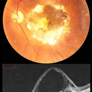
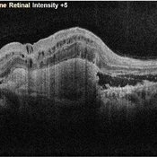
A 75 yrs old male came with the chief complains of DOV in LE since past 20 yrs. His BCVA in RE was 6/9 and in LE, it was CF 1 meter. His IOP was 13 mm of Hg in RE and 15 mm of Hg in LE. Patient is a k/c/o DM type 2 since past 20 yrs and is on regular medication. Patient is a k/c/o solitary kidney. Patient gives h/o ( LE ) Intravitreal injection Avastin 3 times 13 yrs ago i/c/o CNVM. In the LE color photo we can see the scarred CNVM along with altered foveal contour. LE OCT also shows cystic spaces with large elevation and scarring.
Photographer: Dr.Anand Temkar- Vasan Eye Hospital, Tiruchirapalli
Condition/keywords: CNVM, macular edema, scarred cnvm
-
 Massive Subretinal Hemorrhage
Aug 12 2022 by Sashwanthi Mohan
A 70 year old woman with a large area of subretinal hemorrhage secondary topolychoroidal vasculopathy
Photographer: Leo John
Condition/keywords: CNVM, subretinal hemorrhage
-
 Multi-modal Imaging of Type - 1 CNVM
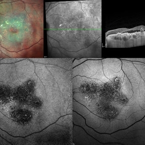
May 30 2025 by Shivankar Sen, MS, FVRS
Multimodal Imaging of a case of Polypoidal Choroidal Vasculopathy Multicolor Reflectance showing multiple green-hyper-fringent lesions in the macular region (Up Left) Infra-red Autofluorescence and Blue Autofluorescence showing hypo-autofluorescent areas correspondingly revealing the exact extent of the sub-RPE Lesion (Down left and right respectively) Optical Coherence Tomography - Enhanced Depth Imaging showing Thumb-shaped Pigment Epithelial Detachment with presence of Sub-retinal fluid and Hyper-reflective foci (Top Right)
Photographer: Dr. Shivankar Sen
Imaging device: Heidelberg Spectralis HRA+OCT
Condition/keywords: Blue autofluroscence, CNVM, multicolor, near infrared autofluorescence (NIRAF), PCV, reflectance
-
 OCT of a Choroidal Neovascular Membrane
Oct 14 2022 by Vaidehi Sathaye
OCT image of a 68 year male patient with a choroidal neovascular membrane in his LE
Photographer: Dr. Vaidehi Sathaye
Imaging device: Mirante
Condition/keywords: CNVM, OCT
-
 Pseudoxanthoma Elasticum
Aug 7 2025 by Alind Murkhe
Fundus photograph of a 42 year-old male with pseudoxanthoma elasticum showing Angiod streak, scarred CNVM, Comet tails lesion.
Photographer: Dr Alind Murkhe, Nandadeep Eye Hospital, Sangli, Maharashtra, India
Condition/keywords: Angiod streaks in Pseudoxanthoma elasticum, CNVM
-
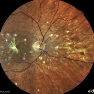 Punctate inner choroidopathy (PIC) with CNVM
Oct 18 2023 by Heitor Nogueira
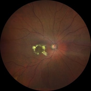
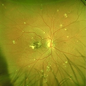
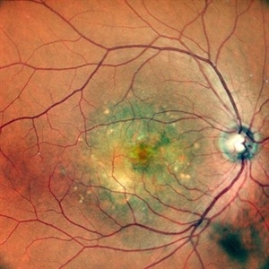
Fundus photograph of a 29-year-old woman with a 2-week history of low visual acuity associated with central scotoma. Ophthalmological history of axial myopia of -4,00D. She denied personal and family history.
Photographer: Heitor Nogueira, Instituto Penido Burnier, Campinas-SP, Brazil.
Imaging device: Eidon True Color
Condition/keywords: CNVM, multifocal chorioretinitis (MCP), punctate inner choroidopathy (PIC)
-
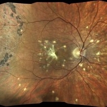 Punctate inner choroidopathy (PIC) with CNVM.
Oct 18 2023 by Heitor Nogueira
Fundus photograph of a 29-year-old woman with a 2-week history of low visual acuity associated with central scotoma. Ophthalmological history of axial myopia of -4,00D. She denied personal and family history.
Photographer: Heitor Nogueira, Instituto Penido Burnier, Campinas-SP, Brazil.
Imaging device: Eidon True Color
Condition/keywords: choroidal neovascularization (CNV), CNVM, multifocal chorioretinitis (MCP), punctate inner choroidopathy (PIC)
-
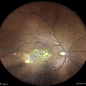 Scarred Choroidal Neovacular Membrane
May 7 2024 by Akansha Sharma
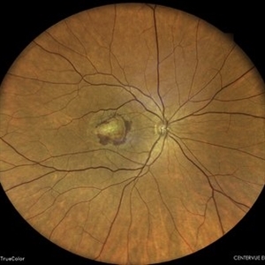
Color fundus photograph of a 75 year old male with scarred choroidal neovascular membrane.
Photographer: Dr. Akansha Sharma, Bharati Eye Hospital
Condition/keywords: choroidal neovascular membrane (CNVM), CNVM
-
 Scarred Choroidal Neovacular Membrane With Large Inferior Horse Shoe Tear
Feb 7 2024 by Akansha Sharma
Color fundus photograph of a 73 year old male with scarred choroidal neovascular membrane with large horse shoe tear inferiorly.
Photographer: Dr. Akansha Sharma, Bharati Eye Hospital
Condition/keywords: atrophic scar, choroidal neovascular membrane (CNVM), CNVM
-
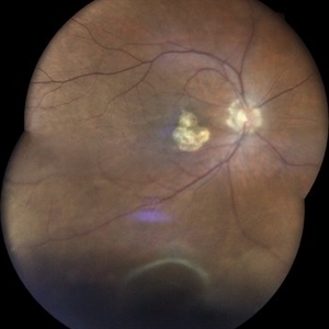
 SCARRED CHOROIDAL NEOVASCULAR MEMBRANE
Oct 15 2022 by Akansha Sharma
FUNDUS PHOTOGRAPH OF AN 18 YEAR OLD MALE WITH A SCARRED CHOROIDAL NEOVASCULAR MEMBRANE
Photographer: Dr. Akansha Sharma-Retina Foundation, Ahmedabad
Condition/keywords: choroidal neovascularization (CNV), CNVM
-
 SUB-RETINAL NEOVASCULAR MEMBRANE
Nov 21 2022 by Akansha Sharma
COLOUR FUNDUS PHOTO OF A 79 YEAR OLD MALE PATIENT WITH SUBRETINAL NEOVASCULAR MEMBRANE
Photographer: Dr. Akansha Sharma-Retina Foundation, Ahmedabad
Condition/keywords: choroidal neovascular membrane (CNVM), CNVM, subretinal neovascularization (SRNV)
-
 SUB-RETINAL NEOVASCULAR MEMBRANE
Nov 21 2022 by Akansha Sharma
COLOUR FUNDUS PHOTOGRAPH OF A 57 YEAR OLD MALE WITH SUBRETINAL NEOVASCULAR MEMBRANE
Photographer: Dr. Akansha Sharma-Retina Foundation, Ahmedabad
Condition/keywords: choroidal neovascularization (CNV), CNVM, subretinal neovascularization (SRNV)
-
 Subretinal Neovascular Membrane
May 8 2023 by Akansha Sharma
Colour fundus photograph of a 60 year old female with subretinal blood suggestive of subretinal neovascular membrane
Photographer: Dr. Urmil Shah, Dr. Denish Patel, Dr. Akansha Sharma, Bharati Eye Clinic, Ahmedabad, Gujarat
Condition/keywords: CNVM, SRNVM, subretinal blood
-
 Subretinal Neovascular Membrane
May 8 2023 by Akansha Sharma
Colour fundus photograph of a 81 year old female with subretinal neovascular memebrane
Photographer: Dr. Urmil Shah, Dr. Denish Patel, Dr. Akansha Sharma, Bharati Eye Clinic, Ahmedabad, Gujarat
Condition/keywords: CNVM, subretinal neovascularization (SRNV)
-
 Subretinal Neovascular Membrane
May 10 2023 by Akansha Sharma
COLOUR FUNDUS PHOTOGRAPH OF A 26 YEAR OLD MALE WITH SUBRETINAL BLEED WITH SUBRETINAL HYPERREFLECTIVE MATERIAL SUGGESTIVE OF SUBRETINAL NEOVASCULAR MEMBRANE
Photographer: Dr. Denish Patel, Dr. Akansha Sharma, Dr. Urmil Shah, Bharati Eye Hospital
Condition/keywords: CNVM, SRNVM, subretinal hemorrhage
 Loading…