Search results (870 results)
-
 RPE - Rest In Peace (RIP)
Dec 17 2025 by SHRADDHA RAJ SHRIVASTAVA
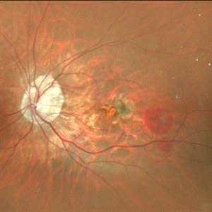
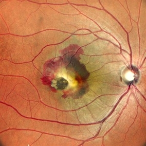
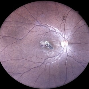
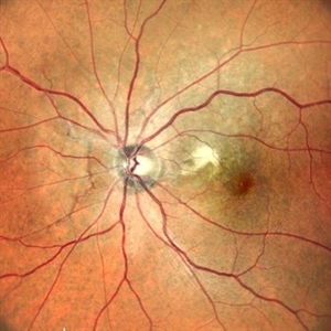
Right eye pseudocolor fundus photo of a 50 year old patient, known case of bilateral familial dominant drusens with right eye CNVM, having undergone multiple intravitreal anti-VEGF injections. Image shows a CDR of 0.3:1, with numerous drusens at macula with residual lipid exudation from CNVM, along the infero-temporal arcade. Temporal to the fovea, we can see a vertical hyperpigmented line corresponding to retracted and redundant torn Retinal pigment epithelium, leaving behind a well circumscribed area of depigmented fundus with bare Bruch's membrane underlying the retina, findings suggestive of an RPE tear post multiple intravitreal injections.
Photographer: Dr. Shraddha Raj Shrivastava
Imaging device: Nidek Mirante SLO/OCT (Confocal scanning/Spectral domain OCT)
Condition/keywords: choroidal neovascular membrane (CNVM), Doyne's Honeycomb, FAMILIAL DOMINANT DRUSEN, lipid exudation, retinal pigment epithelium, RPE Rip
-
 RPE - Rest In Peace (RIP)
Dec 17 2025 by SHRADDHA RAJ SHRIVASTAVA
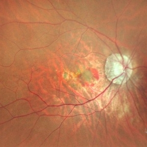
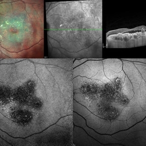
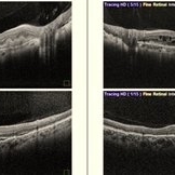
Multimodal imaging of Right eye of a 50 year old patient, known case of bilateral familial dominant drusens with right eye CNVM, having undergone multiple intravitreal anti-VEGF injections. The various imaging modalities highlight the presence of an extrafoveal RPE tear - post multiple intravitreal injections.
Photographer: Dr. Shraddha Raj Shrivastava
Imaging device: Nidek Mirante SLO/OCT (Confocal scanning/Spectral domain OCT)
Condition/keywords: FAMILIAL DOMINANT DRUSEN, multimodal imaging, retinal pigment epithelium, RPE-Rip
-
 RPE - Rest In Peace (RIP)
Dec 17 2025 by SHRADDHA RAJ SHRIVASTAVA
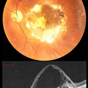
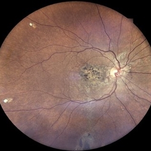
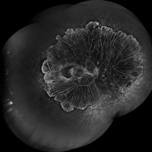
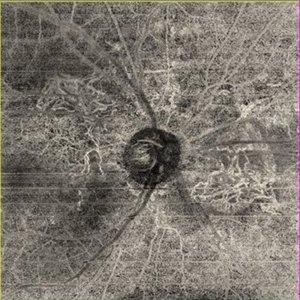
Right eye RETRO mode fundus image of a 50 year old patient, known case of bilateral familial dominant drusens with right eye CNVM, having undergone multiple intravitreal anti-VEGF injections. Among other findings, this novel imaging technique highlights the presence of an extrafoveal RPE tear - post multiple intravitreal injections.
Photographer: Dr. Shraddha Raj Shrivastava
Imaging device: Nidek Mirante SLO/OCT (Confocal scanning/Spectral domain OCT)
Condition/keywords: choroidal neovascular membrane (CNVM), Doyne's Honeycomb, FAMILIAL DOMINANT DRUSEN, lipid exudation, retinal pigment epithelium, RPE Rip
-
 RPE - Rest In Peace (RIP)
Dec 17 2025 by SHRADDHA RAJ SHRIVASTAVA
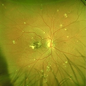
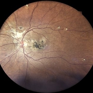
Right eye G-FAF photo of a 50 year old patient, known case of bilateral familial dominant drusens with right eye CNVM, having undergone multiple intravitreal anti-VEGF injections. Fundus autofluorescence better highlights the area of RPE tear in right eye (temporal to fovea), which shows hypoautofluorescence due to lack of RPE and its pigments which accounts for autofluorescence signal. Whereas the linear hyperautofluorescence, represents the torn bunched up retinal pigment epithelium.
Photographer: Dr. Shraddha Raj Shrivastava
Imaging device: Nidek Mirante SLO/OCT (Confocal scanning/Spectral domain OCT)
Condition/keywords: choroidal neovascular membrane (CNVM), Doyne's Honeycomb, FAMILIAL DOMINANT DRUSEN, lipid exudation, retinal pigment epithelium, RPE Rip
-
 Cracking the Angioid Streaks Mystery
Nov 26 2025 by SHRADDHA RAJ SHRIVASTAVA
Left eye pseudocolor fundus photo showing hyperpigmented irregular lines emanating from the disc in a radiating fashion. Surrounding the angioid streaks and at the posterior pole, we can see numerous dot-like hypopigmented deposits along with a grayish-green membrane with exudates at the macula. The image is suggestive of Angioid Streaks with CNVM.
Photographer: Dr. Shraddha Raj Shrivastava
Imaging device: Nidek Mirante SLO/OCT (Confocal scanning/Spectral domain OCT)
Condition/keywords: Angiod streaks in Pseudoxanthoma elasticum, Angioid Streaks, Bruch's membrane, choroidal neovascular membrane (CNVM), color fundus photograph, pseudoxanthoma elasticum (PXE)
-
 Cracking the Angioid Streaks Mystery
Nov 26 2025 by SHRADDHA RAJ SHRIVASTAVA
G-FAF image of left eye better reveals more extensive hypoautofluorescent streaks than might be apparent on standard fundus photo. Characteristic “Para-streak phenomenon” of focal hyperautofluorescent spots are seen along the margins of the dark angioid streaks, corresponding to the areas of pigment clumping seen clinically.
Photographer: Dr. Shraddha Raj Shrivastava
Imaging device: Nidek Mirante SLO/OCT (Confocal scanning/Spectral domain OCT)
Condition/keywords: Angiod streaks in Pseudoxanthoma elasticum, Angioid Streaks, choroidal neovascular membrane (CNVM), fundus autofluorescence (FAF), pseudoxanthoma elasticum (PXE)
-
 Cracking the Angioid Streaks Mystery: Multimodal Mayhem
Nov 26 2025 by SHRADDHA RAJ SHRIVASTAVA
Multimodal imaging of right eye fundus showing Angioid Streaks with scarred CNVM. Color fundus photo shows hyperpigmented irregular lines emanating from the disc in a radiating fashion. Surrounding the angioid streaks and at the posterior pole, we can see numerous dot-like hypopigmented deposits and a disciform scar at macula. G-FAF images better reveal more extensive hypoautofluorescent streaks than are apparent on standard fundus photo. Characteristic “Para-streak phenomenon” of focal hyperautofluorescent spots are seen along the margins of the dark angioid streaks, corresponding to the areas of pigment clumping seen clinically. The para-streak pigment clumps are better delineated on the novel Retro-imaging method, appearing as raised bumps surrounding the angioid streaks.
Photographer: Dr. Shraddha Raj Shrivastava
Imaging device: Nidek Mirante SLO/OCT (Confocal scanning/Spectral domain OCT)
Condition/keywords: Angioid Streaks, Bruch's membrane, disciform scar, fundus autofluorescence (FAF), multimodal imaging, retro mode
-
 Idiopathic CNVM
Sep 30 2025 by T. P . VIGNESH, MBBS,MS
SD-OCT of the left eye of 45 year old man with idiopathic CNVM.
Photographer: Sivanath
Imaging device: Heidelberg Spectralis
Condition/keywords: Idiopathic CNVM
-
 Subretinal Neovascular Membrane
Aug 15 2025 by Akansha Sharma
Color fundus photograph of a 40 year old male with subretinal neovascular membrane.
Photographer: DR. AKANSHA SHARMA
Condition/keywords: choroidal neovascular membrane (CNVM), CNVM, SRNVM, subretinal neovascularization (SRNV), wet age-related macular degeneration (wet AMD)
-
 Subretinal Neovascular Membrane
Aug 15 2025 by Akansha Sharma
Color fundus photograph of a 40 year old male with subretinal neovascular membrane.
Photographer: DR. AKANSHA SHARMA
Condition/keywords: choroidal neovascular membrane (CNVM), CNVM, Myopic CNVM, SRNVM, subretinal neovascularization (SRNV), Wet age related macular degeneration
-
 Macular Mount Everest
Aug 8 2025 by Anand Temkar
A 75 yrs old male came with the chief complains of DOV in LE since past 20 yrs. His BCVA in RE was 6/9 and in LE, it was CF 1 meter. His IOP was 13 mm of Hg in RE and 15 mm of Hg in LE. Patient is a k/c/o DM type 2 since past 20 yrs and is on regular medication. Patient is a k/c/o solitary kidney. Patient gives h/o ( LE ) Intravitreal injection Avastin 3 times 13 yrs ago i/c/o CNVM. In the LE color photo we can see the scarred CNVM along with altered foveal contour. LE OCT also shows cystic spaces with large elevation and scarring.
Photographer: Dr.Anand Temkar- Vasan Eye Hospital, Tiruchirapalli
Condition/keywords: CNVM, macular edema, scarred cnvm
-
 Pseudoxanthoma Elasticum
Aug 7 2025 by Alind Murkhe
Fundus photograph of a 42 year-old male with pseudoxanthoma elasticum showing Angiod streak, scarred CNVM, Comet tails lesion.
Photographer: Dr Alind Murkhe, Nandadeep Eye Hospital, Sangli, Maharashtra, India
Condition/keywords: Angiod streaks in Pseudoxanthoma elasticum, CNVM
-
 When the Macula Decides to Bleed... Artistically (Case of Macular Scar with Subretinal Bleed)
Jun 2 2025 by rohan jain
A case of 42 years old male. Color photograph showing macular scar with subretinal bleed.
Photographer: Dr. ROHAN JAIN
Imaging device: mirante
Condition/keywords: CNVM, macular scar, scar, subretinal hemorrhage, subretinal blood
-
 Multi-modal Imaging of Type - 1 CNVM
May 30 2025 by Shivankar Sen, MS, FVRS
Multimodal Imaging of a case of Polypoidal Choroidal Vasculopathy Multicolor Reflectance showing multiple green-hyper-fringent lesions in the macular region (Up Left) Infra-red Autofluorescence and Blue Autofluorescence showing hypo-autofluorescent areas correspondingly revealing the exact extent of the sub-RPE Lesion (Down left and right respectively) Optical Coherence Tomography - Enhanced Depth Imaging showing Thumb-shaped Pigment Epithelial Detachment with presence of Sub-retinal fluid and Hyper-reflective foci (Top Right)
Photographer: Dr. Shivankar Sen
Imaging device: Heidelberg Spectralis HRA+OCT
Condition/keywords: Blue autofluroscence, CNVM, multicolor, near infrared autofluorescence (NIRAF), PCV, reflectance
-
 Hereditary Macular Dystrophy with CNVM
Apr 4 2025 by Tejaswita Verma
Fundus photo of a 46 y/o male with 6/36 vision, Stargardt's disease with CNVM
Photographer: DR. TEJASWITA VERMA
Imaging device: MIRANTE
Condition/keywords: CNVM, hereditary macular dystrophy, HMD
-
 Angioid Streaks
Mar 18 2025 by T. P . VIGNESH, MBBS,MS
Fundus photograph of a 42-year-old woman with Angioid Streaks and scarred CNVM.
Photographer: Sivanath
Imaging device: EIDON
Condition/keywords: Angioid Streaks
-
 Angioid Streaks
Mar 18 2025 by T. P . VIGNESH, MBBS,MS
Fundus photograph of a 42-year-old woman with Angioid Streaks and scarred CNVM.
Photographer: Sivanath
Imaging device: EIDON
Condition/keywords: Angioid Streaks
-
 Toxoplasma Macular Scar with CNVM
Mar 7 2025 by T. P . VIGNESH, MBBS,MS
Fundus photograph of the right eye of a 28-year-old woman with a macular toxoplasma scar and CNVM.
Photographer: Sivanath
Imaging device: EIDON
Condition/keywords: CNVM, ocular toxoplasmosis
-
 Myopic CNVM
Jan 31 2025 by Thirumalesh Mochi Basavaraj, MD
Widefield image of a 26 year-old male patient with pathologic myopia with history of central scotoma with a sub macular bleed.
Photographer: Puttaswamy N K
Imaging device: Optos Daytona
Condition/keywords: myopic choroidal neovascularization (CNV), Myopic CNVM, pathologic myopia
-
 Choroideremia
Jan 23 2025 by Prashant K Bawankule, M.S.
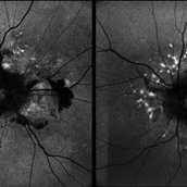
A young male of 25 years, with progressive loss of vision with suspected CNVM. FFA showed 360 degree shutdown with blocked fluorescence in macular region.
Photographer: Prashant Bawankule, Sarakshi Netralaya, Nagpur, Maharashtra , India
Imaging device: Mirante ( by Nidek)
Condition/keywords: Montage of FFA in a case of choroderemia
-
 Pseudoxanthoma Elasticum
Dec 3 2024 by Dr Bilal Mir
This is a fundus picture showing angiod streaks, CNVM, comet lesion.
Photographer: Dr Bilal Ahmed mir MS ophthalmology
Imaging device: Zeiss fundus camera
Condition/keywords: Angiod streaks in Pseudoxanthoma elasticum, Pseudoxanthoma elasticum
-
 Both Eyes OCT in Case of CNVM with Angioid Streaks
Nov 29 2024 by Anand Temkar
A 45 year old male came with chief complaint of blurring vision in right eyes since past 4 days. His vision is 6/12 in right eye and 6/9 in left eye. His vision was 14 mmHg in right eye and 16 mmHg in left eye. He was diagnosed with Angioid Streaks in both eyes about a year ago, then he developed choroidal neovascularization in his left eye 8 months ago, for which he received AntiVEGF injections x 3. Left eye is a stable eye now. Patient presented with right eye choroidal neovascularization in a case of Angioid Streaks on recent follow up. We have advised him right eye AntiVEGF injections x 3. In this image we can see the subretinal hyperreflective material in right eye and in left eye few cystic spaces are noted.
Photographer: Dr.Anand Temkar- Retina Foundation, Ahmedabad
Imaging device: Mirante
Condition/keywords: Angioid Streaks, choroidal neovascular membrane (CNVM)
-
 Both Eyes Fundus Autofluorescence in Case of CNVM with Angioid Streaks
Nov 29 2024 by Anand Temkar
A 45 year old male came with chief complaint of blurring vision in right eyes since past 4 days. His vision is 6/12 in right eye and 6/9 in left eye. His vision was 14 mmHg in right eye and 16 mmHg in left eye. He was diagnosed with Angioid Streaks in both eyes about a year ago, then he developed choroidal neovascularization in his left eye 8 months ago, for which he received AntiVEGF injections x 3. Left eye is a stable eye now. Patient presented with right eye choroidal neovascularization in a case of Angioid Streaks on recent follow up. We have advised him right eye AntiVEGF injections x 3. In this image we can see fundus hypoautofluorescence in right eye due to hemorrhages and angioid streaks and in left eye fundus hypoautofluorescence is noted due to angioid streaks.
Photographer: Dr.Anand Temkar- Retina Foundation, Ahmedabad
Imaging device: Mirante
Condition/keywords: Angioid Streaks, choroidal neovascular membrane (CNVM), fundus autofluorescence (FAF)
-
 RE OCTA (ORCC) in case of CNVM with Angioid Streaks
Nov 29 2024 by Anand Temkar
A 45 year old male came with chief complaint of blurring vision in right eyes since past 4 days. His vision is 6/12 in right eye and 6/9 in left eye. His vision was 14 mmHg in right eye and 16 mmHg in left eye. He was diagnosed with Angioid Streaks in both eyes about a year ago, then he developed choroidal neovascularization in his left eye 8 months ago, for which he received AntiVEGF injections x 3. Left eye is a stable eye now. Patient presented with right eye choroidal neovascularization in a case of Angioid Streaks on recent follow up. We have advised him right eye AntiVEGF injections x 3. In this image we can see the abnormal vessels at outer retina chorio-capillary ( ORCC ) junction in right eye.
Photographer: Dr.Anand Temkar- Retina Foundation, Ahmedabad
Imaging device: Mirante
Condition/keywords: Angioid Streaks, choroidal neovascular membrane (CNVM), OCT Angiography
-
 Left Eye Color Photo With Extrafoveal CNVM (Stable) in Case of Angioid Streaks
Nov 29 2024 by Anand Temkar
A 45 year old male came with chief complaint of blurring vision in right eyes since past 4 days. His vision is 6/12 in right eye and 6/9 in left eye. His vision was 14 mmHg in right eye and 16 mmHg in left eye. He was diagnosed with Angioid Streaks in both eyes about a year ago, then he developed choroidal neovascularization in his left eye 8 months ago, for which he received AntiVEGF injections x 3. Left eye is a stable eye now. Patient presented with right eye choroidal neovascularization in a case of Angioid Streaks on recent follow up. We have advised him right eye AntiVEGF injections x 3. In this image, the left eye color photo shows angioid streaks with extrafoveal CNVM ( stable ) ( status post antiVEGF x 3 )
Photographer: Dr.Anand Temkar- Retina Foundation, Ahmedabad
Imaging device: Mirante
Condition/keywords: Angioid Streaks, choroidal neovascular membrane (CNVM)
 Loading…