Search results (870 results)
-
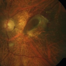
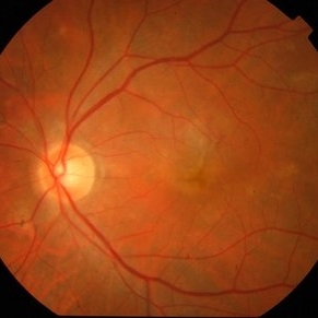 Active CNVM
Jul 11 2016 by Manish Nagpal, MD, FRCS (UK), FASRS
Colour photo showing an active CNVM.
Photographer: pooja barot
Condition/keywords: choroidal neovascular membrane (CNVM), optical coherence tomography (OCT)
-
---thumb.JPG/image-square;max$300,300.ImageHandler) High Myopia with CNVM
High Myopia with CNVM
Dec 1 2013 by Mallika Goyal, MD
22-year-old male with bilateral high myopia with macular degeneration. Left eye has CNVM with bleed.
Photographer: Mallika Goyal, MD, Apollo Hospitals, Hyderabad, India
Condition/keywords: high myopia
-
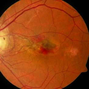
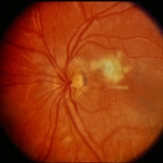
 Peri-Papillary CNVM
Oct 2 2013 by Jerald A. Bovino, MD
There is a peripapillary cnroidal neovascular membrane visible as the hyperfluorescent area temporal to the disk.
Condition/keywords: peripapillary
-
 Angioid Streaks
Oct 8 2012 by David R. Chow, MD, FRCS(C)
Angioid streaks with CNVM
Condition/keywords: angioid streaks
-
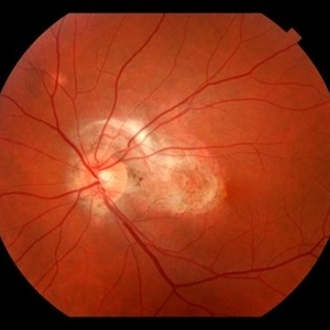
 Peri-Papillary CNVM
Oct 2 2013 by Jerald A. Bovino, MD
There is a peripapillary cnroidal neovascular membrane visible as the yellow-white areas surrounded by hemorrhage temporal to the disk.
Condition/keywords: peripapillary
-
---thumb.JPG/image-square;max$300,300.ImageHandler) High Myopia with CNVM
High Myopia with CNVM
Dec 1 2013 by Mallika Goyal, MD
CNVM regressing and bleed resolved following anti-VEGF therapy.
Photographer: Mallika Goyal, MD, Apollo Hospitals, Hyderabad, India
Condition/keywords: high myopia
-
 Angioid Streaks
Oct 8 2012 by David R. Chow, MD, FRCS(C)
Angioid streaks with CNVM
Condition/keywords: angioid streaks
-
 Peri-Papillary CNVM
Oct 2 2013 by Jerald A. Bovino, MD
There is a peripapillary cnroidal neovascular membrane visible as the hyperfluorescent area temporal to the disk.
Condition/keywords: peripapillary
-
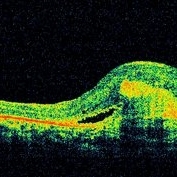
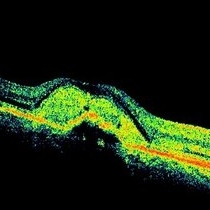
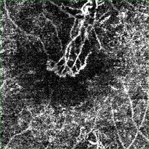 Active CNVM on Angio OCT
Jul 11 2016 by Manish Nagpal, MD, FRCS (UK), FASRS
Angio OCT picture showing neovascularization corresponding to the area of CNVM.
Photographer: pooja barot
Condition/keywords: choroidal neovascular membrane (CNVM), optical coherence tomography (OCT)
-
 Angioid Streaks With CNVM OCT RE
Jun 17 2014 by Neha Goel, MS DNB FRCS (Glasg)
Horizontal OCT scan through the right macula.
Photographer: Neha Goel
Imaging device: RTVue
Condition/keywords: angioid streaks, choroidal neovascularization (CNV)
-
---thumb.JPG/image-square;max$300,300.ImageHandler) Myopia Associated CNVM
Myopia Associated CNVM
Nov 26 2013 by Mallika Goyal, MD
Right eye of a 38-year-old high myope with CNVM at presentation.
Photographer: Mallika Goyal, MD, Apollo Health City, Jubilee Hills, Hyderabad
Condition/keywords: myopia
-
 Peripapillary CNVM / Uveitis
Aug 22 2014 by David Callanan, MD
4-year-old patient with peripapillary CNVM / uveitis.
Condition/keywords: choroidal neovascular membrane (CNVM), peripapillary, uveitis
-
 Myopia Associated CNVM
Dec 8 2013 by Mallika Goyal, MD
OCT of right eye shows CNVM with fluid at presentation in a 38-year-old high myope.
Photographer: Mallika Goyal, MD, Apollo Health City, Hyderabad, India
Condition/keywords: myopia, optical coherence tomography (OCT)
-
 Subfoveal Bleed From Extramacular CNVM
Jul 30 2014 by Mallika Goyal, MD
Subfoveal bleed (and fluid on OCT) resolving 4 weeks after intravitreal avastin for the large extrafoveal CNVM that caused the subfoveal bleed with fluid.
Photographer: Mallika Goyal, MD, Apollo Health City, Jubilee Hills, Hyderabad-500033
Condition/keywords: choroidal neovascular membrane (CNVM)
-
 Choroidal Folds
Nov 28 2014 by Thomas A. Ciulla, MD, MBA, FASRS
This 53-year-old man was noted to have choroidal folds right greater than left. The visual acuity was normal at 20/15. The choroidal folds are visible on OCT, especially on the vertical cuts that image across the horizontal folds. Angiography revealed staining of the folds without CNVM, choroidal mass, or optic nerve edema.
Photographer: Charlotte Harris
Condition/keywords: bilateral chorioretinal folds, choroidal folds
-
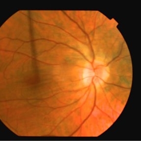
 Subretinal Fibrosis (PPCNVM and POHS) OS
Sep 18 2019 by John S. King, MD
57-year-old white male with history of PPCNVM OS and POHS OU here for a routine visit. History of avastin in 2014, and stable since then. Va OS 20/20. PP scar with macular subretinal fibrosis. No heme or exudates. CR spot supero-nasally.
Photographer: Shelly Blair
Imaging device: Topcon 50
Condition/keywords: choroidal neovascular membrane (CNVM), ocular histoplasmosis syndrome (OHS), peripapillary choroidal neovascularization (PPCNVM), presumed ocular histoplasmosis syndrome (POHS)
-
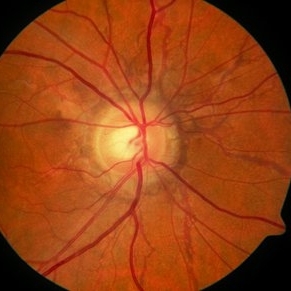
 Active CNVM
Oct 16 2012 by S. Natarajan, MD, FASRS, FRCS (GLASGOW) , FICO, D.Sc, FELA
70-year-old patient presented with hemorrhagic PED.
Photographer: Prof. Dr. S. Natarajan
Condition/keywords: hemorrhagic PED
-
 Myopia Associated CNVM
Dec 8 2013 by Mallika Goyal, MD
OCT of right eye of a 38-year-old high myope shows regressing CNVM and resolving fluid on anti-VEGF therapy.
Photographer: Mallika Goyal, MD, Apollo Health City, Hyderabad, India
Condition/keywords: myopia, optical coherence tomography (OCT)
-
 Nevus With CNVM
Feb 18 2014 by David Callanan, MD
Nevus with CNVM in an 81-year-old patient.
Condition/keywords: nevus
-
---thumb.JPG/image-square;max$300,300.ImageHandler) Perifoveal Telangiectasia with CNVM
Perifoveal Telangiectasia with CNVM
Dec 12 2012 by Mallika Goyal, MD
Left eye of a 45-year-old lady with bilateral perifoveal telangiectasia with scarring with recent onset CNVM and submacular bleed.
Photographer: Mallika Goyal, MD, Apollo Hospitals, Hyderabad, India
Condition/keywords: perifoveal telangiectasia
-
 Co-existing CNVM and CSCR
Dec 16 2012 by Mallika Goyal, MD
OCT of the right eye of a 54-year-old gentleman with co-existing CNVM with CSCR after treatment with avastin and focal photocoagulation for the extrafoveal RPE leak. There is no change in OCT status from presentation. PDT has been performed, and result will be uploaded later.
Photographer: Mallika Goyal, MD, Apollo Health City, Hyderabad, India
Condition/keywords: central serous chorioretinopathy (CSCR)
-
 ARMD / Subfoveal CNVM
Feb 13 2015 by David Callanan, MD
Female patient, ARMD / subfoveal CNVM.
Condition/keywords: age-related macular degeneration (AMD), choroidal neovascular membrane (CNVM), subfoveal choroidal neovascularization
-
 ARMD / Subfoveal CNVM
Feb 13 2015 by David Callanan, MD
Female patient, ARMD / subfoveal CNVM.
Condition/keywords: age-related macular degeneration (AMD), choroidal neovascular membrane (CNVM), subfoveal choroidal neovascularization
-
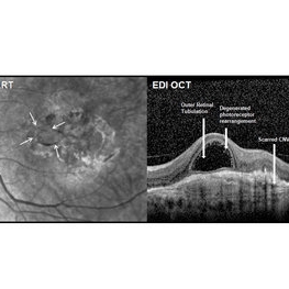 Outer Retinal Tubulation
Mar 27 2018 by Dhaivat Shah
Outer retinal tubulation (ORT) is a feature of photoreceptor rearrangement after chronic retinal damage due to refractory cme, long standing CNVM or old trauma. Photoreceptors lose adhesions to surrounding structures, resulting in outward folding and formation of new lateral contact between photoreceptors to form round structure. They generally remains stable over time. It is important to recognize ORT on OCT because it indicates a refractory state of the pathological condition and poor visual prognosis, and likely not to benefit from any treatment. Here is a case of 62-year-old female with history of 4 previous anti-VEGF injection in left eye for CNVM, with the recent OCT showing formation of ORT with subfoveal scarred membrane.
Photographer: Dr Dhaivat Shah
Condition/keywords: choroidal neovascular membrane (CNVM), outer retinal tubulation
-
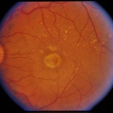
---thumb.JPG/image-square;max$300,300.ImageHandler) Subfoveal CNVM
Subfoveal CNVM
Oct 26 2012 by Mallika Goyal, MD
Fundus photograph of left eye of 65-year-old lady with large subfoveal CNVM
Photographer: Mallika Goyal, MD
Condition/keywords: subfoveal choroidal neovascularization
 Loading…