Search results (1514 results)
-
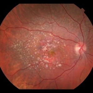
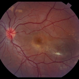
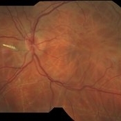
 28-Year-Old Male With Susac's Syndrome
Feb 2 2015 by Gregory J. Mincey, MD, MBA
Recurrent proliferative disease after initial stabilization with PRP.
Photographer: Bill McVerry, Carolina Eye Associates
Imaging device: Topcon
Condition/keywords: Susac's syndrome
-
 5 Watt Blue Laser Pointer, Retinal Hemorrhage
Aug 30 2018 by John S. King, MD
5 watt laser pointer (class 4 laser pointer that can burn skin and material) to eye caused this hemorrhage as a result of injury to the retinal venule (see photo). Seen by Dr. Arnold, who sent this patient to Dr. Ware. Fortunately, fovea spared.
Photographer: Stacy
Imaging device: Topcon
Condition/keywords: laser pointer maculopathy, retinal hemorrhage
-
 Active Neovascular AMD With Disciform Scar
Apr 30 2015 by Mitzy E Torres Soriano, MD
Active neovascular AMD with disciform scar.
Photographer: Mitzy E. Torres Soriano, MD; Centro medico Cagua-Estado Aragua. Venezuela
Imaging device: TOPCON
Condition/keywords: disciform scar, disciform with hemorrhage, neovascular age-related macular degeneration (AMD), wet age-related macular degeneration (wet AMD)
-
 After YAG Laser Treatment
May 24 2016 by Hazem Alaskar, MD, FEBO
Treatment by YAG laser.
Photographer: Hazem Alaskar
Imaging device: Topcon
Condition/keywords: after treatment, laser, laser treatment
-
 Albinotic Fundus
Oct 1 2018 by Rameez N Hussain, MD
Albinotic fundus
Photographer: THAMBI DURAI (Edited by Lafas DE)
Imaging device: TOPCON
Condition/keywords: ocular albinism, oculocutaneous albinism
-
 AMD With Calcified Drusen and Small, Deep IRH
Jul 22 2018 by John S. King, MD
AMD with calcified drusen and small, deep IRH
Photographer: Stacey
Imaging device: Topcon
Condition/keywords: calcified drusen
-
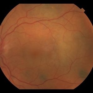
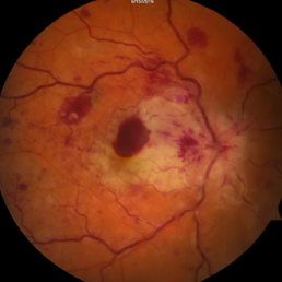
 Anemic Retinopathy in a Young Female
Jan 15 2022 by KRISHNENDU NANDI, MS
Fundus photograph of a 29-year-old female presented with retinal hemorrhages in both eyes with decrease in vision for 1 month. Hemoglobin level was 5.9gm/dl, suggestive of anemic retinopathy in both eyes.
Photographer: Krishnendu Nandi, Netralayam Eye Care Centre, Kolkata, India
Imaging device: Topcon
Condition/keywords: anaemic retinopathy, anemic retinopathy, retinal hemorrhage
-
 ANGIOID STREAKS
Jul 19 2023 by Deepti A Kulkarni, M.B.B.S., D.N.B., F.V.R.
50 YEAR OLD FEMALE WITH NO SYSTEMIC ILLNESS WITH A CLASSICAL PICTURE. VISION REMAINS 6/6. THE FELLOW EYE HAS A SUBFOVEAL SCARRED CHOROIDLA NEOVASCULAR MEMBRANE.
Photographer: DEEPTI KULKARNI, KULKARNI EYE HOSPITAL, MIRAJ, INDIA
Imaging device: TOPCON
Condition/keywords: Angioid Streaks
-
 Angioid Streaks & CNV (Fig 5)
Sep 2 2012 by Hamid Ahmadieh, MD
OCT imaging of a 53-year-old woman with a juxtafoveal CNV secondary to angioid streaks.
Photographer: Hamid Ahmadieh, Ophthalmic Research Center, Labbafinejad Medical Center
Imaging device: Topcon
Condition/keywords: angioid streaks, choroidal neovascularization (CNV), optical coherence tomography (OCT)
-
 Asteroid Hyalosis
Sep 15 2020 by Yoshihiro Yonekawa, MD, FASRS
Fundus photograph of a 70-year-old man with glistening asteroid hyalosis.
Photographer: Alexa Bednar, Mid Atlantic Retina
Imaging device: Topcon
Condition/keywords: asteroid hyalosis
-
 Asteroid Hyalosis
May 26 2025 by Moazzam Parvez
Fundus photograph of a 63 year-old man with extensive asteroid hyalosis in the right eye.
Photographer: Dr Moazzam Parvez
Imaging device: Topcon
Condition/keywords: Asteroid hyalosis, birefringence, yellow dots
-
 Asymptomatic Superior Retinal Detachment
May 5 2016 by Steven J Ryder, MD
38-year-old African American female with moderate myopia (-4.50 Sph OU) and asymptomatic superior retinal detachment in the right eye. Montage fundus photography showing localized retinal detachment superiorly with single full-thickness retinal break at 12:00.
Photographer: Luis Bernhard, Miami VA Healthcare System
Imaging device: Topcon
Condition/keywords: asymptomatic, full thickness retinal hole, myopia, retinal break, retinal detachment with retinal defect
-
 Autosomal Recessive Bestrophinopathy - Color Photo OD
Dec 22 2017 by Tony Tsai, MD, FASRS
11-year-old Asian male with 20/40 vision OU, negative family history for ocular conditions, and bilateral atypical vitelliform deposits and subretinal fluid. EOG confirmed abnormally low Arden ratios OU. Genetic testing revealed homozygous recessive mutation in BEST1 gene (p.L140V:c.418C>G). Also known as p.L80V; Ref: Davidson (2009) Am J Hum Genet 85, 581.
Photographer: San Juanita Zazueta
Imaging device: Topcon
Condition/keywords: Best disease
-
 Autosomal Recessive Bestrophinopathy - color photo OS
Dec 22 2017 by Tony Tsai, MD, FASRS
11-year-old Asian male with 20/40 vision OU, negative family history for ocular conditions, and bilateral atypical vitelliform deposits and subretinal fluid. EOG confirmed abnormally low Arden ratios OU. Genetic testing revealed homozygous recessive mutation in BEST1 gene (p.L140V:c.418C>G). Also known as p.L80V; Ref: Davidson (2009) Am J Hum Genet 85, 581.
Photographer: San Juanita Zazueta
Imaging device: Topcon
Condition/keywords: Best disease
-
 BDUMP
BDUMP
Dec 11 2018 by John S. King, MD
67-year-old white female with normal vision four months ago, consulted for dry AMD. She reported that vision in the left eye had worsened over the last two months and had progressively gotten worse. Denied history of cancer, or her primary eye doctor ever mentioning choroidal nevi. Va cc was 20/30 OD and 20/100 OS. No RAPD. IOP 9-10 OU. Anterior segment had some stellate like pigmented dusting of the endothlium, a/c was quiet, 2+NSC OU. Vitreous quiet; multiple, flat, pigmented choroidal lesions varying in size was seen the in fundus. Area in the temporal macula extending up to the superior arcade in the left eye that was suspicious for a mass; it did have a "giraffe like" pattern on one of the early FA pics; the OCT in this area showed thickening of the choroid without a definite mass lesion, and overlying thickening of the RPE, or exudative like scar, with SRF directly above. Consulted with Dr. Matt Wilson, who confirmed diagnosis, and had patient evaluated by oncology, who diagnosed non-small cell lung cancer.
Photographer: Stacey Coleman
Imaging device: Topcon
Condition/keywords: bilateral diffuse uveal melanocytic proliferation (BDUMP)
-
 Bear Tracks
Apr 28 2018 by xiangbin kong
Fundus photograph of an 24-year-old woman with bear tracks.
Photographer: Yawei Dong
Imaging device: Topcon
Condition/keywords: bear tracks
-
 Bear Tracks
Apr 28 2018 by xiangbin kong
Fundus photograph of an 24-year-old woman with bear tracks.
Photographer: Yawei Dong
Imaging device: Topcon
Condition/keywords: bear tracks
-
 Benign Lobular Inner Nuclear Layer Proliferations (BLIP)
Apr 15 2024 by Virginia Gebhart
29 year old male with multiple flat CHRPE lesions, genetic testing negative for ACP genes associated with Gardner syndrome. Multiple intraretinal amelanotic lesions consistent with Benign Lobular Inner Nuclear Layer Proliferations (BLIP) of the retina
Photographer: Virginia Gebhart
Imaging device: Topcon
Condition/keywords: BLIP Benign Lobular Inner Nuclear Layer Proliferations, CHRPE, congenital hypertrophy of the retinal pigment epithelium (CHRPE)
-
 Benign Nevus
Dec 6 2023 by Virginia Gebhart
71 year old female with benign choroidal nevus with fibrotic PED
Photographer: Virginia Gebhart
Imaging device: Topcon
Condition/keywords: choroidal nevus, fibrotic neovascularization, nevus
-
 Bilateral CRVO and PDR
Nov 4 2021 by Stefanie Palmer
Patient with both PDR and CRVO, 34 year old female, post-COVID.
Photographer: Stefanie Palmer, CRA
Imaging device: Topcon
Condition/keywords: central retinal vein occlusion (CRVO), COVID-19, diabetic retinopathy, proliferative diabetic retinopathy (PDR), venous beading
-
 Bot Fly Larvae
Apr 29 2025 by Daniela Bogenschutz
57 year-old male referred for decreased vision from optometrist. His only complaint was floaters and the letters were moving on the screen. He had never been out of the country, but is a farmer. Upon examination, our retina specialist found a bot fly larvae with numerous tracks made in this patient's retina. Patient was treated with laser to kill the larvae which was successful and he has been monitored yearly.
Photographer: Daniela Bogenschutz, OSC; Retina Consultants of Carolina, P.A.
Imaging device: Topcon
Condition/keywords: Bot Fly Larvae
-
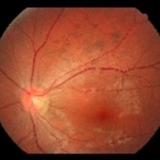
 Branch Retinal Artery Occlusion
Sep 21 2012 by Allen Chiang, MD, FASRS
Fundus photograph of a 27-year old male with a branch retinal artery occlusion. Systemic medical evaluation identified anti-phospholipid antibody syndrome.
Imaging device: Topcon
Condition/keywords: antiphospholipid antibody syndrome, branch retinal artery occlusion (BRAO)
-
 Branch Retinal Artery Occlusion - FA
Sep 21 2012 by Allen Chiang, MD, FASRS
Fluorescein angiogram of a 27-year old male with a branch retinal artery occlusion demonstrates interruption of arterial flow and retrograde venous filling. Systemic medical evaluation identified anti-phospholipid antibody syndrome.
Imaging device: Topcon
Condition/keywords: antiphospholipid antibody syndrome, branch retinal artery occlusion (BRAO)
-
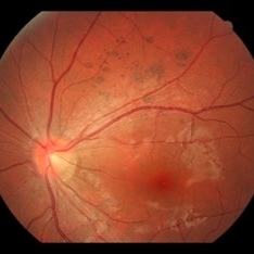
 Branch Retinal Artery Occlusion With Concurrent Central Retinal Vein Occlusion
Oct 5 2016 by Larry M Puthenparambil, MD
Branch retinal artery occlusion with concurrent central retinal vein occlusion.
Photographer: Stacey Groom
Imaging device: Topcon
Condition/keywords: branch retinal artery occlusion (BRAO), central retinal vein occlusion (CRVO)
 Loading…