Search results (68 results)
-
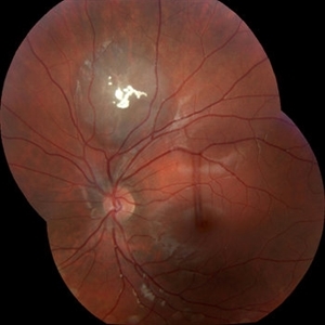
 Macular Star
May 27 2025 by César Adrián Gómez Valdivia, MD
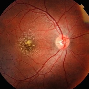
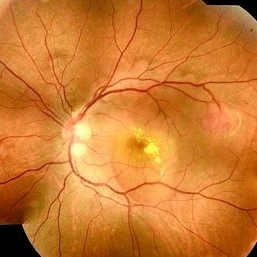
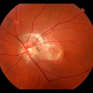
Macular Star found in a 31 year-old male patient with suspected Cat Scratch Disease. Typical intraocular presentations include neuroretinitis with optic nerve edema, macular star formation, and discrete white retinal or choroidal lesions. Findings were unilateral.
Photographer: @eyemissu2
Imaging device: TOPCON TRC-50DX
Condition/keywords: macular star
-
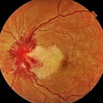
 Choroidal Fracture
Oct 27 2024 by César Adrián Gómez Valdivia, MD
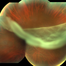
Fundus photograph of a traumatic choroidal fracture & extra-macular sub-retinal hemorrhage.
Photographer: @eyemissu2
Imaging device: TOPCON TRC-50DX
Condition/keywords: Choroidal Fracture
-
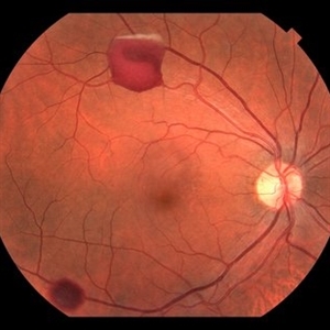
 Hypertensive Retinopathy
Apr 21 2024 by César Adrián Gómez Valdivia, MD
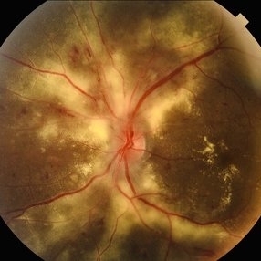
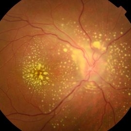
Hypertensive Retinopathy
Photographer: Erika Paulina Ornelas Cazares
Imaging device: TOPCON TRC-50DX
Condition/keywords: hypertensive retinopathy
-
 Melanocytoma of Optic Disc
Nov 3 2023 by Virginia Gebhart
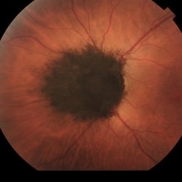
69 year-old female with pigmented lesion that covers the optic nerve. Patient has been aware for over 30 years. Remains stable and unchanged
Photographer: Virginia Gebhart
Imaging device: Topcon
Condition/keywords: benign melanocytoma, Melanocytoma, optic disc melanocytoma
-
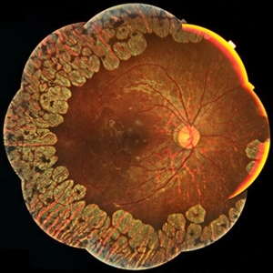
 Gyrate Atrophy
Apr 12 2023 by Ahmed Abbas Hashmi, OD
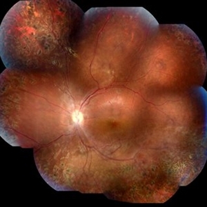
Left eye fundus of a 53-year-old male patient with advanced gyrate atrophy of the choroid and retina with macular sparing. Optic nerve head is healthy.
Photographer: Ahmed Abbas Hashmi
Imaging device: Topcon TRC-NW8F
Condition/keywords: chorioretinal atrophy
-
 Retinal detachment
Apr 12 2023 by Ahmed Abbas Hashmi, OD
Color fundus photograph of the left eye of a 30-year-old man with asymptomatic inferior retinal detachment with pigmented demarcation line. Macula and Disc healthy.
Photographer: Ahmed Abbas Hashmi
Imaging device: Topcon TRC-NW8F
Condition/keywords: Pigmentary demarcation line, Retinal Detachment
-
 Extra-scleral Extension of Choroidal Melanoma
Dec 23 2021 by Jessica Norkus
89-year-old female with extra-scleral extension of choroidal metastatic melanoma. Patient hadn't been seen by any eye doctor in 3 years prior to this visit. Noticed scleral darkening about 6 months ago, with vision loss noted for about 4-5 months. Presented with LP vision. Emergent MRI of brain/orbit showed no extension beyond what is seen at slit lamp. CT C/A/P w/ contrast ordered and found 2 hepatic lesions, concerning for potential mets. Patient referred to medical oncology.
Photographer: Jessica Norkus, COA, OSC
Imaging device: Topcon TRC 50DX
Condition/keywords: external photography, extrascleral extension, metastatic cancer, metastatic lesion
-
 Venous Beading
Nov 4 2021 by Stefanie Palmer
Venous Beading in a patient with both PDR and CRVO.
Photographer: Stefanie Palmer, CRA
Imaging device: Topcon
Condition/keywords: central retinal vein occlusion (CRVO), diabetic retinopathy, proliferative diabetic retinopathy (PDR), venous beading
-
 Retinoschisis
Mar 28 2021 by JEFFERSON R SOUSA, Tecg.º (Biomedical Systems Technology)
A 14-year-old male patient was admitted for visual evaluation. Visual acuity s/c in the right eye and 20/80 in the left eye. According to family members, he reported low vision since childhood. He had already undergone treatment with photocoagulation in another service to which he had a diagnostic hypothesis of Coats' disease. Laboratory tests were requested (HIV, TOXO, TOXOCARIASIS, ECA, VDRL, PPD). In the evaluation it was observed important exudation in the posterior pole, some vascular irregularities in the right eye. In the left eye, there is retinoschisis affecting the entire posterior pole and the region nasal to the optic disc, macula with a characteristic aspect of a cartwheel. Well exemplified by OCT-A (Structrure Deep: IPL - 25, OPL - 25).
Photographer: JEFFERSON R SOUSA - Study Center and Ophthalmological Research Dr. Andre M V Gomes, Institute Dr. Suel Abujamra São Paulo-Brazil
Imaging device: Topcon TRC-50 DX, Imaginet 4.0, angle de 50 graus. Flash 50w-s
Condition/keywords: Coats' disease, retinoschisis
-
 Choroidal Hemangioma
Mar 27 2021 by Paulo Horizonte
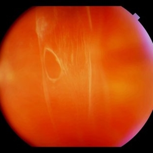
Fundus retinography of a 36-year-old man complaining of low progressive visual acuity in the left eye 2 years ago.
Photographer: Jeferson Sousa
Imaging device: Topcon
Condition/keywords: choroidal hemangioma
-
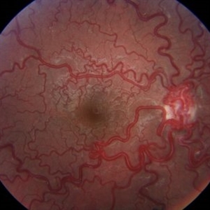 Racemose Hemangioma
Mar 14 2021 by Luiz A Zago, PhD
Racemose hemangioma in a 30-year-old woman with ambliopia in this eye.
Photographer: Luiz Alberto Zago Filho
Imaging device: Topcon 50IX
Condition/keywords: racemose hemangioma
-
 Central Retinal Vein Occlusion with Cilioretinal Artery Occlusion
Oct 21 2020 by Rutul R Patel, MD Ophthalmology
Fundus photograph of left eye of 37-year-old female who presented with sudden painless loss of vision in left eye due to CRVOwith CLRAO.
Photographer: Vidhi Bavishi, Shivjyoti Eye Hospital
Imaging device: TOPCON MAESTRO
Condition/keywords: central retinal vein occlusion (CRVO), cilioretinal artery occlusion
-
 Morning Glory Disc Anomaly
Nov 11 2020 by Yoshihiro Yonekawa, MD, FASRS
Color fundus photograph of a young boy with morning glory disc anomaly. Notice the concavity surrounding the enlarged disc, radial vasculature, and nasally dragged macula. MRI was negative for moyamoya disease, a known association.
Photographer: Alicia Thresher, Mid Atlantic Retina
Imaging device: Topcon
Condition/keywords: disc coloboma, Morning Glory Syndrome, pediatric retina
-
 Torpedo Maculopathy
Jul 29 2020 by Yoshihiro Yonekawa, MD, FASRS
Fundus photograph of a 10-year-old boy with an incidentally identified torpedo maculopathy.
Photographer: Suely Bascope
Imaging device: Topcon
Condition/keywords: macula lesion, pediatric retina, torpedo maculopathy
-
 Retinal Tear
Apr 30 2020 by Giselle DeOliveira
Fundus photograph montage of 32-year-old male with retinal tear after repair.
Photographer: Giselle DeOliveira, University of Miami, Bascom Palmer Eye Institute
Imaging device: Topcon
Condition/keywords: retinal tear
-
 A Motor Vehicle Accident Causing Valsalva Retinopathy OD, While Racing A Side By Side 4 Wheel Off-Road Vehicle
Apr 29 2020 by John S. King, MD
43-year-old white male who was injured while racing a side by side 4-wheel off-road vehicle (see Video: https://imagebank.asrs.org/file/53854/sxs-crash-during-a-race-causing-valsalva-retinopathy-od). He presented about three weeks after the injury. He was being seen by his local eye doctor who wanted an evaluation for the retinal heme and scotoma. His main complaint was a central/parcentral scotoma described as a greyish area in vision. Va 20/50 OD, nomotensive, no APD (by technician), anterior segment u/r; see picture for the fundus exam - of note there are superficial/preretinal heme, with layering of the heme superiorly, and small superficial heme at nasal edge of the optic disc; in the parafoveal region nasally there is some mottling of the RPE that may indicate an area of prior commotio retinae (also possible to have TON), which may account for his scotoma. Really bad accident (video), and amazingly, he had no LOC or injuries other than the right retina. Helmet and racing harness seat belt were used.
Photographer: Asli Ahmed
Imaging device: Topcon 50
Condition/keywords: valsalva retinopathy
-
 Retinal Detachment
Dec 4 2019 by Alexander S Jun, MHA
Fundus photograph of a 47-year-old male with retinal detachment.
Photographer: Alain Bui, Retina Associates of Orange County, California
Imaging device: Topcon TRC-50DX
-
 Retinal Ischemia, Edema, and Hemorrhages on the Infero-Temporal Macula
Aug 26 2019 by Narciso F. Atienza, MD, MBA, FASRS, FPCS, FPAO.
47-year-old female who came in with blurring of vision of the right eye of 2 weeks duration. She is hypertensive with poor control, taking Amlodipine irregularly. Denies any cardiac problem non-diabetic. Vision upon presentation was 20/400 (OD), 20/20 (OS) colored fundus photo of the right eye showing areas of retinal ischemia, edema and hemorrhages on the infero-temporal macula extending to the arcade.
Photographer: Narciso F Atienza, Jr. MD, MBA
Imaging device: Topcon TRC
Condition/keywords: edema, hemorrhage, inferotemporal arcade, retinal ischemia
-
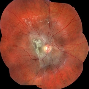
 Subretinal Fibrosis (PPCNVM and POHS) OS
Sep 18 2019 by John S. King, MD
57-year-old white male with history of PPCNVM OS and POHS OU here for a routine visit. History of avastin in 2014, and stable since then. Va OS 20/20. PP scar with macular subretinal fibrosis. No heme or exudates. CR spot supero-nasally.
Photographer: Shelly Blair
Imaging device: Topcon 50
Condition/keywords: choroidal neovascular membrane (CNVM), ocular histoplasmosis syndrome (OHS), peripapillary choroidal neovascularization (PPCNVM), presumed ocular histoplasmosis syndrome (POHS)
-
 Uveitis Posterior
Jul 19 2019 by JEFFERSON R SOUSA, Tecg.º (Biomedical Systems Technology)
A 23-year-old male patient attended the clinic with low vision of the right eye. In the evaluation it presented important fundoscopical alterations like retinal exudations in the posterior pole and nasal retina, aspects of macular star. It was proven that it was a posterior uveitis.
Photographer: JEFFERSON R SOUSA - Study Center and Ophthalmological Research Dr. Andre M V Gomes, Institute Dr. Suel Abujamra São Paulo-Brazil
Imaging device: Topcon TRC-50 DX, Imaginet 4.0, angle de 50 graus. Flash 50w-s
Condition/keywords: uveitis
-
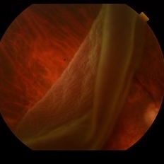
 Weiss Ring
Jan 9 2019 by John S. King, MD
77-year-old white male with ERM and PVD OD; sheet of vitreous with weiss ring in the nasal mid-vitreous cavity.
Photographer: Macey Highfill, RN
Imaging device: Topcon 50
Condition/keywords: posterior vitreous detachment, Weiss ring
-
 Gyrate Atrophy
Jan 6 2019 by Hashim Ali Khan, OD, FAAO
Montage of Multiple Fundus Photographs from the right eye of a 25-year-old woman with gyrate atrophy.
Photographer: Ahmed Abbass
Imaging device: Topcon TRC-NW8F
Condition/keywords: gyrate atrophy, hereditary retinal dystrophy, retinal dystrophy
-
 Astrocytic Hamartoma
Jan 4 2019 by Netan Choudhry, MD, FRCS(C) FASRS
Fundus photograph montage of a 7-year-old boy with an astrocytic hamartoma involving the macula.
Photographer: John Golding BA, Vitreous Retina Macula Specialists of Toronto
Imaging device: Topcon TRC-50 Dx
Condition/keywords: astrocytic hamartoma
-
 Tumor of Retina (Retinocytoma)
Jan 9 2019 by Janet Brazil
Fundus photograph of a 13-year-old male with a bilateral tumor of the retina, with positive RB gene mutation.
Photographer: Janet Atkinson, Eye Associates of New Mexico
Imaging device: TOPCON TRC-50EX
Condition/keywords: RB gene mutation, tumor
-
 Retinal Detachment
Oct 25 2018 by Graciela Nahuelquín Ríos
A 45-year-old patient reported a blow to the right eye 1 month ago, and a week ago he presented with low visual acuity. In retinal mapping and background color photography retinal detachment with giant rupture in temporal arch.
Photographer: Lic. TM. Graciela Nahuelquín Ríos
Imaging device: TRC-50DX - Topcon
 Loading…