Search results (1514 results)
-
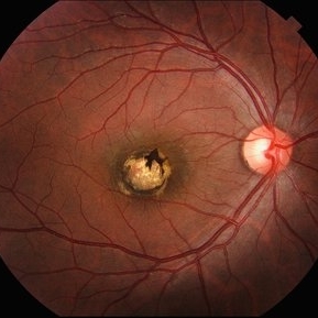
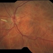
 Double Macular Holes
Jun 26 2025 by Moazzam Parvez
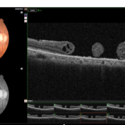
OCT image of a 62 year old man after a blunt trauma by a tennis ball with a vision of CF 3 mt in the right eye.
Photographer: Moazzam Parvez , Netralayam , Kolkata
Imaging device: Topcon Maestro 2
Condition/keywords: double, traumatic macular hole
-
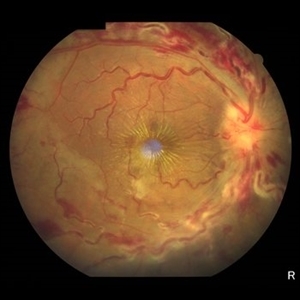
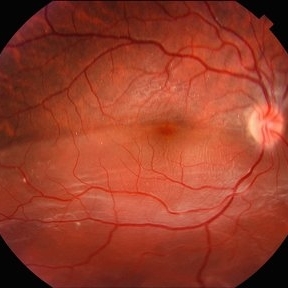
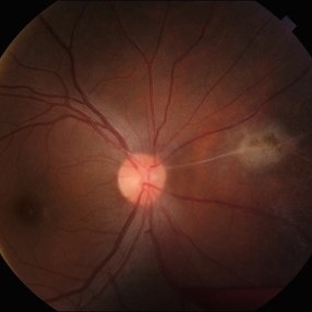
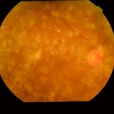
 Two Suns in the Macular Sky
Jun 26 2025 by Moazzam Parvez
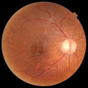
Fundus photograph of a 62 year old gentleman presenting with double adjacent full thickness macular holes in the right eye maintaining a vision of CF 3 mts.
Photographer: Moazzam Parvez ,Netralayam , Kolkata
Imaging device: Topcon Maestro 2
Condition/keywords: double, Macular hole, traumatic macular hole
-
 Epiretinal Membrane
Jun 25 2025 by Kimberly Wakester
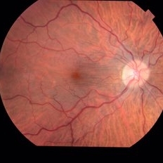
Fundus photograph of a 32-year-old woman with a stable epiretinal membrane in the right eye. Patients vision remains stable. No intervention is required at this time.
Photographer: Kimberly Wakester, COA, OCT-C
Imaging device: Topcon TRC 50DX
Condition/keywords: ERM
-
 Serpiginous Choroidopathy
Jun 23 2025 by César Adrián Gómez Valdivia, MD
Fundus photograph of a 29 year-old female patient diagnosed with Serpiginous Choroidopathy. Finings were bilateral. The most common complication of SC is choroidal neovascularization affecting up to 35% of patients. Other reported complications are subretinal fibrosis, cystoid macular edema, branch vein occlusion, serous retinal detachment, optic disc neovascularization ,and anterior uveitis.
Photographer: @eyemissu2
Imaging device: TOPCON TRC-50DX
Condition/keywords: serpiginous choroiditis
-
 Arcus Retinalis
Jun 21 2025 by Moazzam Parvez
Fundus photograph of a 30 year oiled gentleman with multiple dome shaped sub hyaloid haemorrhage with discrete arches retinals around it. Roth spots are also noted on the retina.
Photographer: Moazzam Parvez , Netralayam , Kolkata
Imaging device: Topcon Maestro 2
Condition/keywords: arcus retinalis, Roth spots, Sub hyaloid haemorrhage
-
 Central Retinal Vein Occlusion
Jun 21 2025 by Moazzam Parvez
Fundus photograph of a 56 year old male presenting with dilated tortuous vessels with adjoining Hard exudates and macular star.
Photographer: Moazzam Parvez , Netralayam , Kolkata
Imaging device: Topcon Maestro 2
Condition/keywords: CRVO with macular edema, hard exudates, macular star
-
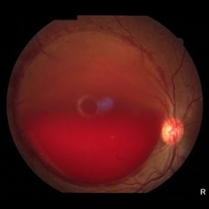 Sub ILM Hemorrhage
Jun 21 2025 by Moazzam Parvez
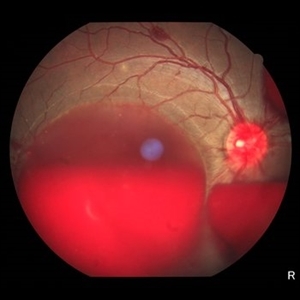
Fundus photograph of a 46 year old female presenting with a massive sharply demarcated, dome shaped bleed in her right eye.
Photographer: Moazzam Parvez , Netralayam , Kolkata
Imaging device: Topcon Maestro 2
Condition/keywords: sub ILM hemorrhage
-
 Pigmented Paravenous Retinochoroidal Atrophy
Jun 18 2025 by César Adrián Gómez Valdivia, MD
Fundus photograph of a 42YO female patient diagnosed with Pigmented Paravenous Retinochoroidal Atrophy. Findings were bilateral. Image shows hypoautofluorescence in the affected areas due to overall loss of RPE cells and thus lower lipofuscin levels.
Photographer: @eyemissu2
Imaging device: TOPCON TRC-50DX
Condition/keywords: Pigmented Paravenous Retinochoroidal Atrophy
-
 Birdshot Retinochoroidopathy
Jun 18 2025 by César Adrián Gómez Valdivia, MD
Fundus photograph of a 86 YO female patient diagnosed with Birdshot Retinochoroidopathy. Characteristically multifocal cream-colored or yellow-orange, oval or round lesions that emerge from around the optic nerve can be appreciated.
Photographer: @eyemissu2
Imaging device: TOPCON TRC-50DX
Condition/keywords: Birdshot Retinochoroidopathy
-
 Macular Coloboma
Jun 5 2025 by César Adrián Gómez Valdivia, MD
Macular Coloboma found in a 28 year-old male patient, visual acuity was 20/60. Resulting due to fusion failure of the optic fissure, colobomas are commonly found in the infero-nasal quadrant. If the retina is involved, it is reduced to glial tissue with no underlying RPE or choroid. This appears as an area of whitening often with pigment deposition at the junction of the coloboma and normal retina. Findings were bilateral.
Photographer: @eyemissu2
Imaging device: TOPCON TRC-50DX
Condition/keywords: coloboma
-
 Retinal Detachment
Jun 5 2025 by César Adrián Gómez Valdivia, MD
Fundus Photograph of a 19 year-old male patient with a RRD due to a Retinal Dialysis. Subretinal fluid and retinal folding can be appreciated.
Photographer: @eyemissu2
Imaging device: TOPCON TRC-50DX
Condition/keywords: retinal detachment
-
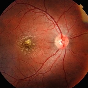
 Wagon-Wheel Lesion
Jun 5 2025 by César Adrián Gómez Valdivia, MD
Wagon-wheel lesion found in a 12 YO male patient diagnosed with congenital toxoplasmosis. Findings were bilateral.
Photographer: @eyemissu2
Imaging device: TOPCON TRC-50DX
Condition/keywords: toxoplasmosis chorioretinitis, Wagon-wheel lesion
-
 Wagon-Wheel Lesion
Jun 5 2025 by César Adrián Gómez Valdivia, MD
Wagon-wheel lesion found in a 12 year-old male patient diagnosed with congenital toxoplasmosis. Findings were bilateral.
Photographer: @eyemissu2
Imaging device: TOPCON TRC-50DX
Condition/keywords: toxoplasmosis, Wagon-wheel lesion
-
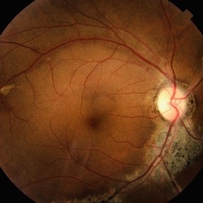
 Franceschetti's Sign
Jun 5 2025 by César Adrián Gómez Valdivia, MD
Franceschetti's sign found in a 22 year-old female patient diagnosed with ocular toxoplasmosis. These bands typically link an old scar to the optic disc, indicative of previous inflammation. Findings were unilateral.
Photographer: @eyemissu2
Imaging device: TOPCON TRC-50DX
Condition/keywords: chorio, Franceschetti's Sign, toxoplasmosis
-
 Parafoveal Telangiectasia Type 2
Jun 3 2025 by Nizamuddin HM Shaik, MD, FRCS
68 years old male patient came for check up. Not Diabetic But known patient of Coronary Artery Disease Post CABG
Photographer: Mahmoud
Imaging device: Topcon Triton OCT
Condition/keywords: parafoveal telangiectasia
-
 Parafoveal Telangiectasia
Jun 3 2025 by Nizamuddin HM Shaik, MD, FRCS
68 years old male patient came for check up. Not Diabetic But known patient of Coronary Artery Disease Post CABG
Photographer: Mahmoud
Imaging device: Topcon Triton OCT
Condition/keywords: parafoveal telangiectasia
-
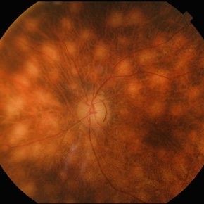
 Multiple evanescent White Dot Syndrome (MEWDS)
May 27 2025 by César Adrián Gómez Valdivia, MD
Fundus photograph of a 21 year-old female patient with suspected Multiple Evanescent White Dot Syndrome (MEWDS). The White Dot Syndromes produce yellow-white retinal lesions classically located at the retinal pigment epithelium or outer retina and are found primarily in young adults. Symptoms of MEWDS include unilateral blurred vision, visual field loss, photopsias, and floaters.
Photographer: @eyemissu2
Imaging device: TOPCON TRC-50DX
Condition/keywords: multiple evanescent white dot syndrome (MEWDS)
-
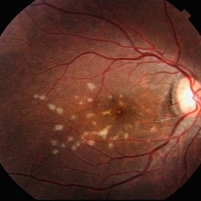
 Macular Star
May 27 2025 by César Adrián Gómez Valdivia, MD
Macular Star found in a 31 year-old male patient with suspected Cat Scratch Disease. Typical intraocular presentations include neuroretinitis with optic nerve edema, macular star formation, and discrete white retinal or choroidal lesions. Findings were unilateral.
Photographer: @eyemissu2
Imaging device: TOPCON TRC-50DX
Condition/keywords: macular star
-
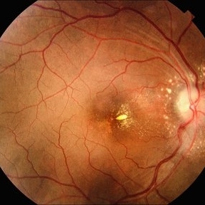 Infectious Neuroretinitis
May 26 2025 by César Adrián Gómez Valdivia, MD
Neuroretinitis found in a 38 year-oldmale patient with IV drugs abuse history. Findings were bilateral. The lipid-rich component of the exudate is able to penetrate into the outer plexiform layer, creating what is clinically seen as a macular star pattern.
Photographer: @eyemissu2
Imaging device: TOPCON TRC-50DX
Condition/keywords: neuroretinitis
-
 Asteroid Hyalosis
May 26 2025 by Moazzam Parvez
Fundus photograph of a 63 year-old man with extensive asteroid hyalosis in the right eye.
Photographer: Dr Moazzam Parvez
Imaging device: Topcon
Condition/keywords: Asteroid hyalosis, birefringence, yellow dots
-
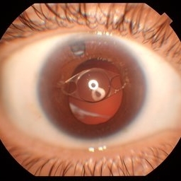 Anterior Iris Claw Artisan Lens
May 14 2025 by Moazzam Parvez
Anterior segment image of a 40 year old gentleman with a anteriorly placed iris claw lens post retinal detachment surgery.
Photographer: Dr Moazzam Parvez, Netralayam , Kolkata
Imaging device: Topcon DC-4
Condition/keywords: Anteriorly placed iris claw lens
-
 BCAMD FP
May 13 2025 by Moazzam Parvez
Fundus photograph of a 40 year old woman with a bulls eye maculopathy
Photographer: Dr Moazzam Parvez, Netralayam , Kolkata
Imaging device: Topcon Maestro 2
Condition/keywords: Benign concentric annular macular dystrophy
-
 BCAMD OCT
May 13 2025 by Moazzam Parvez
OCT image
Photographer: Dr Moazzam Parvez, Netralayam , Kolkata
Imaging device: Topcon Maestro 2
Condition/keywords: Benign concentric annular macular dystrophy
-
 Bot Fly Larvae
Apr 29 2025 by Daniela Bogenschutz
57 year-old male referred for decreased vision from optometrist. His only complaint was floaters and the letters were moving on the screen. He had never been out of the country, but is a farmer. Upon examination, our retina specialist found a bot fly larvae with numerous tracks made in this patient's retina. Patient was treated with laser to kill the larvae which was successful and he has been monitored yearly.
Photographer: Daniela Bogenschutz, OSC; Retina Consultants of Carolina, P.A.
Imaging device: Topcon
Condition/keywords: Bot Fly Larvae
-
 Hourglass in an Eye
Apr 22 2025 by KRISHNENDU NANDI, MS
A twenty-five-year-young male presented with a decrease in vision in the right eye following a blunt trauma with a football. On examination the BCVA in the right eye was CFCF and the left eye was 6/6, N6. The anterior segment was within normal limits. AT was 12 and 10 mm of Hg in the right and left eyes, respectively. Fundus examination reveals subhyaloid haemorrhage in the right eye with an attached retina. The fundus of the left eye was within normal limits. YAG laser hyaloidotomy was done with an energy of 2 mJ in the right eye. After 3 weeks the BCVA in the right eye improved to 6/9, N6.
Photographer: Dr. Krishnendu Nandi
Imaging device: Topcon
Condition/keywords: Trauma, YAG HYALOIDOTOMY, Young Male
 Loading…