Search results (1514 results)
-
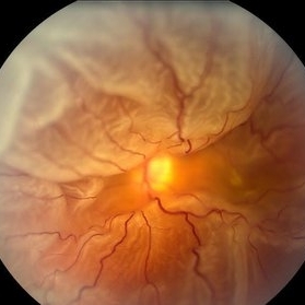
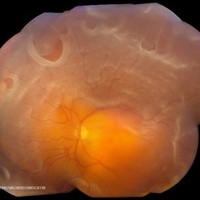
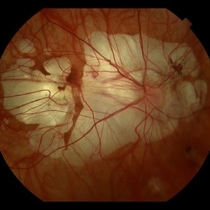
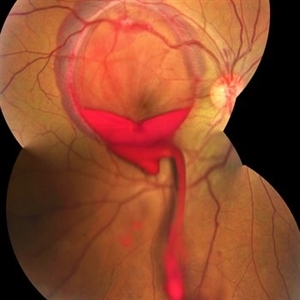 Valsalva Retinopathy
Jan 26 2017 by JEFFERSON R SOUSA, Tecg.º (Biomedical Systems Technology)
Male patient, 23-years-old, with low visual acuity in the right eye. In the ocular examination of the retinography, intense subhyaloidal hemorrhage. 2 minutes after laser application.
Photographer: JEFFERSON R SOUSA - Suel Abujamra Institute - São Paulo - Brazil
Imaging device: Topcon TRC-50 DX, Imaginet, 35 degree field. Flash 36 / Mosaic with four images.
Condition/keywords: subhyaloid hemorrhage, valsalva retinopathy
-
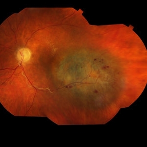
 Tractional Retinal Detachment
Dec 4 2019 by Janet Brazil
Fundus photograph of a 32-year-old female with severe end-stage diabetic tractional retinal detachment.
Photographer: Janet Atkinson, Eye Associates of New Mexico, Albuquerque, NM
Imaging device: Topcon TRC- 50EX
Condition/keywords: diabetes, proliferative diabetic retinopathy (PDR), tractional retinal detachment
-
 Whole Eye OCT
Jan 4 2019 by Netan Choudhry, MD, FRCS(C) FASRS
Swept-Source OCT montage of a 45-year-old male with Alports disease and posterior subcapsular cataract.
Photographer: John Golding BA, Vitreous Retina Macula Specialists of Toronto
Imaging device: Topcon DRI Triton
Condition/keywords: Alports disease, optical coherence tomography (OCT), swept source
-
 Ocular Manifestation of Acute Leukemia
Sep 8 2012 by Hamid Ahmadieh, MD
Color fundus photograph of a 26-year-old man with acute leukemia.
Photographer: Hamid Ahmadieh, MD, Ophthalmic Research Center, Labbafinejad Medical Center, Shahid Beheshti University of Medical Sciences , Tehran
Imaging device: Topcon Fundus Camera
Condition/keywords: acute leukemia, white centered retinal hemorrhage (Roth Spot)
-
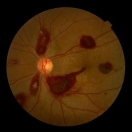
 Retinal Arteriovenous Malformations (Racemose Hemangiomatosis)
Mar 30 2018 by Rameez N Hussain, MD
A 7-years-old Portuguese girl with unilateral retinal arteriovenous malformations composed of dilated, tortuous vessels with normal vision.
Photographer: Thambi Durai. Consultant Optometrist, Orbit Health Care - Dr Agarwal's Eye Hospital, Maputo, Mozambique
Imaging device: TOPCON
Condition/keywords: racemose hemangioma, retinal arteriovenous malformations, Wyburn-Mason
-
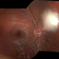
 360 Degree Retinal Detachment
Jun 29 2013 by Jason S. Calhoun
Total retinal detachment in the left eye.
Photographer: Jason S. Calhoun, Mayo Clinic Jacksonville, Florida
Imaging device: TOPCON TRC 50-EX
-
 Malignant Choroidal Melanoma
Dec 4 2015 by Kathy Karsten, COT
Malignant choroidal melanoma and branch retinal vein occlusion in 69-year-old male.
Photographer: Kathy Karsten, COT
Imaging device: Topcon TRC-50 DX
-
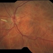
 Proliferative Diabetic Retinopathy
Sep 17 2012 by Michael P. Kelly, FOPS
Retinal fundus photograph of a patient with PDR and NVD.
Photographer: Michael P. Kelly, FOPS Director, Duke Eye Labs, Duke University Hospital, Duke Eye Center
Imaging device: Topcon
Condition/keywords: neovascularization of the disc (NVD)
-
 Tumor of Retina (Retinocytoma)
Jan 9 2019 by Janet Brazil
Fundus photograph of a 13-year-old male with a bilateral tumor of the retina, with positive RB gene mutation.
Photographer: Janet Atkinson, Eye Associates of New Mexico
Imaging device: TOPCON TRC-50EX
Condition/keywords: RB gene mutation, tumor
-
 Venous Beading
Nov 4 2021 by Stefanie Palmer
Venous Beading in a patient with both PDR and CRVO.
Photographer: Stefanie Palmer, CRA
Imaging device: Topcon
Condition/keywords: central retinal vein occlusion (CRVO), diabetic retinopathy, proliferative diabetic retinopathy (PDR), venous beading
-
 Wyburn-Mason Syndrome (Racemose Hemangiomatosis)
Mar 30 2018 by Rameez N Hussain, MD
A 7-year-old Portuguese girl with unilateral retinal arteriovenous malformations composed of dilated, tortuous vessels with normal vision.
Photographer: Thambi Durai
Imaging device: TOPCON
Condition/keywords: arteriovenous malformation, racemose hemangioma, Wyburn-Mason
-
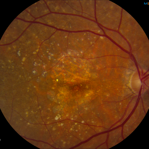 AMD with Calcific Drusen and Geographic Atrophy
Apr 19 2013 by Brandon G. Busbee, MD
AMD with calcific drusen and geographic atrophy.
Photographer: Alecia Camp, CRA - Tennessee Retina - Nashville, TN
Imaging device: Topcon TRC 50-EX
Condition/keywords: geographic atrophy
-
 Retinal Detachment With Multiple Retinal Tears
May 18 2017 by Kamal Kishore, MD, MBBS
77-year-old female presented with a report of gradual decreased vision over the span of one week. Vision finger count. Examination showed retinal detachment with multiple retinal tears and vitreous hemorrhage present.
Photographer: Lindsay Shepard, Illinois Retina and Eye Associates, Peru, IL
Imaging device: Topcon TRC- 50 EX
Condition/keywords: retinal tear
-
 Bilateral CRVO and PDR
Nov 4 2021 by Stefanie Palmer
Patient with both PDR and CRVO, 34 year old female, post-COVID.
Photographer: Stefanie Palmer, CRA
Imaging device: Topcon
Condition/keywords: central retinal vein occlusion (CRVO), COVID-19, diabetic retinopathy, proliferative diabetic retinopathy (PDR), venous beading
-
 Bot Fly Larvae
Apr 29 2025 by Daniela Bogenschutz
57 year-old male referred for decreased vision from optometrist. His only complaint was floaters and the letters were moving on the screen. He had never been out of the country, but is a farmer. Upon examination, our retina specialist found a bot fly larvae with numerous tracks made in this patient's retina. Patient was treated with laser to kill the larvae which was successful and he has been monitored yearly.
Photographer: Daniela Bogenschutz, OSC; Retina Consultants of Carolina, P.A.
Imaging device: Topcon
Condition/keywords: Bot Fly Larvae
-
 Choroidal Fracture
Oct 27 2024 by César Adrián Gómez Valdivia, MD
Fundus photograph of a traumatic choroidal fracture & extra-macular sub-retinal hemorrhage.
Photographer: @eyemissu2
Imaging device: TOPCON TRC-50DX
Condition/keywords: Choroidal Fracture
-
 Hourglass in an Eye
Apr 22 2025 by KRISHNENDU NANDI, MS
A twenty-five-year-young male presented with a decrease in vision in the right eye following a blunt trauma with a football. On examination the BCVA in the right eye was CFCF and the left eye was 6/6, N6. The anterior segment was within normal limits. AT was 12 and 10 mm of Hg in the right and left eyes, respectively. Fundus examination reveals subhyaloid haemorrhage in the right eye with an attached retina. The fundus of the left eye was within normal limits. YAG laser hyaloidotomy was done with an energy of 2 mJ in the right eye. After 3 weeks the BCVA in the right eye improved to 6/9, N6.
Photographer: Dr. Krishnendu Nandi
Imaging device: Topcon
Condition/keywords: Trauma, YAG HYALOIDOTOMY, Young Male
-
 Hypertensive Retinopathy
Feb 25 2013 by Suber S. Huang, MD, MBA, FASRS
32-year-old African American male with Grade IV hypertensive retinopathy and acute renal failure. Vision OD 20/70, OS 20/25. Creatine 7.1. BP: 250/150.
Photographer: Geoffrey Pankhurst, University Hospitals, Eye Institute/Dept. Ophthalmology and Visual Sciences Case Western Reserve University Cleveland, OH
Imaging device: Topcon TRC 50x
Condition/keywords: acute renal failure, disc edema, exudate, hypertension, hypertensive retinopathy, ischemia, macular edema, macular ischemia, optic disc edema
-
 Retinal Tear
Apr 30 2020 by Giselle DeOliveira
Fundus photograph montage of 32-year-old male with retinal tear after repair.
Photographer: Giselle DeOliveira, University of Miami, Bascom Palmer Eye Institute
Imaging device: Topcon
Condition/keywords: retinal tear
-
 Autofluorescence of Choroidal Melanoma
Oct 22 2017 by Daniel Rojas Abatte
Female patient, 53-years-old, diagnosis of choroidal melanoma, already operated in 2009 with brachytherapy.
Photographer: Daniel Rojas
Imaging device: Topcon TRC 50 DX
Condition/keywords: fundus autofluorescence (FAF)
-
 Bilateral CRVO and PDR
Nov 4 2021 by Stefanie Palmer
Patient with both PDR and CRVO, 34 year old female, post-COVID.
Photographer: Stefanie Palmer, CRA
Imaging device: Topcon
Condition/keywords: central retinal vein occlusion (CRVO), COVID-19, diabetic retinopathy, proliferative diabetic retinopathy (PDR), venous beading
-
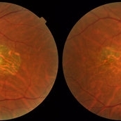 Central Areolar Choroidal Dystrophy
Jul 7 2015 by Hamid Ahmadieh, MD
Color fundus photograph of both eyes of a 58-year-old man with progressive loss of vision. VA OD is 20/60 and VA OS is 20/400.
Photographer: Soulmaz Shahmohammad, Negah Eye Center, Tehran, Iran
Imaging device: Topcon
Condition/keywords: central areolar choroidal dystrophy (CACD), color fundus photograph
-
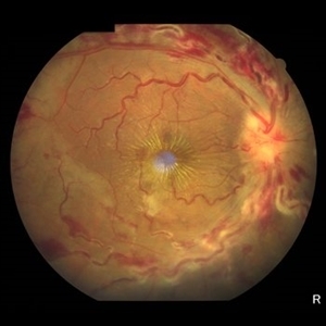 Central Retinal Vein Occlusion
Jun 21 2025 by Moazzam Parvez
Fundus photograph of a 56 year old male presenting with dilated tortuous vessels with adjoining Hard exudates and macular star.
Photographer: Moazzam Parvez , Netralayam , Kolkata
Imaging device: Topcon Maestro 2
Condition/keywords: CRVO with macular edema, hard exudates, macular star
-
 Diffuse Chorioretinal Atrophy
Feb 21 2024 by Virginia Gebhart
61 year male with myopic degeneration and diffuse chorioretinal atrophy. BCVA 20/200.
Photographer: Virginia Gebhart
Imaging device: Topcon TRC 50DX
Condition/keywords: chorioretinal atrophy, myopic degeneration
-
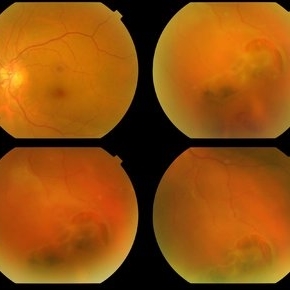 Horseshoe Retinal Break
Apr 3 2018 by Wesam Safwat
Fundus photograph of an 40-year-old woman with lower temporal horseshoe retinal tear associated with lower sub total retinal detachment not involving macula.
Photographer: Wesam Safwat, Elferdaws eye hospital , Zagazig, Egypt.
Imaging device: Topcon
 Loading…