Search results (149 results)
-
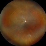
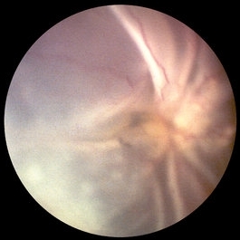
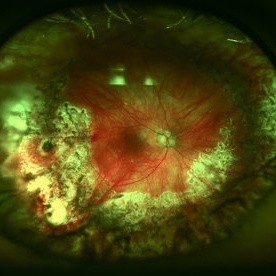
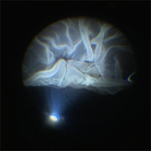 Intraoperative View of a Giant Retinal Tear
Dec 13 2024 by Thirumalesh Mochi Basavaraj, MD
Intraoperative view of 12 year old child with Giant retinal tear with Retinal detachment.
Photographer: Thirumalesh Mochi Basavaraj
Imaging device: Lumera Proveo 8
Condition/keywords: GIANT RETINAL TEAR, PVR, Retinal Detachment
-
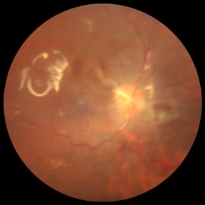
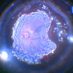
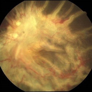 Long-standing RD with PVR
Jan 28 2022 by Gayathri Mohan
Color fundus photograph showing a long standing RD with PVR with fixed retinal folds.
Photographer: Dr Gayathri Mohan
Imaging device: Canon
Condition/keywords: PVR, Retina Folds, Retinal Detachment
-
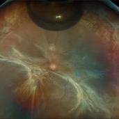
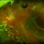
 Proliferative vitreoretinopathy in complex retinal redetachment
Dec 6 2023 by Ma. Guadalupe Perez Guevara
A 58-year-old female underwent phacovitrectomy + scleral buckling by retinal detachment. Silicon oil was used as endo tamponade. In the post-surgical period, the formation of a subretinal PRV is observed with a significant circumferential contraction that generates an inferior redetachment. It was decided to undergo a vitreous cavity lavage to remove membranes + 180° retinectomy.
Photographer: Ma. Guadalupe Pérez-Guevara. Fundación Hospital Nuestra Señora de la Luz I.A.P
Imaging device: Optos
Condition/keywords: PVR, retina surgery complications
-
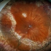
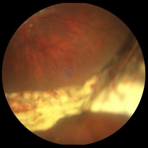
 RD with GRT with Dislocated Cataractous Lens
Nov 9 2025 by SHILPI H NARNAWARE, ICO ( Retina) , FAICO ( Vitreo-Retina)
Young high myope male presented with DOV since 6 months. Examination revealed , Subluxated cataractous lens with RD with PVR with > 270 degrees GRT
Photographer: Shilpi Narnaware, Sarakshi Netralaya , Nagpur, Maharashtra , India
Imaging device: Mirante ( by Nidek)
Condition/keywords: PVR, retinal detachment
-
 Repaired Retinal Detachment with PVR
Mar 25 2025 by Kimberly Wakester
Optomap RGB of a 79-year-old-woman with a repaired retinal detachment with PVR in the right eye. Patient is doing well over 7 months s/p vitrectomy with silicone oil and scleral buckle placement. Retina remains attached on the buckle under oil. Patient is to return in 6 months for follow up exam with repeat imaging.
Photographer: Kimberly Wakester, COA, OCT-C
Imaging device: Optos California
Condition/keywords: PVR, repaired RD, Retinal detachment under Silicon Oil, scleral buckle
-
 Retinal Detachment with PVR
Feb 24 2025 by Kimberly Wakester
Optomap RGB of an 48-year-old man with a retinal detachment with PVR. Patient is 6 weeks s/p RD repair with giant HSRT. Patient has new PVR noted on post op exam causing the retina to re-detach. Patient is having to have a 2nd surgery to remove the scar tissue and have silicone oil placement. Will continue close follow up care.
Photographer: Kimberly Wakester, COA
Imaging device: Optos California
Condition/keywords: gas bubble, PVR, retinal detachment
-
 Retinal Detachment with PVR
Jun 24 2025 by Kimberly Wakester
Optomap RGB of a 61-year-old man with Retinal Detachment with PVR in the right eye. There are multiple small holes present. Surgery was recommended. Patient is to continue follow up care post operatively.
Photographer: Kimberly Wakester, COA, OCT-C
Imaging device: Optos California
Condition/keywords: PVR, RD
-
 S/P Vitreo Retinal Surgery
Feb 27 2025 by Angela Rico
Patient referred to our office for Evaluation and Treatment of re- detached retina following previous repair of RD. 10% gas Bubble- Macular detachment- PVR Temporal Star fold super-temporal - Multiple irregular Tears infero nasal
Photographer: Angela Rico M.D.
Imaging device: California Optos
Condition/keywords: PVR, retinal detachment
-
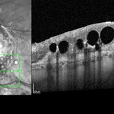 Subretinal PFO
Jun 18 2025 by Korey Starkey
86-year-old patient had history for retinal detachment surgery x2 and intraocular injections for AMD performed elsewhere. Left eye has PVR developing and subretinal PFO. Due to guarded vision, opting to defer any further treatment at this time.
Photographer: Korey Starkey
Imaging device: Heidelberg
Condition/keywords: AMD, Heidelburg Spectralis, OCT, PFO, PVR, retinal detachment, silicone oil
-
Surgery for retinal detachment with starfolds
Oct 24 2022 by Manish Nagpal, MD, FRCS (UK), FASRS
This video show s vitrectomy being done for retinal detachment with starfolds, membranes are removed following which the sequential steps of settling a retinal detachment are carried out
Photographer: Manish Nagpal
Condition/keywords: PVR, RD, starfolds, tears, video, vitrectomy
-
 PVR Detachment Repaired With Primary Segmental
Feb 1 2017 by Jamison R Ridgeley, MD
Fundus photograph of a 50-year-old woman who presented with a chronic mac off RD with starfold formation. She presented with HM vision and is now 20/50.
Photographer: John Stivers, Eye Associates NorthWest
Imaging device: Optos
-
 PVR Related Recurrent RD
May 19 2014 by John W. Kitchens, MD
Recurrent PVR RD (pre-op photo).
Photographer: Ed SLade
Imaging device: Optos 200Tx
Condition/keywords: acute retinal detachment, proliferative vitreoretinopathy (PVR)
-
 PVR Retinal Detachment following Laser Retinopexy Slide 1
Oct 22 2012 by Ronald C. Gentile, MD
Acute onset total retinal detachment with PVR 10 weeks following laser retinopexy.
Photographer: The New York Eye & Ear Infirmary Department of Medical Imaging
Condition/keywords: laser retinopexy, proliferative vitreoretinopathy (PVR)
-
 PVR Retinal Detachment following Laser Retinopexy Slide 2
Oct 22 2012 by Ronald C. Gentile, MD
Fundus drawing during evaluation for a macular pucker following laser retinopexy.
Photographer: The New York Eye & Ear Infirmary Department of Medical Imaging
Condition/keywords: fundus flavimaculatus, macular pucker, proliferative vitreoretinopathy (PVR)
-
 PVR Retinal Detachment with subretinal bands Slide 1
Oct 22 2012 by Ronald C. Gentile, MD
Total retinal detachment with pre-retinal and sub-retinal proliferation. The subretinal bands have a napkin ring configuration posteriorly with the macula folded and dragged above the optic nerve.
Photographer: The New York Eye & Ear Infirmary Department of Medical Imaging
Condition/keywords: proliferative vitreoretinopathy (PVR), subretinal bands
-
 PVR Retinal Detachment with subretinal bands Slide 2
Oct 22 2012 by Ronald C. Gentile, MD
Postoperative fundus photo of the posterior pole with flat retina. As noted by the retinal surface reflex, silicone oil tamponade was used.
Photographer: The New York Eye & Ear Infirmary Department of Medical Imaging
Condition/keywords: proliferative vitreoretinopathy (PVR), subretinal bands
-
 PVR Retinal Detachment with subretinal bands Slide 3
Oct 22 2012 by Ronald C. Gentile, MD
Postoperative fundus photo of the margin of the inferior nasal retinectomy site. The retinectomy was used intra-operatively to flap the retina over and remove the subretinal bands. The scleral buckle effect and acute white endolaser marks are present at the edge of the retinectomy.
Photographer: The New York Eye & Ear Infirmary Department of Medical Imaging
Condition/keywords: proliferative vitreoretinopathy (PVR), subretinal bands
-
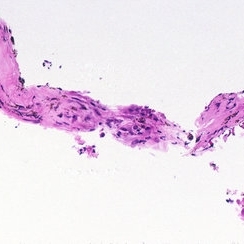 PVR Retinal Detachment with subretinal bands Slide 4
Oct 22 2012 by Ronald C. Gentile, MD
Histo-pathology of the subretinal bands revealed the presence of a fibrocellular membrane. The cells were predominately retinal pigment epithelium cells with myofibroblastic differentiation. Collagen deposition with occasional inflammatory cells and pigment were noted.
Photographer: The New York Eye & Ear Infirmary Department of Pathology and Laboratory Medicine
Condition/keywords: proliferative vitreoretinopathy (PVR), subretinal bands
-
 PVR With Multiple Breaks
Mar 13 2014 by Marcelo Zas, MD PhD
The image show a PVR case with PFCL in the posterior pole, diathermy is used before the retinectomy.
Photographer: Marcelo Zas MD PhD
Condition/keywords: proliferative vitreoretinopathy (PVR)
-
 PVR, Grade B
Aug 7 2015 by H. Michael Lambert, MD
PVR, grade B; surface wrinkling at 7:30.
Condition/keywords: proliferative vitreoretinopathy (PVR)
-
 PVR, Vitreous Hemorrhage, Sub-retinal Debris
Dec 10 2012 by Yale L. Fisher, MD
In this movie formed vitreous hemorrhage (yellow arrow) can be seen moving slowly inside this open-coned retinal detachment, which demonstrates no motion. ; Sub-retinal material (green arrow) with moderate reflectivity is seen moving in an undulating fashion slower than movement usually seen in the vitreous cavity.
Condition/keywords: video
-
 Recurrent RD (Post-Op)
May 19 2014 by John W. Kitchens, MD
PVR RD after repair.
Photographer: Michelle Buck
Imaging device: Optos 200Tx
Condition/keywords: proliferative vitreoretinopathy (PVR)
-
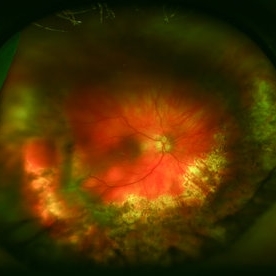
 Traumatic perforation and PVR
May 19 2022 by ALLAN GOMES DA SILVA
PVR, retinal detachment and macular dragging after iatrogenic traumatic perforation during peribulbar block. We can observe the inferior perforation, the brutal formation of PVR and the fovea being pulled.
Photographer: Edimilson Ferreira da Silva
Imaging device: Topcon TRC-50 DX, Imaginet 4.0, angle - 50 degrees
Condition/keywords: posterior perforation, proliferative vitreoretinopathy (PVR)
-
 25G PPV Without Scleral Buckling for RRD, PVR, Giant Breaks
Dec 10 2012 by Yale L. Fisher, MD
Dr. Steve Charles shares his approach to 25G PPV without scleral buckling for RRD, PVR and giant breaks. NOTE: A narration by Dr. Steve Charles will soon be available for this movie- please check back periodically.
Condition/keywords: video
-
 360 Degree Retinectomy
Feb 2 2022 by Manish Nagpal, MD, FRCS (UK), FASRS
Intraoperative photo of a case of retinal detachment with extensive PVR, which underwent 360 degree relaxing retinectomy followed by 360 laser barrage just prior to silicone oil injection.
Photographer: Manish Nagpal, Retina Foundation, Ahmedabad, India
Imaging device: Sony PMW -10 MD surgical camera
Condition/keywords: laser, laser photocoagulation, proliferative vitreoretinopathy (PVR), relaxing retinectomy, retinectomy, silicone oil
 Loading…