Search results (288 results)
-
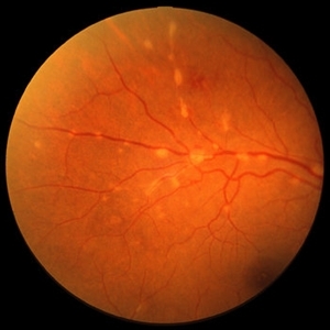
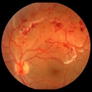 Retinal Vasculitis with Hemorrhages and Cotton Wool Spots
Oct 16 2012 by Jeffrey G. Gross, MD, FASRS
Retinal vasculitis with hemorrhages and cotton wool spots.
Condition/keywords: cotton wool spots, retinal vasculitis
-
 Acute Idiopathic Occlusive Retinal Vasculitis
May 31 2014 by Hamid Ahmadieh, MD
Color fundus photograph of the right eye of a 28-year-old woman with sudden drop of vision due to acute occlusive retinal vasculitis leading to extensive nerve fiber layer infarction and retinal hemorrhages.
Photographer: Naghmeh Nozhat, Negah Eye Center, Tehran
Condition/keywords: color fundus photograph, cotton wool spots, retinal hemorrhage, retinal ischemia
-
 Behcet's Disease
Mar 13 2013 by Hamid Ahmadieh, MD
Color fundus photograph of the right eye of a 23-year-old man with retinal vasculitis and branch retinal vein occlusion (BRVO) due to Behcet's disease .
Photographer: Solmaz Shahmohammad, Negah Eye Center, Tehran
Imaging device: Heidelberg Spectralis
Condition/keywords: branch retinal vein occlusion (BRVO), retinal vasculitis
-
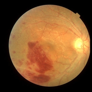
 Behcet's Disease
Nov 25 2012 by Mallika Goyal, MD
Fundus photograph of left eye of a 23-year-old gentleman with Behcet's Disease shows occlusive retinal vasculitis with optic disc pallor and macular ischemia. This eye has no light perception; other eye has similar fundus appearance.
Photographer: Mallika Goyal, MD, Apollo Health City, Hyderabad, India
Condition/keywords: macular ischemia, occlusive vasculitis, optic disc pallor
-
 Sea Fan Neovascularisation
Apr 27 2015 by Neha Goel, MS DNB FRCS (Glasg)
Fluorescein angiography of the left eye of a 40-year-old male.
Photographer: Neha Goel
Imaging device: Zeiss visucam
Condition/keywords: Eales disease, neovascularization elsewhere (NVE), vasculitis
-
 Acute Idiopathic Occlusive Retinal Vasculitis
May 31 2014 by Hamid Ahmadieh, MD
Color fundus photograph of the left eye of a 28-year-old woman with acute drop of vision due to occlusive retinal vasculitis leading to extensive nerve fiber layer infarction and retinal hemorrhages.
Photographer: Naghmeh Nozhat, Negah Eye Center, Tehran
Condition/keywords: color fundus photograph, cotton wool spots, retinal hemorrhage, retinal ischemia
-
 Sarcoid Vasculitis
Oct 11 2012 by Jeffrey G. Gross, MD, FASRS
Sarcoid vasculitis.
Condition/keywords: autoimmunity, sarcoid vasculitis, sarcoidosis
-
 Sarcoid Vasculitis
Oct 11 2012 by Jeffrey G. Gross, MD, FASRS
Sarcoid vasculitis.
Condition/keywords: autoimmunity, sarcoid vasculitis, sarcoidosis
-
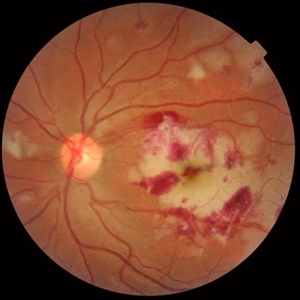
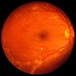
 Idiopathic Retinal Vasculitis, Aneurysms, and Neuroretinitis (IRVAN)
Oct 16 2012 by S. Natarajan, MD, FASRS, FRCS (GLASGOW) , FICO, D.Sc, FELA
Fundus photograph of a young male with IRVAN Syndrome
Photographer: Prof. Dr. S. Natarajan
Condition/keywords: aneurysm, neuroretinitis, retinal vasculitis
-
 Systemic Lupus Erythematosus Vasculitis
Oct 10 2012 by Jeffrey G. Gross, MD, FASRS
SLE vasculitis.
Condition/keywords: systemic lupus erythematosus, systemic lupus erythematosus (SLE) vasculitis
-
 Idiopathic Retinal Vasculitis, Aneurysms, and Neuroretinitis (IRVAN)
Oct 16 2012 by S. Natarajan, MD, FASRS, FRCS (GLASGOW) , FICO, D.Sc, FELA
FFA photograph of a 28-year-old male with IRVAN Syndrome.
Photographer: Prof. Dr. S. Natarajan
Condition/keywords: aneurysm, neuroretinitis, retinal vasculitis
-
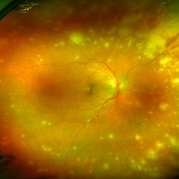
 SLE Retinopathy
Nov 14 2016 by Mitzy E Torres Soriano, MD
25-year-old female patient with systemic lupus erythematosus. Photographs show cotton wool spots, intraretinal hemorrhages and vascular tortuosity. FA demonstrated retinal vasculitis and OCT revealed cystoid macular edema. In this case diagnosis of SLE was made after ocular manifestation.
Photographer: Grupo Laser Vision, Rosario, Argentina
Condition/keywords: cotton wool spots, occlusive retinal vasculitis, occlusive vasculitis, systemic lupus erythematosus, vasculopathy
-
 Systemic Lupus Erythematosus Vasculitis
Oct 9 2012 by Jeffrey G. Gross, MD, FASRS
SLE vasculitis.
Condition/keywords: systemic lupus erythematosus, systemic lupus erythematosus (SLE) vasculitis
-
 Sarcoid Sarcoidosis
Feb 13 2013 by From the Collections of Thomas M. Aaberg, MD and Thomas M. Aaberg Jr., MD
Drawing of retinal sarcoidosis, cultaneous sarcoid granuloma.
Condition/keywords: candle wax dripping, sarcoid granuloma, sarcoidosis, vasculitis
-
 Idiopathic Retinal Vasculitis, Aneurysms, and Neuroretinitis (IRVAN)
Oct 16 2012 by S. Natarajan, MD, FASRS, FRCS (GLASGOW) , FICO, D.Sc, FELA
Fundus photograph of a 28-year-old male with IRVAN Syndrome.
Photographer: Prof. Dr. S. Natarajan
Condition/keywords: aneurysm, neuroretinitis, retinal vasculitis
-
 Lupus Vasculitis
Feb 13 2013 by From the Collections of Thomas M. Aaberg, MD and Thomas M. Aaberg Jr., MD
Obliterative peripheral vasculitis.
Condition/keywords: ischemia, lupus
-
 Idiopathic Retinal Vasculitis, Aneurysms, and Neuroretinitis (IRVAN)
Oct 16 2012 by S. Natarajan, MD, FASRS, FRCS (GLASGOW) , FICO, D.Sc, FELA
Fundus photograph of a 28-year-old male with IRVAN Syndrome.
Photographer: Prof. Dr. S. Natarajan
Condition/keywords: aneurysm, neuroretinitis, retinal vasculitis
-
---thumb.jpg/image-square;max$300,300.ImageHandler) Idiopathic Retinal Vasculitis, Aneurysms, and Neuroretinitis (IRVAN)
Idiopathic Retinal Vasculitis, Aneurysms, and Neuroretinitis (IRVAN)
Oct 16 2012 by S. Natarajan, MD, FASRS, FRCS (GLASGOW) , FICO, D.Sc, FELA
IRVAN
Condition/keywords: aneurysm, neuroretinitis, retinal vasculitis
-
 Idiopathic Retinal Vasculitis, Aneurysms, and Neuroretinitis (IRVAN)
Oct 16 2012 by S. Natarajan, MD, FASRS, FRCS (GLASGOW) , FICO, D.Sc, FELA
FFA photograph of a 28-year-old male with IRVAN Syndrome showing aneurysms around disc.
Photographer: Prof. Dr. S. Natarajan
Condition/keywords: aneurysm, neuroretinitis, retinal vasculitis
-
 Acute Retinal Necrosis (ARN)
Dec 13 2017 by Gabriel Costa Andrade, PhD
Healthy 47-year-old patient presenting with subacute decline in vision, vitritis, periphlebits, and necrotizing retinitis.
Photographer: Gabriel Andrade, RETINA CLINIC, SP
Imaging device: Optos Wide Field Camera
Condition/keywords: acute retinal necrosis, uveitis, vasculitis
-
---thumb.jpg/image-square;max$300,300.ImageHandler) Idiopathic Retinitis, Vasculitis, Aneurysms, and Neuroretinitis (IRVAN)
Idiopathic Retinitis, Vasculitis, Aneurysms, and Neuroretinitis (IRVAN)
Nov 13 2013 by Hamid Ahmadieh, MD
Color fundus photograph of the right eye of a 35-year-old woman with idiopathic retinitis, vasculitis, aneurysms, and neuroretinitis (IRVAN).
Photographer: Solmaz Shahmohammad , Negah Eye Center , Tehran
Condition/keywords: aneurysm, neuroretinitis, retinal vasculitis, retinitis
-
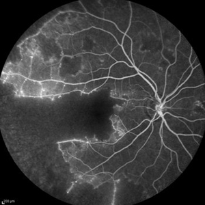 Idiopathic Occlusive Retinal Vasculitis (Late Stage)
May 31 2014 by Hamid Ahmadieh, MD
Late phase FA image of the right eye of a 28-year-old woman with idiopathic occlusive retinal vasculitis 6 months after the onset.
Photographer: Solmaz Shahmohammad, Negah Eye Center, Tehran
Imaging device: Heidelberg Spectralis
Condition/keywords: capillary closure, fluorescein leakage, macular infarction
-
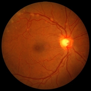
 Retinal Vasculopathy With Retinal Vasculitis and Ischemia
Jul 23 2014 by John S. King, MD
Purtscher's-like retinopathy.
Photographer: UPMC
Condition/keywords: lupus, systemic lupus erythematosus (SLE) retinopathy, systemic lupus erythematosus (SLE) vasculitis
-
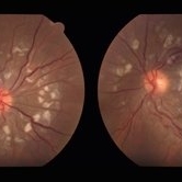
---thumb.JPG/image-square;max$300,300.ImageHandler) Behcet's Disease
Behcet's Disease
Nov 25 2012 by Mallika Goyal, MD
Fundus photograph of right eye of a 23-year-old gentleman with Behcet's Disease shows occlusive retinal vasculitis with optic disc pallor and macular ischaemia. Other eye has similar appearance with no light perception.
Photographer: Mallika Goyal, MD, Apollo Health City, Hyderabad, India
Condition/keywords: macular ischemia, occlusive vasculitis
-
 Idiopathic Retinal Vasculitis, Aneurysms, and Neuroretinitis (IRVAN)
Oct 16 2012 by S. Natarajan, MD, FASRS, FRCS (GLASGOW) , FICO, D.Sc, FELA
FFA photograph of a 28-year-old male with IRVAN Syndrome.
Photographer: Prof. S. Natarajan
Condition/keywords: aneurysm, neuroretinitis, retinal vasculitis
 Loading…