Search results (288 results)
-
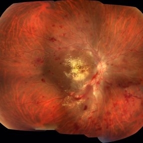
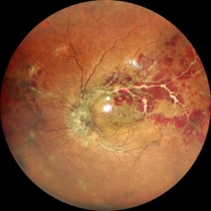
 Active Vasculitis with Proliferative Retinopathy
Jan 30 2021 by Raja Rami P Reddy, MD FRCS FASRS
25-year-old boy with unilateral recent onset visual loss. Fundus shows areas of active vasculitis nasally and large neovascular complexes temporally and on the disc and early fibrous membrane formation. Fellow eye fundus is normal. Further investigations suggested tubercular etiology
Photographer: Raja Rami Reddy P
Imaging device: fundus camera
Condition/keywords: proliferative retinopathy, tuberculosis, vasculitis
-
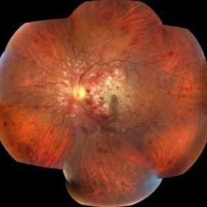
 Acute Necrotizing Retinal Vasculitis as Onset of Systemic Lupus Erythematosus.
Sep 3 2016 by ADRIANO FERREIRA
A 28-year-old white man was referred to the rheumatology clinic with gradually and rapid deterioration of the vision (both eyes). In this picture, we can observe cotton wool spots in the papillomacular area and extensive hemorrhages in posterior polo and in the middle periphery. Hard exudates are present in macular area (macular edema)
Photographer: Claudio Zett Lobo
Imaging device: TRC50DXi TOPCON
Condition/keywords: systemic lupus erythematosus (SLE) vasculitis, vasculitis
-
 Acute Necrotizing Retinal Vasculitis as Onset of Systemic Lupus Erythematosus.
Sep 3 2016 by ADRIANO FERREIRA
A 28-year-old white man was referred to the rheumatology clinic with gradually and rapid deterioration of the vision (both eyes). In this picture we can observe cotton wool spots in the papillomacular area and extensive hemorrhages in the left eye.
Photographer: Claudio Zett Lobo
Imaging device: TRC50DXi TOPCON
Condition/keywords: systemic lupus erythematosus (SLE) vasculitis, vasculitis
-
 Acute Necrotizing Retinal Vasculitis as Onset of Systemic Lupus Erythematosus.
Sep 3 2016 by ADRIANO FERREIRA
28-year-old white man was referred to the rheumatology clinic with gradually and rapid deterioration of the vision (both eyes). In this picture, we can observe vasculitis (leakage from vessels) and diffuse ischemia in the left eye.
Photographer: Claudio Zett Lobo
Imaging device: HRA-Spectralis
Condition/keywords: systemic lupus erythematosus (SLE) vasculitis, vasculitis
-
 Acute Retinal Necrosis (ARN)
Dec 13 2017 by Gabriel Costa Andrade, PhD
Healthy 47-year-old patient presenting with subacute decline in vision, vitritis, periphlebits, and necrotizing retinitis.
Photographer: Gabriel Andrade, RETINA CLINIC, SP
Imaging device: Optos Wide Field Camera
Condition/keywords: acute retinal necrosis, uveitis, vasculitis
-
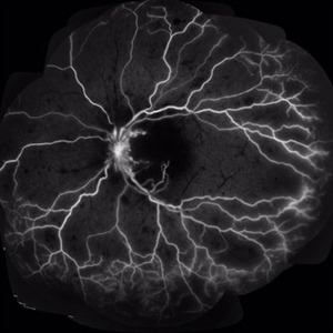
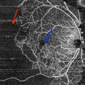
 Angiographic Storm: Fluorescein Leakage in Retinal Vasculitis
Nov 17 2025 by SHRADDHA RAJ SHRIVASTAVA
This left eye montage fundus fluorescein angiography (FFA) image of a 19 year old male with idiopathic retinal vasculitis, having skip vasculitic lesions predominantly involving retinal veins. There are areas of blocked fluorescence due to intraretinal hemorrhages, the involved veins have filling defects and occlusions, leading to formation of numerous collateral channels. The inflamed vessels also show perivascular fuzzy hyperfluorescent stain due to leakage of dye. We can also see multiple peripheral capillary non perfusion (CNP) areas, with a 'hot disc', suggestive of ongoing inflammation.
Photographer: Dr. Shraddha Raj Shrivastava
Imaging device: Nidek Mirante SLO/OCT (Confocal scanning/Spectral domain OCT)
Condition/keywords: FA late phase leakage, Fundus Fluorescein Angiography, idiopathic retinal vasculitis, optic disc leakage, VASCULITIS
-
 Central Retinal Artery Occlusion
Aug 28 2018 by Gabriela Lopezcarasa Hernandez, MD
35-year-old women with CRAO and vasculitis due to systemic lupus.
Photographer: MARCO ANTONIO SAUZA CASTILLEJOS M.D., MEXICO.
Condition/keywords: central retinal artery occlusion (CRAO), vasculitis
-
 Central Retinal Artery Occlusion
Aug 28 2018 by Gabriela Lopezcarasa Hernandez, MD
35-year-old women with CRAO and vasculitis due to systemic lupus.
Photographer: MARCO ANTONIO SAUZA CASTILLEJOS M.D., MEXICO.
Condition/keywords: central retinal artery occlusion (CRAO), vasculitis
-
 CMV
CMV
Apr 25 2013 by Howard Schatz, MD
70-year-old white female, III CMV, 20/40: OK; 5/200: infilesatire, vasculitis viluitis.
Condition/keywords: vasculitis
-
 Disc Edema with Vasculitis
Jul 15 2025 by rohan jain
Case of disc edema with vasculitis.
Photographer: Dr. ROHAN JAIN
Imaging device: mirante
Condition/keywords: disc edema, idiopathic retinal vasculitis, VASCULITIS
-
 Disc Edema with Vasculitis
Jul 15 2025 by rohan jain
Case of disc edema with vasculitis.
Photographer: Dr. ROHAN JAIN
Imaging device: mirante
Condition/keywords: disc edema, idiopathic retinal vasculitis, VASCULITIS
-
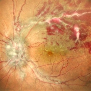
 Extramacular TRD in Idiopathic Occlusive Vasculitis
Dec 5 2024 by Tejaswita Verma
Fundus photo showing extramacular TRD in a 16 year old boy with idiopathic occlusive vasculitis secondary to presumed IOTB. History of taking ATT for 6 months , Mantoux positive previously. Vision was 6/6P,other eye had funnel RD .
Photographer: DR. TEJASWITA VERMA
Imaging device: MIRANTE
Condition/keywords: tractional retinal detachment, vasculitis
-
 FA-ICG
Jan 6 2016 by Jared Watson
FA-ICG
Photographer: Jared Watson COT/CRA
Imaging device: Spectralis
Condition/keywords: necrotizing retina, vasculitis
-
 Fluorescein Angiography Montage of Vasculitis
Nov 27 2020 by Sham Talati, DOMS
A 27-year-old male patient presented with vasculitis in left eye. Fluorescein angiography image showed diffuse vascular leakages.
Photographer: Dr. Sham Talati,Retina Foundation,Ahmedabad
Imaging device: Nidek Mirante
Condition/keywords: diffuse vasculitis, vasculitis
-
 Idiopathic Vasculitis
Feb 4 2023 by Aditya S Kelkar, MS, FRCS, FASRS,FRCOphth
Color fundus photograph of the left eye showing idiopathic retinal vasculitis.
Photographer: Dr. Pranali Surawase. National Institute of Ophthalmology, Pune, India.
Imaging device: Zeiss Clarus 500
Condition/keywords: retinal vasculitis, vasculitis
-
 Idiopathic Vasculitis
Aug 29 2023 by Shobhit Chawla, M.S.
A 28 year old male with acute loss of vision and no history of any illness. Had profound loss of vision with severe vasulitis extending from central fundus to periphery.
Photographer: Ranjit Ray
Imaging device: Clarus
Condition/keywords: VASCULITIS
-
 Idiopathic vasculitis
Aug 29 2023 by Shobhit Chawla, M.S.
A 28 year old male with acute loss of vision and no history of any illness. Had profound loss of vision with severe vasulitis extending from central fundus to periphery.
Photographer: Ranjit Ray
Imaging device: Clarus
Condition/keywords: VASCULITIS
-
 Idiophatic vasculitis OD
Jul 9 2023 by Luiz A Zago, PhD
Idiophatic vasculitis
Photographer: Luiz Zago, PhD.
Imaging device: Topcon 50IX
Condition/keywords: intraretinal hemorrhage, leaky parafoveal capillaries, optic disc leakage, vasculitis
-
 Inflammatory BRVO
Apr 27 2015 by Neha Goel, MS DNB FRCS (Glasg)
Montage fundus photograph of the right eye of a 24-year-old male.
Photographer: Neha Goel
Imaging device: Zeiss visucam
Condition/keywords: inflammatory retinal vein occlusion, vasculitis
-
 NVE in a Patient With Vasculitis
Nov 5 2018 by awaneesh m upadhyay, MBBS, DNB
FFA image of a 22-year-old male vasculitis patient with NVE.
Photographer: Hiteshwar Saikia
Condition/keywords: neovascularization elsewhere (NVE), tuberculosis, vasculitis
-
 OCTA in Vasculitis
Sep 3 2019 by Manish Nagpal, MD, FRCS (UK), FASRS
OCTA in a case of vasculitis highlighting the capillary non perfusion areas closing in on macula.
Photographer: Gayathri Mohan
Condition/keywords: capillary nonperfusion, vasculitis
-
 OLD ISCHEMIC CRVO
Jun 11 2022 by Nivesh Gupta
FA montage image of an 36 year old man with a old central retinal vein occlusion showing 360 degree capillary non perfusion areas.
Photographer: DR. NIVESH GUPTA, RETINA FELLOW , RETINA FOUNDATION, AHMEDABAD
Imaging device: NIDEK MIRANTE
Condition/keywords: ischemic CRVO, VASCULITIS
-
 Oral Ulcers in Behcet's Disease
Apr 10 2017 by Deepak Bhojwani, MS
A 19-year-old boy presented with with recurrent oral and genital ulcers along with blurring of vision. Systemically he was HLA B-51 Positive suggesting Behcet's Disease. The photograph depicts the classic aphthous mouth ulcers seen in the Behcet's disease.
Photographer: DEEPAK BHOJWANI, RAGHUDEEP EYE HOSPITAL, AHMEDABAD.
Condition/keywords: Behcet's uveitis, vasculitis
-
 Retinal Vasculitis
Aug 22 2023 by Karen Santamaría
Fluorescein angiography of patient of a 23 year-old man diagnosed with retinal vasculitis and juvenile glaucoma.
Photographer: Karen Santamaría, Hospital Militar de Especialidades Oftalmológicas - Servicio de Glaucoma, Ciudad de México
Imaging device: Optos California
Condition/keywords: juvenile glaucoma, retina, vasculitis
-
 Sarcoid Sarcoidosis
Feb 13 2013 by From the Collections of Thomas M. Aaberg, MD and Thomas M. Aaberg Jr., MD
Drawing of retinal sarcoidosis, cultaneous sarcoid granuloma.
Condition/keywords: candle wax dripping, sarcoid granuloma, sarcoidosis, vasculitis
 Loading…