Search results (288 results)
-
 Central Retinal Artery Occlusion
Aug 28 2018 by Gabriela Lopezcarasa Hernandez, MD
35-year-old women with CRAO and vasculitis due to systemic lupus.
Photographer: MARCO ANTONIO SAUZA CASTILLEJOS M.D., MEXICO.
Condition/keywords: central retinal artery occlusion (CRAO), vasculitis
-
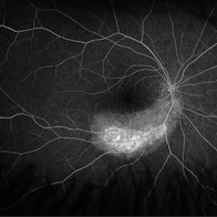
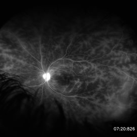 Retinal Vasculitis in Behcet's OS
Jun 29 2018 by Gareth Lema, MD, PhD
IVFA at 7 minutes showing retinal vasculitis, cystoid macular edema, and disc staining.
Photographer: Ross Eye Institute, University at Buffalo Jacobs School of Medicine, Buffalo. NY
Imaging device: Optos
Condition/keywords: Behcet's Disease, cystoid macular edema (CME), disc staining, retinal vasculitis
-
 Acute Retinal Necrosis (ARN)
Jul 3 2025 by Heitor Nogueira
Fundus photograph of an 63-year-old woman who reported unilateral visual acuity loss for 10 days associated with ocular pain. He presented conjunctival hyperemia with temporal and nasal nodular scleritis, anterior chamber reaction 2+/4+, Koeppe nodules, granulomatous PKs, vitreitis 2+/4+, multiple areas of vasculitis in the arcades and periphery, associated with hemorrhages and necrotizing retinitis in the temporal, inferior and nasal periphery. Positive serology for Herpes Virus
Photographer: Heitor Nogueira, Penido Burnier Institute, Campinas, São Paulo, Brazil
Imaging device: Optos Daytona
Condition/keywords: ARN complications, Herpes, progressive outer retinal necrosis (PORN), Uveitis
-
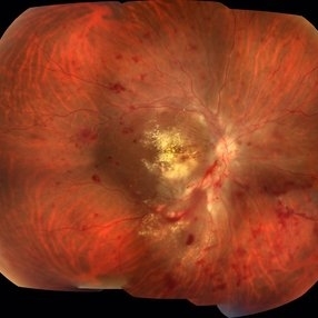
 Acute Idiopathic Occlusive Retinal Vasculitis
May 31 2014 by Hamid Ahmadieh, MD
Color fundus photograph of the right eye of a 28-year-old woman with sudden drop of vision due to acute occlusive retinal vasculitis leading to extensive nerve fiber layer infarction and retinal hemorrhages.
Photographer: Naghmeh Nozhat, Negah Eye Center, Tehran
Condition/keywords: color fundus photograph, cotton wool spots, retinal hemorrhage, retinal ischemia
-
 Cat Scratch Disease
Mar 29 2021 by Gabriel Costa Andrade, PhD
Fundus photograph of an 36-year-old woman with a macular vasculitis, pre retinal hemorrhage and exudation due to Bartonella henselae infection.
Photographer: Gabriel Andrade
Condition/keywords: cat scratch retinitis
-
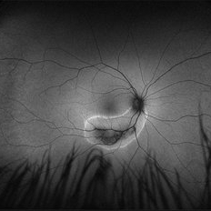
 Acute Idiopathic Occlusive Retinal Vasculitis
May 31 2014 by Hamid Ahmadieh, MD
Mid- phase fluorescein angiogram of the left eye of a 28-year-old woman with acute drop of vision due to occlusive retinal vasculitis leading to extensive capillary nonperfusion and macular infarction.
Photographer: Naghmeh Nozhat, Negah Eye Center, Tehran
Imaging device: Heidelberg Spectralis
Condition/keywords: capillary nonperfusion, retinal vasculitis
-
 Acute Idiopathic Occlusive Retinal Vasculitis
May 31 2014 by Hamid Ahmadieh, MD
Color fundus photograph of the left eye of a 28-year-old woman with acute drop of vision due to occlusive retinal vasculitis leading to extensive nerve fiber layer infarction and retinal hemorrhages.
Photographer: Naghmeh Nozhat, Negah Eye Center, Tehran
Condition/keywords: color fundus photograph, cotton wool spots, retinal hemorrhage, retinal ischemia
-
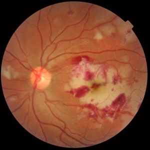
 Acute Necrotizing Retinal Vasculitis as Onset of Systemic Lupus Erythematosus.
Sep 3 2016 by ADRIANO FERREIRA
A 28-year-old white man was referred to the rheumatology clinic with gradually and rapid deterioration of the vision (both eyes). In this picture, we can observe cotton wool spots in the papillomacular area and extensive hemorrhages in posterior polo and in the middle periphery. Hard exudates are present in macular area (macular edema)
Photographer: Claudio Zett Lobo
Imaging device: TRC50DXi TOPCON
Condition/keywords: systemic lupus erythematosus (SLE) vasculitis, vasculitis
-
 Acute Retinal Necrosis
Jul 3 2025 by Heitor Nogueira
Fundus photograph of an 53-year-old woman with patient who reported unilateral visual acuity loss for 10 days associated with ocular pain. She presented conjunctival hyperemia with temporal and nasal nodular scleritis, anterior chamber reaction 2+/4+, Koeppe nodules, granulomatous PKs, vitritis 2+/4+, multiple areas of vasculitis in arcades and periphery, associated with hemorrhages and necrotizing retinitis in temporal, inferior and nasal periphery. patient who reported unilateral visual acuity loss for 10 days associated with ocular pain. He presented conjunctival hyperemia with temporal and nasal nodular scleritis, anterior chamber reaction 2+/4+, Koeppe nodules, granulomatous PKs, vitreitis 2+/4+, multiple areas of vasculitis in the arcades and periphery, associated with hemorrhages and necrotizing retinitis in the temporal, inferior and nasal periphery. Positive serology for Herpes Virus.
Photographer: Heitor Nogueira, Penido Burnier Institute and CHOV, Campinas, São Paulo, Brazil
Imaging device: Optos Daytona
Condition/keywords: ARN complications, Herpes, progressive outer retinal necrosis (PORN)
-
 Acute Zonal Occult Outer Retinopathy (AZOOR) FA, Fluorescein Angiography, Peripheral Vasculitis
Jan 19 2022 by James B. Soque, CRA, OCT-C, COA, FOPS
Acute Zonal Occult Outer Retinopathy (AZOOR). Peripheral Vasculitis OD. Fluorescein angiography showing vasculitis in the far right periphery 8-10 o'clock. 46-year-old white male, VA CC 20/16, 20/12.5, has had recurrent vasculitis for 11 years. No treatment.
Photographer: James Soque, CRA, OCT-C, COA, FOPS, Island Retina, Shirley, NY
Imaging device: Optos California
Condition/keywords: acute zonal occult outer retinopathy (AZOOR), FA early phase, fluorescein angiogram (FA), Peripheral Vasculitis, ultra-wide field imaging
-
 Acute Zonal Occult Outer Retinopathy, (AZOOR) FAF, Fundus Autofluorescence
Jan 19 2022 by James B. Soque, CRA, OCT-C, COA, FOPS
Acute Zonal Occult Outer Retinopathy, FAF, Fundus Auto Fluorescence, OD. 46-year-old white male, VA CC 10/16, 20/12.5, has had recurrent vasculitis for 11 years. No treatment.
Photographer: James Soque, CRA, OCT-C, COA, FOPS, Island Retina, Shirley, NY
Imaging device: Optos California
Condition/keywords: acute zonal occult outer retinopathy (AZOOR), fundus autofluorescence (FAF), ultra-wide field imaging
-
 AMPPE Ret Vasculitis
Jul 29 2013 by H. Michael Lambert, MD
AMPPLE ret vasculitis.
Condition/keywords: acute posterior multifocal placoid pigment epitheliopathy (APMPPE)
-
 Behcet's Disease
Mar 13 2013 by Hamid Ahmadieh, MD
Wide field FA of the left eye of a 23-year-old man with retinal vasculitis due to Behcet's disease .
Photographer: Solmaz Shahmohammadi , Negah Eye Center, Tehran
Imaging device: Heidelberg Spectralis
Condition/keywords: retinal vasculitis
-
 Behcet's Disease
Mar 13 2013 by Hamid Ahmadieh, MD
Wide field FA of the right eye of a 23-year-old man with retinal vasculitis and branch retinal vein occlusion (BRVO) due to Behcet's disease .
Photographer: Solmaz Shahmohammad, Negah Eye Center, Tehran
Imaging device: Heidelberg Spectralis
Condition/keywords: branch retinal vein occlusion (BRVO), retinal vasculitis
-
 Behcet's Syndrome
Apr 1 2016 by Nichole Lewis
Behcet's Syndrome
Photographer: Nichole Lewis - Pennsylvania Retina Specialists, Camp Hill, PA
Condition/keywords: Behcet's Disease, obliterative peripheral vasculitis
-
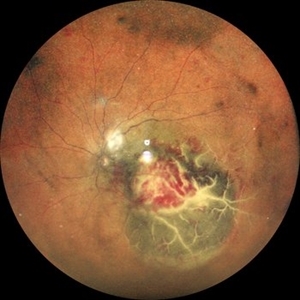 Choroidal Abscess with occlusive retinal vasculitis
Jan 28 2023 by Anjana Mirajkar, MS Ophthalmology
Widefield color image of LE of a 55 year old male a case of Choroidal abscess with occlusive retinal vasculitis.
Photographer: Dr. Anjana Mirajkar -Retina Foundation, Ahmedabad
Condition/keywords: choroidal abscess
-
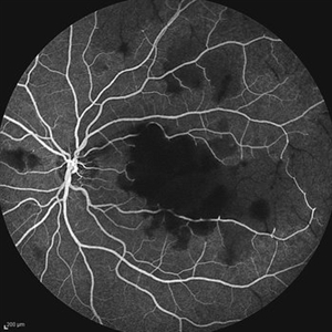
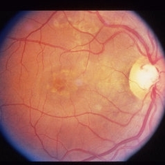
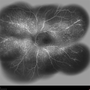
 Idiopathic retinal vasculitis, aneurysms and neuroretinitis
Apr 24 2022 by Aniruddha K Agarwal, MD
Ultra-wide field fundus fluorescein angiography (FFA) of the left eye from an asymptomatic, healthy 33-year-old woman who was referred to the retina clinic from a refractive surgery unit due to the presence of vascular anomalies and hard exudates in both eyes. FFA revealed the characteristic sacular aneurysms at the bifurcation of retinal arterioles in the posterior pole, together with microvascular anomalies and capillary closure peripherally.
Photographer: Julio J GONZALEZ-LOPEZ, MD, PhD, FEBO and Teresa GONZALEZ-LOMAS, RN
Imaging device: Optos California
Condition/keywords: IRVAN Syndrome, IUSG, neuroretinitis, retinal vasculitis, uveitis
-
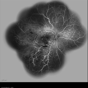
 Idiopathic Retinal Vasculitis, Aneurysms, and Neuroretinitis (IRVAN)
Oct 16 2012 by S. Natarajan, MD, FASRS, FRCS (GLASGOW) , FICO, D.Sc, FELA
FFA photograph of a 28-year-old male with IRVAN Syndrome.
Photographer: Prof. Dr. S. Natarajan
Condition/keywords: aneurysm, neuroretinitis, retinal vasculitis
-
 Idiopathic Retinal Vasculitis, Aneurysms, and Neuroretinitis (IRVAN)
Oct 16 2012 by S. Natarajan, MD, FASRS, FRCS (GLASGOW) , FICO, D.Sc, FELA
Fundus photograph of a young male with IRVAN Syndrome
Photographer: Prof. Dr. S. Natarajan
Condition/keywords: aneurysm, neuroretinitis, retinal vasculitis
-
 Idiopathic Vasculitis
Feb 4 2023 by Aditya S Kelkar, MS, FRCS, FASRS,FRCOphth
Color fundus photograph of the left eye showing idiopathic retinal vasculitis.
Photographer: Dr. Pranali Surawase. National Institute of Ophthalmology, Pune, India.
Imaging device: Zeiss Clarus 500
Condition/keywords: retinal vasculitis, vasculitis
-
 Laser Induced BRAO in IRVAN Syndrome
May 3 2019 by Deependra Vikram Singh, MD FASRS
Fundus photograph of a 26-year-old man with IRVAN syndrome referred for vitreous surgery in OS for secondary rhegmatogenous retinal detachment. OD has received laser photocoagulation for capillary nonperfusion areas and retinal artery macroaneurysm associated with retinal vasculitis. Fundus photograph of OD shows laser induced nasal BRAO. Case re-emphasizes why laser for macroaneurysm should be avoided in cases with IRVAN.
Photographer: Deependra V Singh, Eye-Q Superspecialty Eye Hospitals. Gurugram, India
Imaging device: Zeiss Visucam 500
Condition/keywords: arteriolar macroaneurysm, branch retinal artery occlusion (BRAO), laser photocoagulation
-
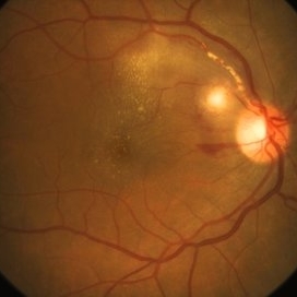
 NVE in a Patient With Vasculitis
Nov 5 2018 by awaneesh m upadhyay, MBBS, DNB
FFA image of a 22-year-old male vasculitis patient with NVE.
Photographer: Hiteshwar Saikia
Condition/keywords: neovascularization elsewhere (NVE), tuberculosis, vasculitis
-
 Obliterative Peripheral Vasculitis
Apr 3 2019 by HECTOR LUIS VILLARROEL GUIZAR, MD, RETINA FELLOW
Ultra-wide angle retinal angiography of an 27-year-old with retinal vasculitis with late stain of the vascular wall and obliteration of the peripheral vasculature.
Photographer: HECTOR VILLARROEL, HOSPITAL DE LA LUZ, MEXICO CITY
Imaging device: OPTOS
Condition/keywords: obliterative peripheral vasculitis
-
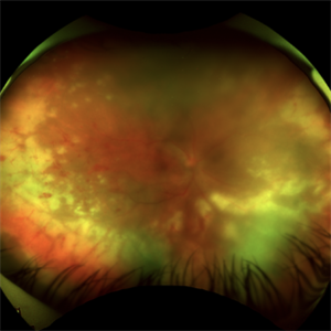
 Occlusive Retinal Vasculitis
Oct 3 2024 by Logan ryzenga
4 view ultra-widefield Optos fluorescein angiogram in the left eye of a 39 year old woman occlusive retinal vasculitis with four quadrant Kyrieleis plaques. This is a showcase of a suspected, rarely reported, and atypical presentation of Behcet's Syndrome.
Photographer: Logan Ryzenga
Imaging device: Optos California
Condition/keywords: Behcet's Disease, Behcet's uveitis, Fluorescein angiography, fluorescein leakage, kyrieleis plaques, non-perfusion, OPTOS, OPTOS CALIFORNIA, ultra-wide field imaging, Uveitis
-
 Oral Ulcers in Behcet's Disease
Apr 10 2017 by Deepak Bhojwani, MS
A 19-year-old boy presented with with recurrent oral and genital ulcers along with blurring of vision. Systemically he was HLA B-51 Positive suggesting Behcet's Disease. The photograph depicts the classic aphthous mouth ulcers seen in the Behcet's disease.
Photographer: DEEPAK BHOJWANI, RAGHUDEEP EYE HOSPITAL, AHMEDABAD.
Condition/keywords: Behcet's uveitis, vasculitis
 Loading…