Search results (141 results)
-
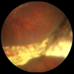
 Exposed Scleral Buckle, with Exposed Suture, Infection - Infero View
Feb 4 2013 by James B. Soque, CRA, OCT-C, COA, FOPS
External photograph of a 66-year-old WM with Hx of SBOD in 2009, graft attempt failed, infection resulted. Scheduled for removal of SBOD.
Photographer: James Soque CRA COA
Imaging device: External Photo, Topcon TRC 50 DX, MERGE software
Condition/keywords: exposed scleral buckle, exposed suture, infection
-
---thumb.jpg/image-square;max$300,300.ImageHandler) C3F8 gas bubble after retinal detachment surgery
C3F8 gas bubble after retinal detachment surgery
Feb 1 2013 by Sharon Fekrat, MD FACS FASRS
63 year old man s/p encircling scleral buckle and 23g pars plana vitrectomy for a macula off phakic rhegmatogenous retinal detachment. This fundus photograph shows the effect of the encircling buckle and the residual C3F8 intravitreal gas bubble in the right eye.
Photographer: Tiffanie Keaton, Duke Eye Imaging, Duke University Eye Center, Durham, NC
Imaging device: Optos
Condition/keywords: intravitreal gas bubble, vitrectomy
-
 UWF of Retinal Detachment Corrected with Scleral Buckle
Aug 29 2017 by Carolyn Daley
An ultra wide field fundus photograph of a 57-year-old male who has a past history of retinal detachment corrected with scleral buckle and three treated retinal tears.
Photographer: Carolyn Daley
Imaging device: Optos Imaging
Condition/keywords: cryo-retinal tear, cryotherapy, Optos, retinal tear, scleral buckle, ultra-wide field imaging
-
 Exposed Scleral Buckle, with Exposed Suture, Infection - Infero Nasal View, Upgaze
Feb 4 2013 by James B. Soque, CRA, OCT-C, COA, FOPS
External Photograph of a 66-year-old WM with Hx of SBOD in 2009, graft attempt failed, infection resulted. Scheduled for removal of SBOD.
Photographer: James Soque CRA COA
Imaging device: External Photo, Topcon TRC 50 DX, MERGE software
Condition/keywords: scleral buckle, suture exposed
-
 Scleral Buckle and Cryo Color
Dec 29 2012 by Barbara Parolini, MD
Panoramic fundus photograph of a 55-year-old man after episcleral sugary for retinal detachment. An encircling scleral buckle and a superotemporal cryotherapy scar are visible.
Photographer: Barbara Parolini, MD
Imaging device: Daytona
Condition/keywords: scleral buckle
-
 Exposed Scleral Buckle, with Exposed Suture, Infection - Infero Temporal View
Feb 4 2013 by James B. Soque, CRA, OCT-C, COA, FOPS
External Photograph of a 66-year-old WM with Hx of SBOD in 2009, graft attempt failed, infection resulted. Scheduled for removal of SBOD.
Photographer: James Soque CRA COA
Imaging device: External Photo, Topcon TRC 50 DX, MERGE software
Condition/keywords: exposed suture, scleral buckle
-
 Extruded Scleral Buckle
Oct 19 2012 by Larry Halperin, MD
Extruded scleral buckle
Condition/keywords: extruded scleral buckle
-
 Aphakic Retinal Detachment
Nov 9 2012 by Norman Byer
This 75-year-old woman had a scleral buckling operation on this aphakic eye two months previously. Two months following surgery she again had new symptoms of blurred vision and was found to have a redetachment caused by this small tractional retinal tear which may have been missed at the time of her original operation.
Condition/keywords: aphakic eye, scleral buckle, tractional retinal tear
-
 Exposed Scleral Buckle, with Infection - Infero Nasal View
Feb 4 2013 by James B. Soque, CRA, OCT-C, COA, FOPS
External photograph of a 66-year-old WM with Hx of SBOD in 2009, graft attempt failed, infection resulted. Scheduled for removal of SBOD.
Photographer: James Soque CRA COA
Imaging device: External Photo, Topcon TRC 50 DX, MERGE software
Condition/keywords: scleral buckle, suture exposed
-
 Optos Picture With Speculum: Dislocated Natural Lens
Oct 9 2018 by John S. King, MD
55-year-old white female with history of pathologic myopia+, lattice (laser), SB OU (1990s), and dislocated natural lenses OU that had been watched for years. In the fellow eye she developed phacolytic glaucoma and a PPV, PPL was performed. Plan for both eyes are monitoring. I wanted to get a good picture of her lens today with the optos machine, as the other pics had artifact from the lower lid. It worked out well to use a speculum in the left eye. Vision cc is 20/400 J1+ OD and 20/40 J2 OS; aphakic OU; vitreous clear OD; dislocated lens OS (see pic); retinas attached.
Photographer: Maisee Yang
Imaging device: Optos California
Condition/keywords: dislocated crystalline lens, pathologic myopia, scleral buckle, staphyloma
-
 Scleral Buckle
Oct 2 2013 by Jerald A. Bovino, MD
The elevation from a scleral buckle is noticeable in this mildly high buckle.
Condition/keywords: scleral buckle
-
 Scleral Buckle Suture
Jul 14 2013 by Jason S. Calhoun
Scleral buckle suture temporal quadrant.
Photographer: Jason S. Calhoun, Department of Ophthalmology, Mayo Clinic Jacksonville, Florida
Imaging device: TOPCON D-90 SL NIKON CAMERA
Condition/keywords: scleral buckle
-
 Encircling Scleral Buckle Axial View
Dec 10 2012 by Yale L. Fisher, MD
The buckle is an encircling element. the anterior posterior view shows a cross section above and below on the screen. Rotation vertically demonstrates a superior and inferior cross-section of a highly elevated scleral buckle. There is movement of the separated formed vitreous and far anterior (visible at the left of the screen). Optic nerve shadow is visible as the nerve moves vertically.
Condition/keywords: B scan ultrasound, scleral buckle, video
-
 Vitreous Base Avulsion
Sep 19 2019 by Anfisa Ayalon, MD
Fundus picture of a 34-year-old patient with left eye vitreous base avulsion three months after rhegmatogenous retinal detachment repair with circular scleral buckle implantation. Note bucket handle sign and 360 degrees scleral buckle indentation with a flat retina.
Photographer: Anfisa Ayalon, MD., Meir Medical Center, Kfar Saba, Israel.
Imaging device: California, Optos 200 DTX
Condition/keywords: avulsed vitreous base, behind the vitreous base, scleral buckle
-
 Exposed Scleral Buckle, Infection - Temporal View
Feb 4 2013 by James B. Soque, CRA, OCT-C, COA, FOPS
External Photograph of a 66-year-old WM with Hx of SBOD in 2009, graft attempt failed, infection resulted. Scheduled for removal of SBOD.
Photographer: James Soque CRA COA
Imaging device: External Photo, Topcon TRC 50 DX, MERGE software
Condition/keywords: scleral buckle, suture exposed
-
 Retinal Tear
Oct 12 2012 by Jeffrey G. Gross, MD, FASRS
Retinal tear, on radial scleral buckle.
Condition/keywords: radial scleral buckle, retinal tear
-
 Retinal Detachment Repair With Silicone Oil and Scleral Buckle, Fourteen Years Later, With Visual Acuity of 20/25
Sep 12 2016 by Timothy S Fuller, MD
65-year-old woman s/p scleral buckle 14 years ago. Two weeks later, the retina re-detached, and vitrectomy, laser, and silicone oil procedure was performed. Patient remains 20/25 with correction fourteen years later. The cornea is clear, there is no oil emulsification, and there is a stable, moderately inferiorly subluxated PCIOL (as it was prior to RD surgery). IOP is 17 on Cosopt BID.
Photographer: Nicholas Hesse, Texas Retina Associates
Imaging device: Optos
Condition/keywords: laser, scleral buckle, silicone oil
-
 Retinal Tear on Radial Scleral Buckle
Oct 12 2012 by Jeffrey G. Gross, MD, FASRS
Retinal tear on radial scleral buckle.
Condition/keywords: radial scleral buckle, retinal tear
-
 Giant Retinal Tear Slide 3
Oct 22 2012 by Ronald C. Gentile, MD
Following repair of the retinal detachment with vitrectomy and scleral buckle the edge of the giant retinal tear can be seen to be lying flat on the indentation of the scleral buckle.
Photographer: The New York Eye & Ear Infirmary Department of Medical Imaging
Condition/keywords: retinal tear, vitrectomy
-
 Horseshoe Tear With Scleral Buckle
Oct 31 2013 by Jason S. Calhoun
Patient had a retinal detachment with retinal tear superior temporally. Underwent surgery and had a scleral buckle placed with good support of the tear. VA is count fingers and will return in 2-months for follow up.
Photographer: Jason S. Calhoun, Ophthalmic Photographer, Department of Ophthalmology, Mayo Clinic Jacksonville
Imaging device: TOPCON TRC 50-EX
Condition/keywords: retinal tear, scleral buckle
-
---thumb.JPG/image-square;max$300,300.ImageHandler) Scleral Buckle Suture 1
Scleral Buckle Suture 1
Jul 14 2013 by Jason S. Calhoun
Scleral buckle suture temporal quadrant.
Photographer: Jason S. Calhoun, Department of Ophthalmology, Mayo Clinic Jacksonville, Florida
Imaging device: TOPCON D-90 SL NIKON CAMERA
Condition/keywords: scleral buckle
-
 Scleral Buckle Protrusion
Feb 23 2015 by Matt Poe, COA
This was taken with a Nikon D80 camera to document the protrusion of the scleral buckle.
Photographer: Matt Poe, COA. Northwest Arkansas Retina Associates, Springdale, AR.
Condition/keywords: external, extruded scleral buckle, scleral buckle
-
 PVR Retinal Detachment with subretinal bands Slide 3
Oct 22 2012 by Ronald C. Gentile, MD
Postoperative fundus photo of the margin of the inferior nasal retinectomy site. The retinectomy was used intra-operatively to flap the retina over and remove the subretinal bands. The scleral buckle effect and acute white endolaser marks are present at the edge of the retinectomy.
Photographer: The New York Eye & Ear Infirmary Department of Medical Imaging
Condition/keywords: proliferative vitreoretinopathy (PVR), subretinal bands
-
 Giant Retinal Tear Slide 2
Oct 22 2012 by Ronald C. Gentile, MD
Following repair of the retinal detachment with vitrectomy and scleral buckle the retina is attached and the patient's vision improved.
Photographer: The New York Eye & Ear Infirmary Department of Medical Imaging
Condition/keywords: retinal tear, vitrectomy
-
 Scleral Buckle and Cryoptherapy Scar
Dec 29 2012 by Barbara Parolini, MD
Panoramic autofluorescence photograph of a 55-year-old man after episcleral sugary for retinal detachment. An encircling scleral buckle, a superotemporal cryotherapy scar and the demarcation line of hyperautofluorescence in the previous detachment area are visible.
Photographer: Barbara Parolini, MD
Imaging device: Daytona
Condition/keywords: scleral buckle
 Loading…