Search results (26 results)
-
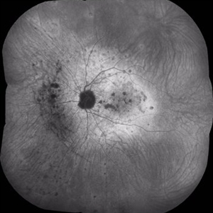
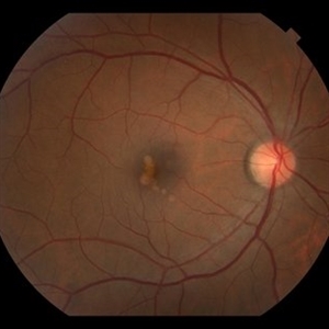
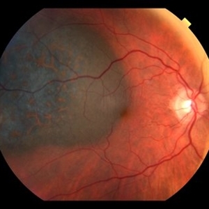 Orange Pigment Overlying a Lesion Suspicious for a Choroidal Melanoma
Jan 16 2019 by John S. King, MD
76-year-old white male saw his eye doctor with a three week complaint of photopsias and a shadow in his vision. Found to have a 10.5/12.5/2.5 (medium reflectivity) pigmented, choroidal mass associated with SRF and orange pigment (hyper-autofluorescence of lipofuscin), and without drusen or halo. See photo
Photographer: Stacey Coleman
Imaging device: Topcon 50
Condition/keywords: lipofuscin, orange pigment
-
 Acute Macular Neuroretinopathy OD
Mar 28 2013 by John S. King, MD
19-year-old patient c ADHD and acute photopsia following flu-like illness; inferior scotoma OD. Ellipsoid region abn c characteristic parafoveal lesions on voxel.
Photographer: Wayne A. Ladlee Jr., OcuSight Eye Care Center, Rochester, NY
Imaging device: Cirrus
Condition/keywords: acute macular neuroretinopathy
-
 Acute Macular Neuroretinopathy OS
Mar 28 2013 by John S. King, MD
19-year-old patient c ADHD and acute photopsia following flu-like illness; inferior scotoma OD. Ellipsoid region abn c characteristic parafoveal lesions on voxel.
Photographer: Wayne A. Ladlee Jr., OcuSight Eye Care Center, Rochester, NY
Imaging device: Cirrus
Condition/keywords: acute macular neuroretinopathy
-
 Retinal Tear
Jan 8 2018 by John S. King, MD
Acute floaters/photopsias
Imaging device: Optos
Condition/keywords: bridge of tissue between tears, full thickness retinal tear
-
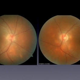
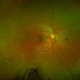
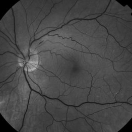
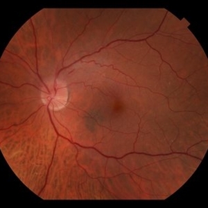 NAION, ERM
Sep 7 2018 by John S. King, MD
70-year-old white male with background history of fovea involving RD repaired with PPV 5 months ago, and history of HTN. Some recent photopsias, mild scotoma. Focal ERM superiorly and swollen superior part of the optic disc.
Photographer: Macey
Imaging device: Topcan
Condition/keywords: drusen, epiretinal membrane (ERM), ischemic optic neuropathy
-
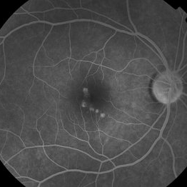
 Hyper-autofluorescence of Orange Pigment Overlying a Lesion Suspicious for a Choroidal Melanoma
Jan 16 2019 by John S. King, MD
76-year-old white male saw his eye doctor with a three week complaint of photopsias and a shadow in his vision. Found to have a 10.5/12.5/2.5 (medium reflectivity) pigmented, choroidal mass associated with SRF and orange pigment (hyper-autofluorescence of lipofuscin, see image), and without drusen or halo.
Photographer: Stacey Coleman
Imaging device: Topcon 50
Condition/keywords: lipofuscin, orange pigment
-
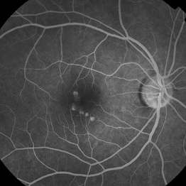
 BSCR/HLA-A29+
Jul 1 2014 by John S. King, MD
BSCR/HLA-A29+, MA male, photopsia on p/c, small hypopigmented round to oval lesions mainly peripapillary and inf-nasal to nerve, controlled on cellcept and cyclosporine.
Photographer: Wayne A Ladlee Jr
Condition/keywords: birdshot chorioretinopathy
-
 MEWDS
MEWDS
Feb 25 2018 by Armando L. Oliver, MD
Young male college student with unilateral photopsias. Fundus findings were compatible with MEWDS. The condition resolved without therapy.
Photographer: Moises Castro
Imaging device: Optos California Red Green Photos
Condition/keywords: multiple evanescent white dot syndrome (MEWDS)
-
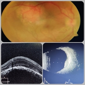 Circumscribed Choroidal Hemangioma
Jul 3 2020 by Dhaivat Shah
A 30-year-old young male presented with drop in vision in right eye since 1 year (6/60). Fundus examination revealed choroidal hemangioma superotemporal to macula. Choroidal hemangioma is an unusual benign vascular tumor of the choroid. It can be circumscribed solitary or diffuse tumor with the later having other systemic associations. Circumscribed choroidal hemangiomas (CCHs) are usually unilateral, unifocal hamartomatous vascular tumor affecting people in second to fourth decade. It appers as round to oval, orangish-red mass in posterior pole with smooth homogenous surface mostly present in macular and peripapillary area. Hyperopic shift is seen in sub-foveal tumors in contrast to para-foveal ones which are usually asymptomatic or present with metamorphopsia or photopsia and diminished vision secondary to exudative retinal detachment. B-scan shows highly reflective tumor without any shadowing or acoustic solidity with high anterior A scan spike. EDI-OCT here depicts a smooth gently sloping choridal mass with compressed choriocapillaries and enlarged medium and large choroidal vessels. Over a period of time structural abnormalities of the outer retina can be visualised. Ancillary testing using Fluorescein Angiography shows lacy hyper-fluorescence during early arterial phase followed by increased hyper-fluorescence due to progressive profuse leakage from pin point foci during arterial and venous phase. Indocyanine green angiography shows lacy diffuse fluorescent tumor in early phase followed by hypo-fluorescent tumor due to dye wash out in late phase. Intrinsic auto-fluorescence is also seen in CCHs from lipofuscin and fresh sub-retinal fluid. Tumor is relatively hyper-intense with respect to vitreous in T1-weighted images in iso-intense in T2-weighted images of MRI. Asymptomatic cases need no treatment, while patients showing vision loss with presence or absence of exudative retinal detachment can be treated with photodynamic therapy which is preferred treatment due to site specific action. Selective occlusion of choroidal neovascularization can be achieved while the neurosensory retinal layers and Bruch membrane are almost unaffected, leaving retinal function intact. Green or rarely red wavelength laser photocoagulation is used to create a chorioretinal adhesion and resolve the SRF. Other treatment modalities include Transpupilary thermotherapy, external beam irradiation, proton beam therapy, brachytherapy and gamma knife.
Photographer: Miss Deepika Nagle
Imaging device: Zeiss
Condition/keywords: B scan ultrasound, choroidal hemangioma, fundus photograph, optical coherence tomography (OCT), photodynamic therapy
-
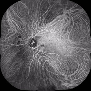 Multifocal Choroiditis
Aug 16 2018 by FELIPE PEREIRA
Mid-phase indocyanine green angiography of a 25-year-old woman with sudden central vision loss and photopsias for 7 days. The hypofluorescent lesions in the macula and nasal to the disc correspond to the yellow-white deep lesions in the fundus examination. No leakage is observed at any stage of the exam
Photographer: Claudio Zett Lobos
Imaging device: HEIDELBERG SPECTRALIS HRA
Condition/keywords: indocyanine green (ICG) angiography, multifocal choroiditis, white dot syndrome
-
 MEWDS
MEWDS
Feb 25 2018 by Armando L. Oliver, MD
Young male college student with unilateral photopsias. Fundus findings were compatible with MEWDS. The condition resolved without therapy.
Photographer: Moises Castro
Imaging device: Optos California FAF
Condition/keywords: multiple evanescent white dot syndrome (MEWDS)
-
 NAION, ERM
Sep 7 2018 by John S. King, MD
70-year-old white male with background history of fovea involving RD repaired with PPV 5 months ago, and history of HTN. Some recent photopsias, mild scotoma. Focal ERM superiorly and swollen superior part of the optic disc.
Photographer: Macey
Imaging device: Topcon
Condition/keywords: drusen, epiretinal membrane (ERM), ischemic optic neuropathy
-
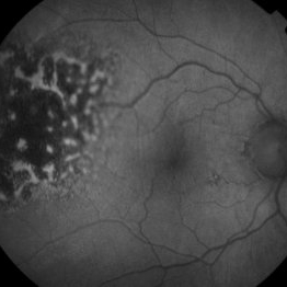
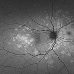
 Multiple Evanescent White Dot Syndrome
Feb 10 2021 by Cláudia Farinha
Ultra-widefield color and autofluorescence of a 30-year-old myopic female with decreased visual acuity, photopsias, and temporal scotomata.
Photographer: Claudia Farinha, MD
Imaging device: Optomap, Optos
Condition/keywords: multiple evanescent white dot syndrome (MEWDS)
-
 CMV Retinitis
Jan 10 2019 by Rahul Komati, MD
63-year-old male with history of plasma cell leukemia, presenting with photopsias and 20/25 vision. Fundus photograph shows superior area of retinitis, intraretinal hemorrhage, and vessel sclerosis. Retinitis regressed with systemic valganciclovir and 5 intravitreal foscarnet injections over 3 weeks.
Photographer: Pamela Hulvey, University of Chicago
Imaging device: Optos
Condition/keywords: CMV retinitis
-
 MEWDS
MEWDS
Feb 25 2018 by Armando L. Oliver, MD
Young male college student with unilateral photopsias. Fundus findings were compatible with MEWDS. The condition resolved without therapy.
Photographer: Moises Castro
Imaging device: Optos California IVFA
Condition/keywords: multiple evanescent white dot syndrome (MEWDS)
-
 Multifocal Choroiditis
Dec 22 2018 by FELIPE PEREIRA
Late phase ICG exam of a 25-year-old woman with 7 days history of central vision loss and photopsias. The hypofluorescent dots corresponds to the classically yellow-white lesion in the retinography. In the late fases the hypofluorescent lesions acquire a hiperfluorescent ring of staining. This image demonstrate more lesion than is possible to see in the clinical exam.
Photographer: Claudio Zett Lobos
Imaging device: HEIDELBERG SPECTRALIS HRA
Condition/keywords: indocyanine green (ICG) angiography, multifocal choroiditis, white dot syndrome
-
 MEWDs
MEWDs
Feb 28 2022 by Sruthi Arepalli, MD
Fundus photograph of a 37 year old woman presenting with photopsias and ellipsoid zone disruptions on OCT with findings consistent with MEWDS on autofluorescence. These areas of autofluorescence improved with oral steroids.
Condition/keywords: Autoflourescence, multiple evanescent white dot syndrome (MEWDS)
-
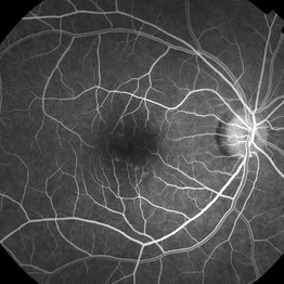
 Unilateral PIC Following Recent Influenza Vaccine
Jan 6 2019 by John S. King, MD
FA 5 minute 42-year-old African American female with high myopia and type 2 diabetes, presented to her eye doctor with distortion in the right eye "that looked like seeing through a Coke bottle." She denied any photopsias or other symptoms. She received an influenza vaccine two weeks before onset of metamorphopsia. I saw her about a month after symptoms began. Va cc 20/30 OD J1 (20/15 J1+ OS); a/c and vitreous without cell or flare. Posterior pole OD showed yellowish, rounded small to medium RPE pigment alterations without heme or exudes (OS U/R). FA showed early focal areas of hyperfluorescence that stained in the later frames without CNVM or CME; rare MA inferiorly. The OCT showed some focal PEDs with some possible overlying SRHRM. We discussed options and decided to try a medrol dose pack. A few weeks later she was 20/20 J1, with minimal to no symptoms; OCT shows near total resolution of PEDs.
Photographer: Kay Dalby
Imaging device: Topcon 50
Condition/keywords: punctate inner choroidopathy (PIC), white dot syndrome
-
 Unilateral PIC Following Recent Influenza Vaccine
Jan 6 2019 by John S. King, MD
42-year-old African American female with high myopia and type 2 diabetes, presented to her eye doctor with distortion in the right eye "that looked like seeing through a Coke bottle." She denied any photopsias or other symptoms. She received an influenza vaccine two weeks before onset of metamorphopsia. I saw her about a month after symptoms began. Va cc 20/30 OD J1 (20/15 J1+ OS); a/c and vitreous without cell or flare. Posterior pole OD showed yellowish, rounded small to medium RPE pigment alterations without heme or exudes (OS U/R). FA showed early focal areas of hyperfluorescence that stained in the later frames without CNVM or CME; rare MA inferiorly. The OCT showed some focal PEDs with some possible overlying SRHRM. We discussed options and decided to try a medrol dose pack. A few weeks later she was 20/20 J1, with minimal to no symptoms; OCT shows near total resolution of PEDs.
Photographer: Kay Dalby
Imaging device: Topcon 50
Condition/keywords: punctate inner choroidopathy (PIC), white dot syndrome
-
 AMPPPE
Mar 26 2019 by Gary R. Cook, MD, FACS
Left eye of the same 31-year-old white female with a history of photopsias OS X 3 months. VA= 20/20.
Condition/keywords: acute multifocal placoid pigment epitheliopathy (AMPPE), photopsia
-
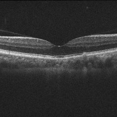 Unilateral PIC Following Recent Influenza Vaccine
Jan 6 2019 by John S. King, MD
OCT few weeks after initial visit. 42-year-old African American female with high myopia and type 2 diabetes, presented to her eye doctor with distortion in the right eye "that looked like seeing through a Coke bottle." She denied any photopsias or other symptoms. She received an influenza vaccine two weeks before onset of metamorphopsia. I saw her about a month after symptoms began. Va cc 20/30 OD J1 (20/15 J1+ OS); a/c and vitreous without cell or flare. Posterior pole OD showed yellowish, rounded small to medium RPE pigment alterations without heme or exudes (OS U/R). FA showed early focal areas of hyperfluorescence that stained in the later frames without CNVM or CME; rare MA inferiorly. The OCT showed some focal PEDs with some possible overlying SRHRM. We discussed options and decided to try a medrol dose pack. A few weeks later she was 20/20 J1, with minimal to no symptoms; OCT shows near total resolution of PEDs.
Photographer: Kay Dalby
Imaging device: Topcon 50
Condition/keywords: punctate inner choroidopathy (PIC), white dot syndrome
-
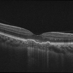 Unilateral PIC Following Recent Influenza Vaccine
Jan 6 2019 by John S. King, MD
Initial OCT. 42-year-old African American female with high myopia and type 2 diabetes, presented to her eye doctor with distortion in the right eye "that looked like seeing through a Coke bottle." She denied any photopsias or other symptoms. She received an influenza vaccine two weeks before onset of metamorphopsia. I saw her about a month after symptoms began. Va cc 20/30 OD J1 (20/15 J1+ OS); a/c and vitreous without cell or flare. Posterior pole OD showed yellowish, rounded small to medium RPE pigment alterations without heme or exudes (OS U/R). FA showed early focal areas of hyperfluorescence that stained in the later frames without CNVM or CME; rare MA inferiorly. The OCT showed some focal PEDs with some possible overlying SRHRM. We discussed options and decided to try a medrol dose pack. A few weeks later she was 20/20 J1, with minimal to no symptoms; OCT shows near total resolution of PEDs.
Photographer: Kay Dalby
Imaging device: Topcon 50
Condition/keywords: punctate inner choroidopathy (PIC), white dot syndrome
-
 MEWDS
MEWDS
Feb 25 2018 by Armando L. Oliver, MD
Young male college student with unilateral photopsias. Fundus findings were compatible with MEWDS. The condition resolved without therapy.
Photographer: Moises Castro
Imaging device: Optos California IVFA
Condition/keywords: multiple evanescent white dot syndrome (MEWDS)
-
 Unilateral PIC Following Recent Influenza Vaccine
Jan 6 2019 by John S. King, MD
FA 2 minute 42-year-old African American female with high myopia and type 2 diabetes, presented to her eye doctor with distortion in the right eye "that looked like seeing through a coke bottle". She denied any photopsias or other symptoms. She received an influenza vaccine two weeks before onset of metamorphopsia. I saw her about a month after symptoms began. Va cc 20/30 OD J1 (20/15 J1+ OS); a/c and vitreous without cell or flare. Posterior pole OD showed yellowish, rounded small to medium RPE pigment alterations without heme or exudes (OS U/R). FA showed early focal areas of hyperfluorescence that stained in the later frames without CNVM or CME; rare MA inferiorly. The OCT showed some focal PEDs with some possible overlying SRHRM. We discussed options and decided to try a medrol dose pack. A few weeks later she was 20/20 J1, with minimal to no symptoms; OCT shows near total resolution of PEDs.
Photographer: Kay Dalby
Imaging device: Topcon 50
Condition/keywords: punctate inner choroidopathy (PIC), white dot syndrome
-
 Unilateral PIC following recent influenza vaccine
Jan 6 2019 by John S. King, MD
FA 22 seconds 42-year-old African American female with high myopia and type 2 diabetes, presented to her eye doctor with distortion in the right eye "that looked like seeing through a Coke bottle." She denied any photopsias or other symptoms. She received an influenza vaccine two weeks before onset of metamorphopsia. I saw her about a month after symptoms began. Va cc 20/30 OD J1 (20/15 J1+ OS); a/c and vitreous without cell or flare. Posterior pole OD showed yellowish, rounded small to medium RPE pigment alterations without heme or exudes (OS U/R). FA showed early focal areas of hyperfluorescence that stained in the later frames without CNVM or CME; rare MA inferiorly. The OCT showed some focal PEDs with some possible overlying SRHRM. We discussed options and decided to try a medrol dose pack. A few weeks later she was 20/20 J1, with minimal to no symptoms; OCT shows near total resolution of PEDs.
Photographer: Kay Dalby
Imaging device: Topcon 50
Condition/keywords: punctate inner choroidopathy (PIC), white dot syndrome
 Loading…