 Initializing download.
-
 By FELIPE PEREIRA
UNIFESP
Co-author(s): Claudio Zeet Lobos - Uploaded on Dec 22, 2018.
- Last modified by Caroline Bozell on Jan 3, 2019.
- Rating
- Appears in
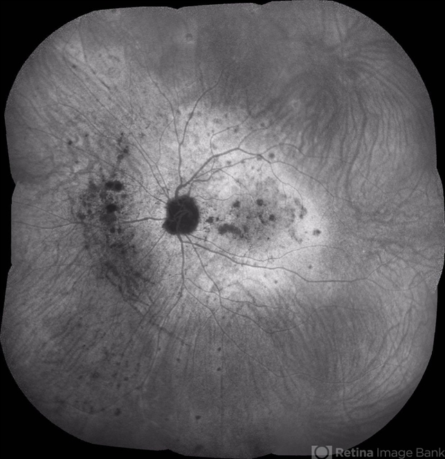
- Multifocal Choroiditis
- Condition/keywords
- multifocal choroiditis, white dot syndrome, indocyanine green (ICG) angiography
- Photographer
- Claudio Zett Lobos
- Imaging device
- HEIDELBERG SPECTRALIS HRA
- Description
- Late phase ICG exam of a 25-year-old woman with 7 days history of central vision loss and photopsias. The hypofluorescent dots corresponds to the classically yellow-white lesion in the retinography. In the late fases the hypofluorescent lesions acquire a hiperfluorescent ring of staining. This image demonstrate more lesion than is possible to see in the clinical exam.
")


")
")
")
")

")