Search results (63 results)
-
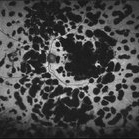
---thumb.jpg/image-square;max$300,300.ImageHandler) Multifocal Choroiditis
Multifocal Choroiditis
Feb 26 2013 by Henry J. Kaplan, MD
Multifocal choroiditis, MFC, inactive scars in the periphery.
Condition/keywords: multifocal choroiditis
-
 Toxoplasma chorioretinitis 1
Jan 11 2013 by Alex P. Hunyor, MD
Toxoplasmosis 1 - chorioretinal scar from previous toxoplasma chorioretinitis. See image 2 - recurrent todo adjacent to this scar
Condition/keywords: inactive toxoplasmosis, ocular toxoplasmosis, toxoplasmosis, toxoplasmosis retinitis
-
---thumb.jpg/image-square;max$300,300.ImageHandler) Multifocal Choroiditis and Panuveitis Syndrome
Multifocal Choroiditis and Panuveitis Syndrome
Feb 26 2013 by Henry J. Kaplan, MD
Multifocal choroiditis and panuveitis: left eye. Acute stage: haziness of the media due to vitritis and multiple active yellow and also inactive choroidal lesions are present.
Condition/keywords: multifocal choroiditis
-
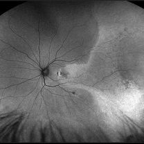
 Serpiginous Choroiditis
Feb 25 2013 by Henry J. Kaplan, MD
Serpiginous choroiditis, right eye. Both active and inactive lesions clearly visible; active lesions are the yellowish subretinal area most prominant nasal to optic nerve head and also around the inferior arcade and temporal to the macular lesion.
Condition/keywords: serpiginous choroiditis
-
 Serpiginous Choroiditis - Fundus Autofluorescence
Sep 20 2014 by Rameez N Hussain, MD
Fundus autofluorescence of serpiginous choroiditis showing decreased autofluorescence area corresponding to the inactive lesion (RPE atrophy) and increased autofluorescence area corresponding to active lesion.
Photographer: Dr.Rameez N Hussain, MD, Central Imaging Center, Vitreo Retinal Services, Giridhar Eye Institute, Cochin, India
Imaging device: Heidelberg Blue Peak Autofluorescence imaging.
Condition/keywords: serpiginous choroiditis
-
 Angioid Streaks with Macular Fibrosis Secondary to Inactive CNV
Oct 11 2012 by Gabriela Lopezcarasa Hernandez, MD
50-year-old female with decrease in VA in right eye
Photographer: Ricardo Montoya, Mexico
Imaging device: Zeiss FF4
Condition/keywords: angioid streaks
-
 Congenital Toxoplasmosis RE
Apr 29 2015 by Neha Goel, MS DNB FRCS (Glasg)
Fundus photograph of the right eye of a 25-year-old male with decreased vision since early childhood.
Photographer: Neha Goel
Imaging device: Zeiss visucam
Condition/keywords: congenital toxoplasmosis, inactive toxoplasmosis, toxoplasmosis
-
 Ocular Toxoplasmosis
Nov 20 2015 by Ahmad B. Tarabishy, MD
28-year-old male with active toxoplasmosis chorioretinitis OS and a large macular toxoplasmosis scar OD. Vision is 20/25 OU.
Photographer: Phaedra Lund, Retina Specialists of Tampa
Imaging device: Zeiss Cirrus OCT
Condition/keywords: inactive toxoplasmosis, macula lesion, toxoplasmosis
-
 OcularToxoplasmosis
Feb 25 2013 by Henry J. Kaplan, MD
Toxoplasmosis, right eye: congenital typical macular scar with peripheral hyperpigmentation.
Condition/keywords: inactive toxoplasmosis, toxoplasmosis
-
 Inactive Toxoplasmosis
Nov 9 2012 by Norman Byer
This 28-year-old man had inactive toxoplasmosis and presented with acute symptoms caused by this tractional retinal tear adjacent to a retinochorodial scar. He also had an acute posterior vitreous detachment which had torn this retinal operculum completely free. The next slide shows the same lesion. Note the early rolled edge on the left side of the tear.
Condition/keywords: acute posterior vitreous detachment, inactive toxoplasmosis, operculum, rolled edges of retina, tractional retinal tear
-
 Toxoplasma Scar
Sep 22 2018 by Hashim Ali Khan, OD, FAAO
Fundus photograph of a 17-year-old male with inactive macular toxoplasma scar.
Condition/keywords: inactive toxoplasmosis, toxoplasmosis chorioretinitis
-
 Inactive Photocoagulated Diabetic Retinopathy
Apr 5 2017 by Linda A Cernichiaro- Espinosa, MD
Extensive pan-retinal photocoagulation in a Mexican patient that was required to inactivate the disease.
Photographer: Linda A Cernichiaro, E
Imaging device: Optos Daytona
Condition/keywords: diabetes, laser photocoagulation
-
 Inactive Toxoplasmosis
Nov 9 2012 by Norman Byer
This is the same case as in the previous photograph showing the very large free operculum torn from the retina.
Condition/keywords: acute posterior vitreous detachment, free operculum, inactive toxoplasmosis, tractional retinal tear
-
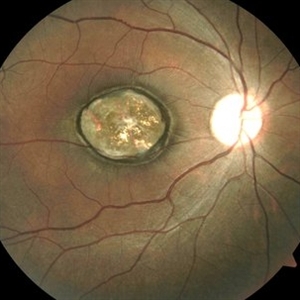
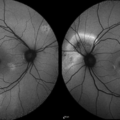
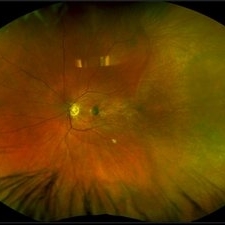
 Congenital Toxoplasmosis
Dec 18 2019 by Yoshihiro Yonekawa, MD, FASRS
Widefield fundus image of a teenage girl's right eye with an inactive congenital toxoplasmosis macular lesion. Her vision is 20/400 in this eye.
Photographer: Netanya Lerner, COA, Wills Eye Hospital/Mid Atlantic Retina
Imaging device: Optos California
Condition/keywords: congenital toxoplasmosis, pediatric retina
-
 Congenital Toxoplasmosis LE
Apr 29 2015 by Neha Goel, MS DNB FRCS (Glasg)
Fundus photograph of the left eye of a 25-year-old male with decreased vision since early childhood.
Photographer: Neha Goel
Imaging device: Zeiss visucam
Condition/keywords: congenital toxoplasmosis, inactive toxoplasmosis, toxoplasmosis
-
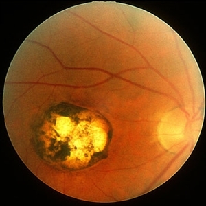
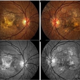
 Congenital Toxoplasmosis
Apr 8 2019 by Gary R. Cook, MD, FACS
Left eye of the same 38-year-old female with congenital toxoplasmosis lesion; V.A. = 20/40 due to temporal location of the Toxo scar.
Imaging device: Topcon VT-50
Condition/keywords: chorioretinal scar, congenital toxoplasmosis, inactive toxoplasmosis, macular scar, ocular toxoplasmosis
-
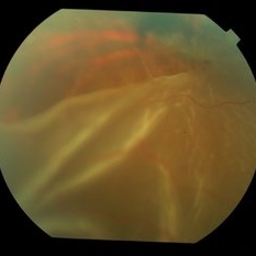
 Macula Off Retinal Detachment with CNV
Nov 11 2019 by Olivia Rainey
Ultra-wide field pseudocolor photograph of a 42-year-old female with a long-standing, macula-off retinal detachment affecting her left eye. Patient was unaware of vision loss until testing her visual acuity and she denied seeing flashing lights. Patient decided to proceed with scleral buckling. The CNV is potentially secondary the retinal detachment, but may be myopic related or idiopathic. The CNV appears fibrotic and inactive. The patient was warned that this will absolutely limit how much vision she recovers once the retina is reattached.
Photographer: Olivia Rainey
Imaging device: Optos California
Condition/keywords: choroidal neovascularization (CNV), chronic retinal detachment, fundus autofluorescence (FAF), left eye, montage, Optos, retinal detachment of the macula, ultra-wide field imaging
-
 Central Serous Chorioretinopathy
Jan 25 2022 by Olivia Rainey
Widefield fundus autofluorescence of a 60-year-old male with Central Serous Chorioretinopathy affecting both eyes. Chronic history of CSR followed with observation without treatment prior to presenting at our office. The physician noted significant findings on exam and imaging with multifocal areas of inactive and active changes in the right eye and subfoveal subretinal fluid with recent visual decline in the left eye. There are hyper and hypoautofluorescent changes, consistent with CSR.
Photographer: Olivia Rainey, OCT-C, COA
Imaging device: Heidelberg Spectralis
Condition/keywords: 55-degrees, central serous chorioretinopathy (CSCR), central serous retinopathy (CSR), chronic central serous chorioretinopathy (CSCR), fundus autofluorescence (FAF), heidelberg spectralis, left eye
-
 Heavy Focal Laser FAF Photograph - OS
Jul 20 2018 by Hosam Attia, MD
65-year-old, African American, woman with inactive PDR, S/P multiple PRP/ heavy focal OU, now receiving simultaneous Ozurdex/ Eylea injection OS, on regular basis w/ long standing poor vision 20/200-20/400 OS, since 2016 - patient was and currently being treated by another physician.
Imaging device: Optos California
Condition/keywords: focal laser, fundus autofluorescence (FAF), proliferative diabetic retinopathy (PDR)
-
 Macula Off Retinal Detachment with CNV
Nov 11 2019 by Olivia Rainey
Ultra-wide field pseudocolor photograph of a 42-year-old female with a long-standing, macula-off retinal detachment affecting her left eye. Patient was unaware of vision loss until testing her visual acuity and she denied seeing flashing lights. Patient decided to proceed with scleral buckling. The CNV is potentially secondary the retinal detachment, but may be myopic related or idiopathic. The CNV appears fibrotic and inactive. The patient was warned that this will absolutely limit how much vision she recovers once the retina is reattached.
Photographer: Olivia Rainey
Imaging device: Optos California
Condition/keywords: choroidal neovascularization (CNV), left eye, montage, Optos, pseudocolor, retinal detachment of the macula, ultra-wide field imaging
-
 Angioid Streaks
May 11 2016 by Andrea Arriola-Lopez, MD MSc
64-year-old man, VA CF AO. Inactive neovascularization. Color fundus and red free photograph.
Photographer: Andrea E. Arriola-Lopez MD MSc
Imaging device: Visucam lite Zeiss
Condition/keywords: angioid streaks, color fundus photograph, neovascularization (NV), red-free
-
 CMV retinitis
Oct 31 2012 by Mallika Goyal, MD
Retinal detachment in an eye with inactive CMV retinitis. Large retinal break adjacent to areas of healed retinitis seen.
Photographer: Mallika Goyal, MD
-
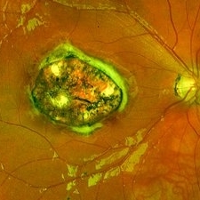
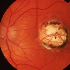
 Toxoplasma Macular Scar
Sep 22 2018 by Hashim Ali Khan, OD, FAAO
Fundus Photographs of a 17-year-old male with inactive macular toxoplasma scar.
Condition/keywords: inactive toxoplasmosis, toxoplasmosis chorioretinitis
-
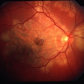
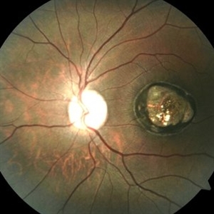
 Ocular Toxoplasmosis
May 26 2016 by Sam Kanavati
A blue autofluorescence image of an inactive well-demarcated chorioretinal scar involving the right optic nerve head secondary to ocular toxoplasmosis in a 65 year-old female patient. This scar dates back to 1971 without any recurrence. Although the scar is extensive, visual acuity was 6/6 but with a corresponding visual field defect.
Photographer: Sam Kanavati, University Hospital Southampton NHS Foundation Trust, UK
Imaging device: Heidelberg Spectralis
Condition/keywords: inactive toxoplasmosis
-
 Relentless Placoid Chorioretinitis
Jan 22 2021 by Renata Garcia Franco, Md
20-year-old male with reduction of vision in both eyes, scotoma and metamorphopsia. Widespread multiple chorioretinal lesions with RPE hyperplasia, which appear from posterior pole to peripheral retina and inactive choroidal neovascular membrane.
Photographer: Fatima Hernandez, Instituto de la Retina del Bajio SC
Imaging device: Zeiss
Condition/keywords: chorioretinitis
 Loading…