Search results (63 results)
-
 Retinocoroiditis Inactiva Por Toxoplasmosis
Apr 28 2025 by Paulina Araujo
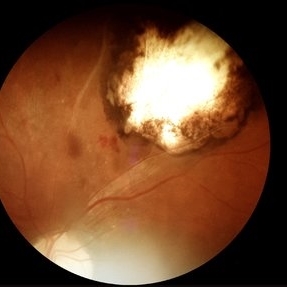
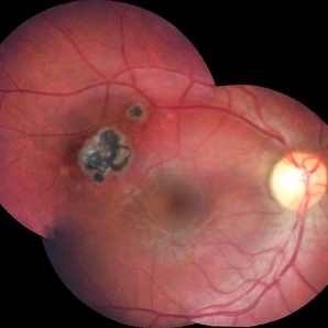
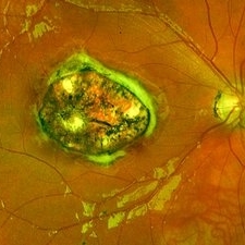
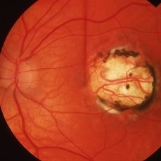
Fundus photography demonstrates a 2-disc-diameter chorioretinal scar in the superior temporal arcade, consistent with inactive toxoplasmic retinochoroiditis. The lesion exhibits pigmented borders and central atrophy, with adjacent splinter hemorrhages and vascular sheathing. No vitreous inflammation or active satellite lesions are present.
Photographer: Paulina D.Araujo Martínez, Asociación para Evitar la Ceguera en México I.A.P., Hospital Dr Luis Sánchez Bulnes.
Condition/keywords: toxoplasmosis chorioretinitis
-
 Toxocara Granuloma
Apr 18 2025 by Chellarani Kumarasamy, MD
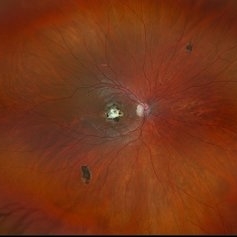
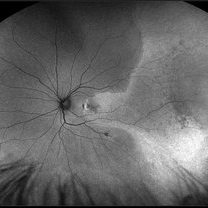
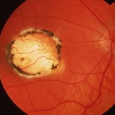
10 year old girl referred for starbismus and red free image showed well demarcted inactive lesion.
Condition/keywords: toxocara granuloma
-
 Inactive Chorioretinal Scars
Dec 11 2024 by Virginia Gebhart
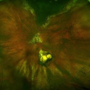
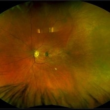
30 year old female with chorioretinal and macula scars. Appears post-infectious, most likely toxoplasmic. No active inflammatory changes or choroidal neovascularization. Will continue to monitor. Central vision limited by macula scar, BCVA 20/100
Photographer: Virginia Gebhart, Retina Consultants of Carolina
Imaging device: Optos California
Condition/keywords: chorioretinal scar, inactive toxoplasmosis
-
 Hidden Mickey / Toxo Scar
Feb 29 2024 by Virginia Gebhart
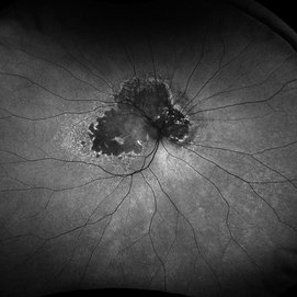
87 year old female with inactive toxoplasmosis chorioretinitis inferior. Stable regressed malignant neoplasm of choroid superior (s/p brachytherapy 2011).
Photographer: Virginia Gebhart
Imaging device: Optos California
Condition/keywords: inactive toxoplasmosis, toxo chorioretinitis, toxoplasmosis chorioretinitis
-
 Idiopathic Peripapillary CNV
Jan 4 2024 by Virginia Gebhart
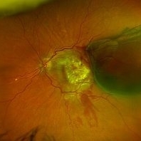
13 year old female with inactive CNV. Increased pigment 360 at 1 year follow up. No inflammation or SRF, pt remains asymptomatic
Photographer: Virginia Gebhart
Imaging device: Optos California
Condition/keywords: choroidal neovascularization (CNV), peripapillary choroidal neovascularization (PPCNVM)
-
 Geographic Atrophy
Nov 16 2023 by Virginia Gebhart
67 year old female with Neovascular AMD with inactive CNV. Extensive geographic atrophy with minimal foveal sparing. Extensive ectopic CNV just superiorly to ON remains inactive. Discussed with pt treating with Syfovre to slow down GA progression
Photographer: Virginia Gebhart
Imaging device: Optos
Condition/keywords: advanced geographic atrophy, age-related macular degeneration (AMD), dry age-related macular degeneration (dry AMD), geographic atrophy
-
 Peripheral VPT Pre and Post Treatment
Aug 14 2023 by Joseph Juliano, MD
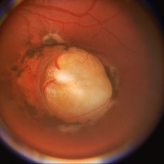
Peripheral VPT with surrounding exudation in a 35 year old woman (Top Left). Three weeks after cryotherapy there are small areas of hemorrhage along the posterior margin of the VPT (Top Right). Four months after cryotherapy, the lesion has significantly less exudation and the areas of hemorrhage have resolved (Bottom Left). Nine months after cryotherapy, there is no further exudation and the VPT is inactive with surrounding cryotherapy scarring (Bottom Right).
Condition/keywords: Vasoproliferative Tumor, VPT
-
 Multifocal choroiditis secondary to sarcoidosis- Quiescent
May 6 2023 by Niloofar Piri, MD
Montage fundus photograph of the left eye in a patient with sarcoidosis demonstrating peripheral inactive multifocal chorioretinal scars after systemic immunomodulatory therapy.
Photographer: Sean Kelso, Saint Lousi University
Condition/keywords: multifocal chorioretinitis (MCP), multifocal choroiditis, sarcoid uveitis, sarcoidosis
-
 Ocular Toxoplasmosis
May 6 2022 by Eder Díaz Dorado
Fundus photograph of an 31-year-old man with a macular scar of toxoplasmosis and a review with OCT
Photographer: Eder Díaz Dorado, Hospital Central Militar, Ciudad de México
Imaging device: Heidelberg Spectralis /Smartphone Photography
Condition/keywords: inactive toxoplasmosis, ocular toxoplasmosis, toxoplasmosis chorioretinitis
-
 Recurrence of Ocular Toxoplasmosis
Apr 11 2022 by Aniruddha K Agarwal, MD
Inactive toxoplasmosis lesion with active lesion at the inferior edge. The active lesion appears "fuzzy". Note also the overlying vitritis
Photographer: Debra A. Goldstein, MD, FRCSC Magerstadt Professor of Ophthalmology Director, Uveitis Service Director, Uveitis Fellowship Department of Ophthalmology Northwestern University Feinberg School of Medicine
Condition/keywords: IUSG, recurrence, retinochoroiditis, toxoplasmosis chorioretinitis, uveitis
-
 Central Serous Chorioretinopathy
Jan 25 2022 by Olivia Rainey
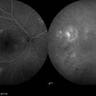
Late phase widefield fluorescein angiography of a 60-year-old male with Central Serous Chorioretinopathy. Chronic history of CSR followed with observation without treatment prior to presenting at our office. The physician noted significant findings on exam and imaging with multifocal areas of inactive and active changes OD. FA shows superotemporal macular leakage, subtle inferonasal macular leakage and staining as well as multifocal hypercyanescence on ICG. Fortunately foveal sparing and thus observation is recommended at this time OD.
Photographer: Olivia Rainey, OCT-C, COA
Imaging device: Heidelberg Spectralis
Condition/keywords: 55-degrees, central serous chorioretinopathy (CSCR), central serous retinopathy (CSR), chronic central serous chorioretinopathy (CSCR), fluorescein angiogram (FA), fluorescein leakage, heidelberg spectralis, indocyanine green (ICG) angiography, late phase
-
 Central Serous Chorioretinopathy
Jan 25 2022 by Olivia Rainey
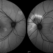
Widefield fundus autofluorescence of a 60-year-old male with Central Serous Chorioretinopathy affecting both eyes. Chronic history of CSR followed with observation without treatment prior to presenting at our office. The physician noted significant findings on exam and imaging with multifocal areas of inactive and active changes in the right eye and subfoveal subretinal fluid with recent visual decline in the left eye. There are hyper and hypoautofluorescent changes, consistent with CSR.
Photographer: Olivia Rainey, OCT-C, COA
Imaging device: Heidelberg Spectralis
Condition/keywords: 55-degrees, central serous chorioretinopathy (CSCR), central serous retinopathy (CSR), chronic central serous chorioretinopathy (CSCR), fundus autofluorescence (FAF), heidelberg spectralis, left eye
-
 Toxoplasmosis Scar
Aug 24 2021 by Andrea Arriola-Lopez, MD MSc
Pigmented retinochoroidal scar temporal to fovea.
Condition/keywords: inactive toxoplasmosis, ocular toxoplasmosis
-
 Hemorrhagic Choroidal Detachment
Jul 6 2021 by Kristen Wagner
Hemorrhagic choroidal detachment with overlying retinal detachment, neovascular AMD with inactive scar.
Photographer: Kristen Wagner COT, Tennessee Retina, Nashville TN
Imaging device: optos
Condition/keywords: choroidal detachment, hemorrhage, wet age-related macular degeneration (wet AMD)
-
 Relentless Placoid Chorioretinitis
Jan 22 2021 by Renata Garcia Franco, Md
20-year-old male with reduction of vision in both eyes, scotoma and metamorphopsia. Widespread multiple chorioretinal lesions with RPE hyperplasia, which appear from posterior pole to peripheral retina and inactive choroidal neovascular membrane.
Photographer: Fatima Hernandez, Instituto de la Retina del Bajio SC
Imaging device: Zeiss
Condition/keywords: chorioretinitis
-
 Multifocal Choroiditis
Jul 19 2020 by Aditya S Kelkar, MS, FRCS, FASRS,FRCOphth
Fundus photograph of 29-year-old male with both eyes inactive multifocal choroiditis.
Photographer: Dr. Sayali Tidke
Imaging device: CLARUS 500
Condition/keywords: multifocal choroiditis
-
 Congenital Toxoplasmosis
Dec 18 2019 by Yoshihiro Yonekawa, MD, FASRS
Widefield fundus image of a teenage girl's right eye with an inactive congenital toxoplasmosis macular lesion. Her vision is 20/400 in this eye.
Photographer: Netanya Lerner, COA, Wills Eye Hospital/Mid Atlantic Retina
Imaging device: Optos California
Condition/keywords: congenital toxoplasmosis, pediatric retina
-
 Macula Off Retinal Detachment with CNV
Nov 11 2019 by Olivia Rainey
Ultra-wide field pseudocolor photograph of a 42-year-old female with a long-standing, macula-off retinal detachment affecting her left eye. Patient was unaware of vision loss until testing her visual acuity and she denied seeing flashing lights. Patient decided to proceed with scleral buckling. The CNV is potentially secondary the retinal detachment, but may be myopic related or idiopathic. The CNV appears fibrotic and inactive. The patient was warned that this will absolutely limit how much vision she recovers once the retina is reattached.
Photographer: Olivia Rainey
Imaging device: Optos California
Condition/keywords: choroidal neovascularization (CNV), left eye, montage, Optos, pseudocolor, retinal detachment of the macula, ultra-wide field imaging
-
 Macula Off Retinal Detachment with CNV
Nov 11 2019 by Olivia Rainey
Ultra-wide field pseudocolor photograph of a 42-year-old female with a long-standing, macula-off retinal detachment affecting her left eye. Patient was unaware of vision loss until testing her visual acuity and she denied seeing flashing lights. Patient decided to proceed with scleral buckling. The CNV is potentially secondary the retinal detachment, but may be myopic related or idiopathic. The CNV appears fibrotic and inactive. The patient was warned that this will absolutely limit how much vision she recovers once the retina is reattached.
Photographer: Olivia Rainey
Imaging device: Optos California
Condition/keywords: choroidal neovascularization (CNV), chronic retinal detachment, fundus autofluorescence (FAF), left eye, montage, Optos, retinal detachment of the macula, ultra-wide field imaging
-
 Fingolimob Associated Macular Edema (FAME)?
Jun 1 2019 by John S. King, MD
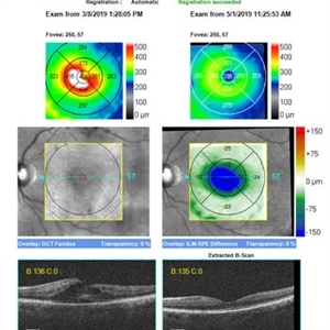
60 year old caucasian female with two week history of decreased vision in the left eye. Background history includes multiple sclerosis for which she uses Gileyna for the past five years, and no history of uveitis or recent MS relapse. Her vision in the left eye was 20/100 J5. The eye appeared overall "quiet" with the exception of rare cells in the anterior vitreous. The fundus appearance and FA can be seen in the images provided. OCT shows CME and SRF in OS only (left image). STK was administered and neurologist was able to discontinue the Gileyna. STK has been reported to be effective in FAME in patients who continue Gileyna (see below). 10 days later the CME had decreased significantly. 8 weeks later the edema had resolved as seen in the OCT images of the initial and latest appearance. Of note, this is a late presentation for FAME, and there was some rare debris in the anterior vitreous; it is possible, although no history of uveitis and the MS was inactive, that the CME may be related to other causes like uveitis (if there is recurrence of CME while patient is off Gileyna, then further work-up will be performed) Minuk A, Belliveau MJ, Almeida DR, Dorrepaal SJ, Gale JS. Fingolimod-associated macular edema: resolution by sub-tenon injection of triamcinolone with continued fingolimod use. JAMA ophthalmol 2013; 131(6): 802–804.
Photographer: Kay Dalby
Imaging device: Cirrus
Condition/keywords: cystoid macular edema (CME), Gilenya, macular edema
-
 Congenital Toxoplasmosis
Apr 8 2019 by Gary R. Cook, MD, FACS
Left eye of the same 38-year-old female with congenital toxoplasmosis lesion; V.A. = 20/40 due to temporal location of the Toxo scar.
Imaging device: Topcon VT-50
Condition/keywords: chorioretinal scar, congenital toxoplasmosis, inactive toxoplasmosis, macular scar, ocular toxoplasmosis
-
 Congenital Toxoplasmosis
Apr 8 2019 by Gary R. Cook, MD, FACS
Right eye of a 38-year-old female with bilateral congenital toxoplasmosis lesions; V.A. = 20/70 OD
Imaging device: Topcon VT-50
Condition/keywords: chorioretinal scar, congenital toxoplasmosis, inactive, inactive toxoplasmosis, macular scar, ocular toxoplasmosis
-
 Congenital Toxoplasmosis Scar
Apr 8 2019 by Gary R. Cook, MD, FACS
5-year-old white male with a typical, deep, pigmented chorioretinal scar secondary to congenital toxoplasmosis OS.
Condition/keywords: chorioretinal scar, congenital toxoplasmosis, inactive toxoplasmosis, macular scar, ocular toxoplasmosis
-
 Recurrent Toxoplasmosis
Apr 8 2019 by Gary R. Cook, MD, FACS
17-year-old male with recurrent toxoplasmosis lesion adjacent to an earlier, inactive toxoplasmosis chorioretinal scar superior to the optic disc OS. The recurrent lesion is becoming less inflamed and 'harder' in appearance, indicating resolution
Condition/keywords: toxo chorioretinitis, toxoplasmosis, toxoplasmosis chorioretinitis, toxoplasmosis reactivation
-
 Congenital Toxoplasmosis
Apr 8 2019 by Gary R. Cook, MD, FACS
23-year-old with congenital toxoplasmosis; view of optic disc and macular scar OS.
Condition/keywords: chorioretinal scar, congenital toxoplasmosis, inactive toxoplasmosis, macular scar, ocular toxoplasmosis
 Loading…