 Initializing download.
-
 By Rameez N Hussain, MD
C CARE WELLKIN HOSPITAL, MAURITIUS
Co-author(s): Dr A Giridhar MD, Mahesh G MD FRCS, Thomas Thachil MD, Bindu Rajesh MD. - Uploaded on Sep 20, 2014.
- Last modified by Caroline Bozell on Sep 22, 2014.
- Rating
- Appears in
- Macular Imaging
- Condition/keywords
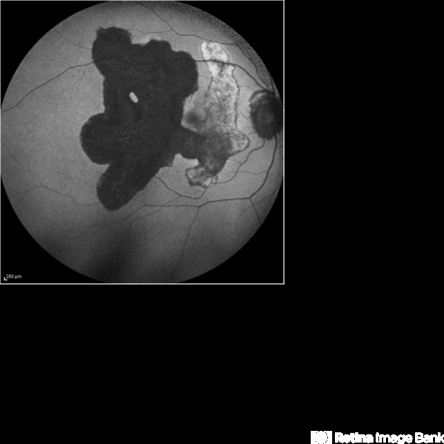
- serpiginous choroiditis
- Photographer
- Dr.Rameez N Hussain, MD, Central Imaging Center, Vitreo Retinal Services, Giridhar Eye Institute, Cochin, India
- Imaging device
-
Scanning laser ophthalmoscope
Heidelberg Blue Peak Autofluorescence imaging. - Description
- Fundus autofluorescence of serpiginous choroiditis showing decreased autofluorescence area corresponding to the inactive lesion (RPE atrophy) and increased autofluorescence area corresponding to active lesion.


