Search results (167 results)
-
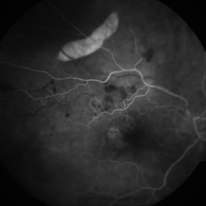
 "Smoke Stack" Hyperfluorescence in Central Serous Chorioretinopathy
Mar 2 2014 by Homayoun Tabandeh, MD, FASRS
"Smoke Stack" hyperfluorescence in central serous chorioretinopathy.
Condition/keywords: central serous chorioretinopathy (CSCR)
-
 Multiple Evanescent White Dot Syndrome (MEWDS)
Oct 20 2012 by Hyung-Woo Kwak, MD
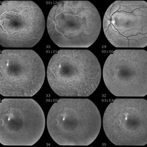
Numerous small deep ill-defined, grey-white dot were seen at the posterior pole and mid-periphery. Some lesions showed mild hyperfluorescence in autofluorescence (AF) but were of limited diagnostic value. ICG showed more numerous hypofluorescent spots than are apparent clinically or on AF/FA
Condition/keywords: hypofluorescent spots, multiple evanescent white dot syndrome (MEWDS)
-
 WET Age Related Macular Degeneration (WET AMD)
Sep 8 2012 by Ratimir Lazic, MD, PhD
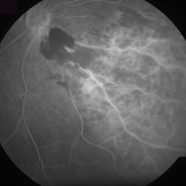
FAG image of a 65 - year- old male. In early venous phase hyperfluorescence due to feeling of occult CNV can be observed
Photographer: Ratimir Lazic, PhD MD
Imaging device: Zeis Visucam Lite 2
Condition/keywords: fundus photograph
-
 Multiple Evanescent White Dot Syndrome (MEWDS)
Oct 20 2012 by Hyung-Woo Kwak, MD
Numerous small deep ill-defined, grey-white dot were seen at the posterior pole and mid-periphery. Some lesions showed mild hyperfluorescence in autofluorescence (AF) but were of limited diagnostic value. ICG showed more numerous hypofluorescent spots than are apparent clinically or on AF/FA
Condition/keywords: hypofluorescent spots, multiple evanescent white dot syndrome (MEWDS)
-
---thumb.jpg/image-square;max$300,300.ImageHandler) Retinoblastoma To Chemothermotherapy
Retinoblastoma To Chemothermotherapy
Oct 4 2013 by Maurice F. Rabb
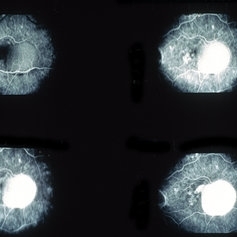
A 7 week old girl with a family history of retinoblastoma was found to have a small retinoblastoma in each eye. In the right eye the tumor was adjacent to the optic disc in the papillomacular bundle and measured 2 X 2 X 2 mm. Its temporal margin was 1.0 mm from the foveola and it overhung 20% of the optic disc surface. There was not clinical or ultrasonographic evidence of vitreous seeking or optic nerve invation. In the left eye there was a solitary tumor 1mm superonasal to the optic disc. The tumor measured 1 X 1 X 1 mm. The foveal reflex was normal in both eyes. Both tumors showed a fluorescein angiographic pattern compatible with retinoblastoma with rapid filling and late hyperfluorescence.
Condition/keywords: retina
-
---thumb.jpg/image-square;max$300,300.ImageHandler) Frosted Branch Angiitis
Frosted Branch Angiitis
Feb 26 2013 by Henry J. Kaplan, MD
Frosted branch angiitis patient. Left eye: F/A leakage seen as hyperfluorescence along involved vessels. #2
Condition/keywords: frosted branch angiitis
-
 Fibrovascular PED
May 2 2013 by Henry J. Kaplan, MD
Fluorescein angiogram of the fibrovascular PED in the same patient; early homogeneous hyperfluorescence in the PED area which is increased in fluorescence to the late phase with a granular hyperfluorescence adjacent to PED in the foveal side which starts in the mid-phase of the F/A and is increased later; #2.
Condition/keywords: exudative age-related macular degeneration, fibrovascular pigment epithelial detachment (PED)
-
 Optic Pit FA
Jul 4 2012 by John T. Thompson, MD
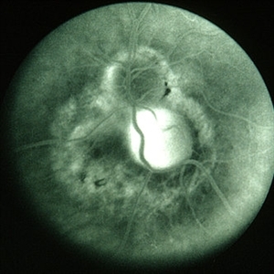
Hyperfluorescence in optic pit due to fluorescein leakage
Imaging device: Zeiss FF4
Condition/keywords: fluorescein leakage, optic disc pit
-
---thumb.jpg/image-square;max$300,300.ImageHandler) Retinoblastoma To Chemothermotherapy
Retinoblastoma To Chemothermotherapy
Oct 4 2013 by Maurice F. Rabb
A 7 week old girl with a family history of retinoblastoma was found to have a small retinoblastoma in each eye. In the right eye the tumor was adjacent to the optic disc in the papillomacular bundle and measured 2 X 2 X 2 mm. Its temporal margin was 1.0 mm from the foveola and it overhung 20% of the optic disc surface. There was not clinical or ultrasonographic evidence of vitreous seeking or optic nerve invation. In the left eye there was a solitary tumor 1mm superonasal to the optic disc. The tumor measured 1 X 1 X 1 mm. The foveal reflex was normal in both eyes. Both tumors showed a fluorescein angiographic pattern compatible with retinoblastoma with rapid filling and late hyperfluorescence.
Condition/keywords: retina
-
 Toxocara Granuloma
Feb 25 2013 by Henry J. Kaplan, MD
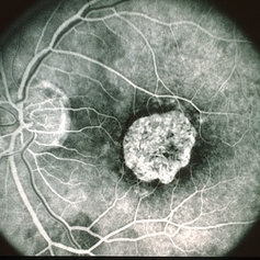
Toxocara granuloma of ON, late stage F/A. #3 Late hyperfluorescence in the granuloma due to staining.
Condition/keywords: ocular toxoplasmosis, toxocara granuloma, toxocariasis
-
---thumb.jpg/image-square;max$300,300.ImageHandler) Coats Disease
Coats Disease
Oct 30 2012 by Lihteh Wu, MD
FA frame showing blocked fluorescence from the massive lipid exudation. There is also hyperfluorescence secondary to vascular leakage and hypofluorescence from the hyperplastic RPE. Superotemporal to the fovea there are areas of telangiectasia.
Condition/keywords: massive lipid exudation, retinal pigment epithelium, retinal telangiectasia
-
 Hemicentral Retinal Vein Occlusion - Fluorescein Angiogram
Aug 23 2012 by Gerardo Garcia-Aguirre, MD
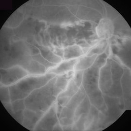
Fluorescein angiogram in late phase showing wide areas of capillary nonperfusion and perivascular hyperfluorescence secondary to vascular incompetence.
Photographer: Noemí Hernández, Asociación para Evitar la Ceguera en México
Condition/keywords: capillary nonperfusion, hemicentral retinal vein occlusion, vascular incompetence
-
 Inferonasal BRVO - Fluorescein Angiogram
Aug 23 2012 by Gerardo Garcia-Aguirre, MD
Fluorescein angiogram showing hypofluorescence secondary to intraretinal hemorrhages, and perivascular hyperfluorescence secondary to vascular incompetence.
Photographer: Noemí Hernández, Asociación para Evitar la Ceguera en México
Condition/keywords: branch retinal vein occlusion (BRVO), intraretinal hemorrhage, vascular incompetence
-
 Central areolar atrophy
May 2 2013 by Henry J. Kaplan, MD
Fluorescein angiogram demonstrates early hyperfluorescence due to window defect; the patient is young with GA like lesion and vision of 20/200 which is compatible with central areolar atrophy#1
Condition/keywords: central areolar choroidal dystrophy (CACD)
-
 Multiple Evanescent White Dot Syndrome (MEWDS)
Oct 20 2012 by Hyung-Woo Kwak, MD
Numerous small deep ill-defined, grey-white dot were seen at the posterior pole and mid-periphery. Some lesions showed mild hyperfluorescence in autofluorescence (AF) but were of limited diagnostic value. ICG showed more numerous hypofluorescent spots than are apparent clinically or on AF/FA.
Condition/keywords: hypofluorescent spots, multiple evanescent white dot syndrome (MEWDS)
-
---thumb.jpg/image-square;max$300,300.ImageHandler) Acute Posterior Multifocal Placoid Pigment Epitheliopathy
Acute Posterior Multifocal Placoid Pigment Epitheliopathy
Feb 27 2013 by Henry J. Kaplan, MD
APMPPE. F/A .Late hyperfluorescence and staining of the lesions apparent #3.
Condition/keywords: acute posterior multifocal placoid pigment epitheliopathy (APMPPE), white dot syndrome
-
 Central Serous Retinopathy (CSR)
Sep 8 2012 by Ratimir Lazic, MD, PhD
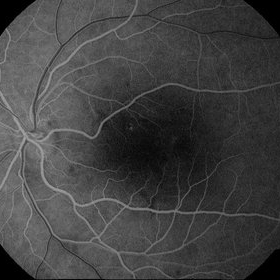
FAG image of a 31 - year - old male. In early venous phase a hyperfluorescence spot is seen in the upper para foveal region
Photographer: Ratimir Lazic, PhD MD
Imaging device: Zeis Visucam Lite 2
Condition/keywords: central serous retinopathy (CSR)
-
 Drusen of Optic Disc
Mar 6 2018 by JEFFERSON R SOUSA, Tecg.º (Biomedical Systems Technology)
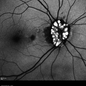
Female patient, 37 years old, Caucasian, with complaint of low lateral stroke in abos the eyes. In the retinal mapping examination and retinography, important alterations in the optic nerve head suggestive of DRUSAS DE PAPILA were observed. After being confirmed in the Autofluorescence examination, we observed Autohyperfluorescence compatible with deposits of calcified hyaline material, as well as another complementary exam such as USG and OCT.
Photographer: JEFFERSON R SOUSA - Study Center and Ophthalmological Research Dr. Andre M V Gomes, Institute Dr. Suel Abujamra, Clinic Marco Antonio Albhy Ophthalmology / São Paulo-Brazil
Imaging device: Heidelberg - HRA Angiograph, Autofluorescence com 30 degrees.
Condition/keywords: calcified drusen, drusen of optic disc
-
 Uveitis With Exudative Retinal Detachment
May 3 2014 by Mallika Goyal, MD
Fluorescein angiogram of an elderly patient with bilateral posterior uveitis shows punctate hyperfluorescence and inferior RD. He responded well to oral steroids with complete resolution of the uveitis and RD.
Photographer: Mallika Goyal, MD, Apollo Health City, Jubilee Hills, Hyderabad, India
Condition/keywords: exudative retinal detachment, uveitis
-
 Circumscribed Choroidal Hemangioma
Oct 20 2012 by Hyung-Woo Kwak, MD
Fundus and OCT examination showed an oval mass at the posterior pole with indistinct margins that blend with surrounding choroid. FA early phase showed hyperfluorescence.
-
 Extrafoveal PED with RPE rip FA1
Dec 23 2012 by Alex P. Hunyor, MD
80-year-old female with subfoveal occult CNV and large extrafoveal PED which underwent spontaneous RPE rip. Early phase FA showing intense hyperfluorescence in the area of acute absence of RPE.
Condition/keywords: pigment epithelial detachment (PED), retinal pigment epithelium (RPE) tear
-
---thumb.jpg/image-square;max$300,300.ImageHandler) Adult Vitelliform Dystrophy
Adult Vitelliform Dystrophy
Aug 7 2013 by From the Collections of Thomas M. Aaberg, MD and Thomas M. Aaberg Jr., MD
Typical FA in adult vitelliform dystrophy shows central hypofluorescene with a ring of hyperfluorescence, left eye #2.
Condition/keywords: adult vitelliform dystrophy
-
 Uveitis With Exudative Retinal Detachment
May 3 2014 by Mallika Goyal, MD
Fluorescein angiogram of an elderly patient with bilateral posterior uveitis shows punctate hyperfluorescence and inferior RD. He responded well to oral steroids with complete resolution of the uveitis and RD.
Photographer: Mallika Goyal, MD, Apollo Health City, Jubilee Hills, Hyderabad, India
Condition/keywords: exudative retinal detachment, uveitis
-
 Retina
May 31 2014 by ruth pav
A 32-year-old woman with a history of drug abuse was admitted due to acute manifestation of multiple infarcts, including acute stroke, splenic and renal infarcts, and multiple cutaneous hematomas. Due to decreased vision in her left eye the patient was referred for ophthalmic evaluation. On exam, visual acuity was 6/10 in the right eye and no light perception in her left eye. Ophthalmoscopic examination was normal in the right eye but showed pallor of the optic nerve head with attenuated retinal vessels in the left eye. Fluorescein angiography showed an oval area of hyperfluorescence from from non-perfusion involving the macular center with staining of overlying retinal capillaries.
Photographer: Ruth Pav, Rambam medical center,Hifa Israel.
Imaging device: Zeiss FF4
Condition/keywords: retina
-
 chronic central serous chorioretinopathy
Oct 31 2012 by Mallika Goyal, MD
Late phase fluorescein angiogram of chronic CSCR shows extensive hyperfluorescence and ill-defined leaks.
Photographer: Mallika Goyal, MD
Condition/keywords: central serous chorioretinopathy (CSCR), chronic central serous chorioretinopathy (CSCR)
 Loading…