Search results (167 results)
-
 Choroidal Hemangioma 4 Ways
Mar 13 2025 by Virginia Gebhart
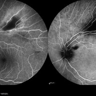
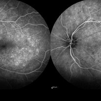
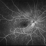
Color fundus, FAF, late FA, late ICG of 64 year old male with choroidal hemangioma. Early hyperfluorescence with late leakage on FA, early hypercyanescence with late washout (25 min) on ICG.
Photographer: Virginia Gebhart, Retina Consultants of Carolina
Imaging device: Optos California
Condition/keywords: autofluorescence imaging, choroidal hemangioma, FA late phase, Fluorescein angiography, hemangioma, indocyanine green (ICG) angiography
-
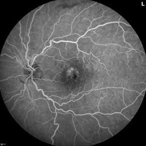
 FA/ICG Choroidal Melanoma
Mar 10 2025 by Virginia Gebhart
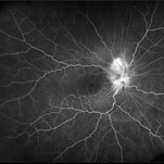
Side by Side comparison of late FA/ICG on choroidal melanoma. FA showed early lacy hyperfluorescence with late leakage, ICG showed late Hypocyanescence.
Photographer: Virginia Gebhart, Retina Consultants of Carolina
Imaging device: Optos California
Condition/keywords: FA, Fluorescein angiography, fluorescein leakage, indocyanine green (ICG) angiography
-
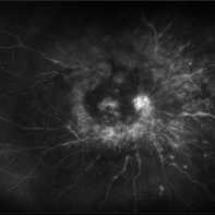
 Indocyanine Green (ICG) of Circumscribed Choroidal Hemangioma (CCH)
Feb 6 2025 by Jack B Margines, MD, MHCI
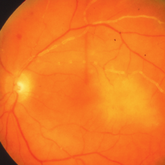
Peripheral patchy hyperfluorescence is seen on this early image of ICG-A on a 53-year-old asymptomatic with an extramacular circumscribed choroidal hemangioma.
Photographer: W Ryan Miliam, CRA, OCT-C, University of California, Irvine Gavin Herbert Eye Institute
Imaging device: Optos
Condition/keywords: choroidal hemangioma, indocyanine green (ICG) angiography
-
 Stargardt Disease (FA)
Jan 22 2025 by Virginia Gebhart
Fluorescein angiogram of 19 year old female with confirmed Stargardt Disease. Hyperfluorescence in the macula with staining defect and silent choroid.
Photographer: Virginia Gebhart, Retina Consultants of Carolina
Imaging device: Optos California
Condition/keywords: fluorescein angiogram (FA), Silent Choroid, Stargardt disease
-
 Toxoplasmosis
Dec 5 2024 by Tejaswita Verma
26 year old male with 6/18 vision , anterior chamber reaction, vitritis and retinitis lesion along the superotemporal arcade with full thickness involvement on OCT . FFA showing hypofluorescence with surrounding hyperfluorescence characterstic of toxoplasma retinitis . ICGA shows hypocyanescence.
Photographer: DR. TEJASWITA VERMA
Imaging device: MIRANTE
Condition/keywords: Fundus Fluorescein Angiography, indocyanine green (ICG) angiography, toxoplasmosis
-
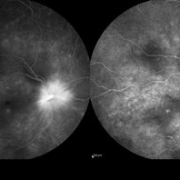
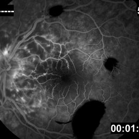
 Fluorescein and Indocyanine Green Angiography in Right Eye in Case of Choroidal Hemangioma
Nov 29 2024 by Anand Temkar
Right eye Fluorescein and Indocyanine green angiography of a 42 year old male in case of Choroidal hemangioma. Choroidal hemangioma have a unique pattern of circulation where the large blood vessels produce a “COARSE VASCULAR PATTERN.” Fluorescein angiography of circumscribed choroidal hemangiomas typically reveals very early hyperfluorescence of larger-caliber choroidal blood vessels either before or simultaneously with the initial filling of the retinal arterioles. Indocyanine green angiography typically shows filling of the intralesional vascular channels, intense hypercyanescence of the lesion by the intermediate frames (peaks around 3-4 minutes) and late washout of the central portion of the lesion.
Photographer: Dr.Anand Temkar- Retina Foundation, Ahmedabad
Imaging device: Mirante
Condition/keywords: Choroidal Hemangioma, FLUORESCEIN ANGIOGRAPHY, indocyanine green (ICG) angiography
-
 Branch Retinal Vein Occlusion with Multifactorial Macular Edema and Epiretinal Membrane
Oct 3 2024 by Logan ryzenga
Fluorescein angiogram of a 62 year old woman with cystoid macular edema from concurrent Epiretinal Membrane and Branch Retinal Vein occlusion. She has an extensive history of anti-VEGF injections with stable but unresolved macular edema. Following angiography, it was determined that an epiretinal membrane peel would be indicated in an attempt to achieve resolution of macular edema.
Photographer: Logan Ryzenga
Imaging device: Heidelberg Spectralis
Condition/keywords: 55-degrees, branch retinal vein occlusion (BRVO), cystoid macular edema (CME), epiretinal membrane (ERM), Fluorescein angiography, heidelberg spectralis, hyperfluorescence, leakage, left eye, OS, wide angle imaging
-
 Active Proliferative Diabetic Retinopathy
Jul 12 2024 by Korey Starkey
Fluorescein angiogram performed on 35 year old female with active proliferative diabetic retinopathy. Patient has peripapillary vascular loop and history of PRP treatment in both eyes. Patients left eye vision measured at Dcc20/200-1 at this visit.
Photographer: Korey Starkey
Imaging device: Optos
Condition/keywords: FLUORESCEIN ANGIOGRAPHY, hyperfluorescence, laser scarring, Optos, proliferative diabetic retinopathy (PDR), sea fan, ultra-wide field imaging, vascular loop
-
 Panuveitis
Jul 12 2024 by Korey Starkey
Ultra widefield Optos FA of 59 year old female presents with panuveitis in both eyes. Patients vision was VA OS: Dcc20/60-2 at time of visit.
Photographer: Korey Starkey
Imaging device: Optos
Condition/keywords: FLUORESCEIN ANGIOGRAPHY, hyperfluorescence, Optos, Panuveitis, ultra-wide field imaging, Uveitis
-
 Posterior Uveitis with Macular Edema
Jul 9 2024 by Korey Starkey
Ultra-wide field angiography of a 70 year old female with cystoid macular edema secondary to posterior uveitis. Patient's vision was Dcc20/200 at time of visit.
Photographer: Korey Starkey
Imaging device: Optos
Condition/keywords: cystoid macular edema (CME), fluorescein angiogram (FA), FLUORESCEIN ANGIOGRAPHY, hyperfluorescence, posterior uveitis, ULTRA WIDE FIELD, ultra-widefield image, vitreous debris
-
 Classic presentation of PEHCR in an elderly Asian female
Apr 15 2024 by David A Reichstein, MD
(A) Ultra-widefield color fundus photograph demonstrating a large, localized area of subretinal fluid surrounded by lipid exudation at its superior and posterior borders. (B) Posterior segment B-scan ultrasonography demonstrates that the lesion is hollow, suggestive of localized subretinal fluid. (C) Early-stage ultra-widefield FA demonstrates an absence of early fluorescence. (D) Late-stage ultra-widefield FA demonstrates late hyperfluorescence.
Condition/keywords: peripheral exudative hemorrhagic chorioretinopathy (PEHCR)
-
 Choroidal Melanoma
Jan 4 2024 by Virginia Gebhart
57 year old female with new choroidal melanoma. Early hyperfluorescence with vascularity and minimal late leakage on FA.
Photographer: Virginia Gebhart
Imaging device: Optos California
Condition/keywords: FA, FA early phase, fluorescein angiogram (FA), Fluorescein angiography
-
 VKH
VKH
Sep 29 2023 by Anjana Mirajkar, MS Ophthalmology
Late frame of FA+ICG of RE of a 41 year old female showing disc leakage with hyperfluorescence suggestive of leakage with hypofluoroscence on FA and ICG in a case of VKH.
Photographer: Dr. Anjana Mirajkar -Retina Foundation, Ahmedabad.
Imaging device: Heidelberg
Condition/keywords: Vogt-Koyanagi-Harada
-
 VKH
VKH
Sep 29 2023 by Anjana Mirajkar, MS Ophthalmology
Late frame of FA+ICG of LE of a 41 year old female showing disc leakage with hyperfluorescence suggestive of leakage with hypofluoroscence on FA and ICG in a case of VKH.
Photographer: Dr. Anjana Mirajkar -Retina Foundation, Ahmedabad.
Imaging device: Heidelberg
Condition/keywords: vkh
-
 Combined Tractional and Rhegmatogenous Retinal Detachment
Jan 30 2023 by Olivia Rainey
Ultra-widefield fluorescein angiography of a combined tractional and rhegmatogenous retinal detachment repair affecting the left eye. The retina is attached following silicone oil placement during most recent surgery. The patient was seeing CF at the time the image was taken.
Photographer: Olivia Rainey, OCT-C, COA
Imaging device: Optos California
Condition/keywords: diabetes, diabetic macular edema, diabetic retinopathy, fluorescein angiogram (FA), hyperfluorescence, right eye, scleral buckle, silicone oil, tractional retinal detachment, ultra-wide field imaging, ultra-widefield image
-
 Acute syphilitic posterior placoid chorioretinitis
Apr 24 2022 by Aniruddha K Agarwal, MD
Green-light fundus autofluorescence (FAF) of the right eye from a 55-year-old man with risk factors for sexually trasnmitted diseases who presented to the retina clinic for a central scotoma. Funduscopy revealed a placoid lesion in the posterior pole. FAF highlights a hyperautofluorescent placoid lesion involving the macula with granular hyperfluorescence. The patient tested positive for syphilis and received intravenous penicillin treatment.
Photographer: Esther CIANCAS, MD, PhD, Gema CRESPO-RODRÍGUEZ, RN
Imaging device: Zeiss Clarus fundus camera
Condition/keywords: chorioretinitis, IUSG, syphilis, uveitis
-
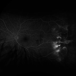
 Purtscher's Retinopathy
Mar 22 2021 by Marco Antonio Sauza
FA in late phases in the same patient with Purtscher's retinopathy, but in this stage we can see multiple places in the posterior pole vessels and around the optic nerve with hyperfluorescence.
Photographer: Marco Sauza
Imaging device: Zeiss
Condition/keywords: Purtscher's retinopathy
-
 Coats' Disease
Feb 2 2021 by Niloofar Piri, MD
#4 Recirculation phase fluorescein angiography of the same patient demonstrating increased hyperfluorescence and leakage from abnormal vascular lesions in temporal periphery. Note the capillary non perfusion area anteriorly.
Condition/keywords: Coats' disease, Leber's miliary aneurysm
-
 Serous Retinal Detachment and Retinal Infiltrate due to B. Hensele, Cat-Scratch Disease
Dec 21 2020 by John S. King, MD
64-year-old female had at least a two week history of blurry vision in the right eye. She was being followed for a CRVO in the right eye, and as vision worsened, was referred to our clinic, and saw Dr. Zocchi. Vision in the right eye was CF; there was 1+ cell in the A/C; 1+ vitreous cell was present; disc edema with surrounding SRF as well as a small, white, retinal infiltrate just superior to the optic disc; vessel tortuosity was present as well as a few IRHs (left eye was u/r). There was sub-foveal and PP SRF on OCT. FA in the early to mid phase showed optic disc hyperfluorescence and early filling into the subretinal space. In the later frames there was disc leakage, staining/leakage of the retinal infiltrate, and filling into the subretinal space (See OCT - Left Image is initial visit). .... Multiple tests were done, she was started on doxycycline 100 mg BID, and Bartonella serology test came back positive. One week later vision improved to 20/100, a/c cell present, disc edema improved and the SRF was resolving (See OCT - Right Image is latest visit).
Imaging device: Zeiss Cirrus
Condition/keywords: Bartonella bacteria, cat scratch retinitis
-
 Serous Retinal Detachment and Retinal Infiltrate due to B. Hensele, Cat-Scratch Disease
Dec 19 2020 by John S. King, MD
64-year-old female had at least a two week history of blurry vision in the right eye. She was being followed for a CRVO in the right eye, and as vision worsened, was referred to our clinic, and saw Dr. Zocchi. Vision in the right eye was CF; there was 1+ cell in the A/C; 1+ vitreous cell was present; disc edema with surrounding SRF as well as a small, white, retinal infiltrate just superior to the optic disc; vessel tortuosity was present as well as a few IRHs (left eye was u/r). There was sub-foveal and PP SRF on OCT. FA in the early to mid phase showed optic disc hyperfluorescence and early filling into the subretinal space. In the later frames there was disc leakage, staining/leakage of the retinal infiltrate, and filling into the subretinal space (See Image). Multiple tests were done, she was started on doxycycline 100 mg BID, and Bartonella serology test came back positive. One week later vision improved to 20/100, a/c cell present, disc edema improved and the SRF was resolving. (will add more photos next visit)
Photographer: Shelly Blair
Imaging device: Optos CA
Condition/keywords: cat scratch retinitis
-
 Serous Retinal Detachment and Retinal Infiltrate due to B. Hensele, Cat-Scratch Disease
Dec 19 2020 by John S. King, MD
64-year-old female had at least a two week history of blurry vision in the right eye. She was being followed for a CRVO in the right eye, and as vision worsened, was referred to our clinic, and saw Dr. Zocchi. Vision in the right eye was CF; there was 1+ cell in the A/C; 1+ vitreous cell was present; disc edema with surrounding SRF as well as a small, white, retinal infiltrate just superior to the optic disc; vessel tortuosity was present as well as a few IRHs (See Image) (left eye was u/r). There was sub-foveal and PP SRF on OCT. FA in the early to mid phase showed optic disc hyperfluorescence and early filling into the subretinal space. In the later frames there was disc leakage, staining/leakage of the retinal infiltrate, and filling into the subretinal space. Multiple tests were done, she was started on doxycycline 100 mg BID, and Bartonella serology test came back positive..... One week later vision improved to 20/100, a/c cell present, disc edema improved and the SRF was resolving. (will add more photos next visit)
Photographer: Shelly Blair
Imaging device: Optos CA
Condition/keywords: cat scratch retinitis
-
 Serous Retinal Detachment and Retinal Infiltrate due to B. Hensele, Cat-Scratch Disease
Dec 19 2020 by John S. King, MD
64-year-old female had at least a two week history of blurry vision in the right eye. She was being followed for a CRVO in the right eye, and as vision worsened, was referred to our clinic, and saw Dr. Zocchi. Vision in the right eye was CF; there was 1+ cell in the A/C; 1+ vitreous cell was present; disc edema with surrounding SRF as well as a small, white, retinal infiltrate just superior to the optic disc; vessel tortuosity was present as well as a few IRHs (left eye was u/r). There was sub-foveal and PP SRF on OCT. FA in the early to mid phase showed optic disc hyperfluorescence and early filling into the subretinal space (See Image). In the later frames there was disc leakage, staining/leakage of the retinal infiltrate, and filling into the subretinal space. Multiple tests were done, she was started on doxycycline 100 mg BID, and Bartonella serology test came back positive. One week later vision improved to 20/100, a/c cell present, disc edema improved and the SRF was resolving. (will add more photos next visit)
Photographer: Shelly Blair
Imaging device: Optos CA
Condition/keywords: cat scratch retinitis
-
 Serous Retinal Detachment and Retinal Infiltrate due to B. Hensele, Cat-Scratch Disease
Dec 19 2020 by John S. King, MD
64-year-old female had at least a two week history of blurry vision in the right eye. She was being followed for a CRVO in the right eye, and as vision worsened, was referred to our clinic, and saw Dr. Zocchi. Vision in the right eye was CF; there was 1+ cell in the A/C; 1+ vitreous cell was present; disc edema with surrounding SRF as well as a small, white, retinal infiltrate just superior to the optic disc; vessel tortuosity was present as well as a few IRHs (left eye was u/r). There was sub-foveal and PP SRF on OCT. FA in the early to mid phase showed optic disc hyperfluorescence and early filling into the subretinal space. In the later frames there was disc leakage, staining/leakage of the retinal infiltrate, and filling into the subretinal space (See Image). Multiple tests were done, she was started on doxycycline 100 mg BID, and Bartonella serology test came back positive. One week later vision improved to 20/100, a/c cell present, disc edema improved and the SRF was resolving. (will add more photos next visit)
Photographer: Shelly Blair
Imaging device: Optos CA
Condition/keywords: cat scratch retinitis
-
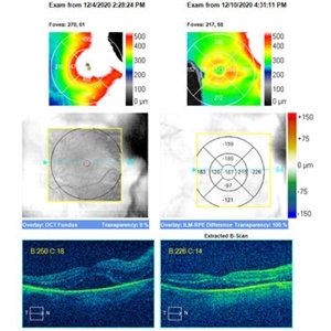
 Bilateral Calcific Retina Arteriolar Occlusions in a Patient with Metastatic Ovarian Carcinoma
Dec 10 2020 by McGill University Health Centre
47-year-old female with cough and fever. Imaging showed a right pulmonary infiltrate. Transbronchial needle biopsy revealed lymphangitic spread of papillary adenocarcinoma with psammoma bodies. MRI of thyroid, CT of abdomen and pelvis were negative. gynecologic evaluation negative at that time . The patient had bilateral floaters, VA: 20/40 OD and 20/20 OS. Fundus examination showed retinal arteriolar sheathing and a flat choroidal lesion OS and vitritis OD. Fluorescein angiogram showed staining of left superior temporal retinal arterioles and bilateral midperipheral patchy hyperfluorescence at RPE The patient vision in the OD deteriorated to 20/400, and in the OS 20/50. Four months later a new choroidal lesion was diagnosed OS. An abdominal mass consistent with a cystadenoma of the ovary was diagnosed. After a year patient developed systemic metastasis. Autopsy: Metastatic adenocarcinoma to the lung, both adrenals, para-aortic lymph nodes, left hip, right breast, occipital skin, serosal surface of liver, pituitary. In almost all metastatic lesions psammoma bodies were found. Presumptive diagnosis is a primary tumor of the ovary.
Condition/keywords: bilateral, calcification, metastatic adenocarcinoma, retinal arteriolar occlusion
 Loading…