Search results (167 results)
-
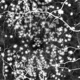
 Coats' Disease
Feb 2 2021 by Niloofar Piri, MD
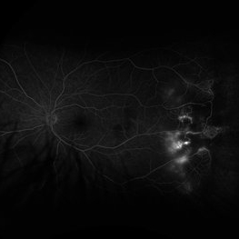
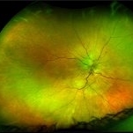
#4 Recirculation phase fluorescein angiography of the same patient demonstrating increased hyperfluorescence and leakage from abnormal vascular lesions in temporal periphery. Note the capillary non perfusion area anteriorly.
Condition/keywords: Coats' disease, Leber's miliary aneurysm
-
 Acute syphilitic posterior placoid chorioretinitis
Apr 24 2022 by Aniruddha K Agarwal, MD
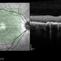
Green-light fundus autofluorescence (FAF) of the right eye from a 55-year-old man with risk factors for sexually trasnmitted diseases who presented to the retina clinic for a central scotoma. Funduscopy revealed a placoid lesion in the posterior pole. FAF highlights a hyperautofluorescent placoid lesion involving the macula with granular hyperfluorescence. The patient tested positive for syphilis and received intravenous penicillin treatment.
Photographer: Esther CIANCAS, MD, PhD, Gema CRESPO-RODRÍGUEZ, RN
Imaging device: Zeiss Clarus fundus camera
Condition/keywords: chorioretinitis, IUSG, syphilis, uveitis
-
 Branch Retinal Vein Occlusion with Multifactorial Macular Edema and Epiretinal Membrane
Oct 3 2024 by Logan ryzenga
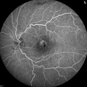
Fluorescein angiogram of a 62 year old woman with cystoid macular edema from concurrent Epiretinal Membrane and Branch Retinal Vein occlusion. She has an extensive history of anti-VEGF injections with stable but unresolved macular edema. Following angiography, it was determined that an epiretinal membrane peel would be indicated in an attempt to achieve resolution of macular edema.
Photographer: Logan Ryzenga
Imaging device: Heidelberg Spectralis
Condition/keywords: 55-degrees, branch retinal vein occlusion (BRVO), cystoid macular edema (CME), epiretinal membrane (ERM), Fluorescein angiography, heidelberg spectralis, hyperfluorescence, leakage, left eye, OS, wide angle imaging
-
 Drusen
Mar 29 2018 by JEFFERSON R SOUSA, Tecg.º (Biomedical Systems Technology)
Male patient 27-years-old, with complaint of low vision in both eyes. The fundoscopic evaluation found the presence of drusen topography in the posterior pole with foveal. Fluorescein angiography shows the typical pattern of hyperfluorescence of drusen in the first minute of angiography.
Photographer: JEFFERSON R SOUSA - Study Center and Ophthalmological Research Dr. Andre M V Gomes, Institute Dr. Suel Abujamra São Paulo-Brazil
Imaging device: Topcon TRC-50 DX, Imaginet 5.0, angle de 50 graus. Flash 150.
Condition/keywords: colloidal drusen, drusen
-
 Indocyanine Green (ICG) of Circumscribed Choroidal Hemangioma (CCH)
Feb 6 2025 by Jack B Margines, MD, MHCI
Peripheral patchy hyperfluorescence is seen on this early image of ICG-A on a 53-year-old asymptomatic with an extramacular circumscribed choroidal hemangioma.
Photographer: W Ryan Miliam, CRA, OCT-C, University of California, Irvine Gavin Herbert Eye Institute
Imaging device: Optos
Condition/keywords: choroidal hemangioma, indocyanine green (ICG) angiography
-
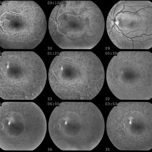
 Posterior Uveitis with Macular Edema
Jul 9 2024 by Korey Starkey
Ultra-wide field angiography of a 70 year old female with cystoid macular edema secondary to posterior uveitis. Patient's vision was Dcc20/200 at time of visit.
Photographer: Korey Starkey
Imaging device: Optos
Condition/keywords: cystoid macular edema (CME), fluorescein angiogram (FA), FLUORESCEIN ANGIOGRAPHY, hyperfluorescence, posterior uveitis, ULTRA WIDE FIELD, ultra-widefield image, vitreous debris
-
 Retina
May 31 2014 by ruth pav
A 32-year-old woman with a history of drug abuse was admitted due to acute manifestation of multiple infarcts, including acute stroke, splenic and renal infarcts, and multiple cutaneous hematomas. Due to decreased vision in her left eye the patient was referred for ophthalmic evaluation. On exam, visual acuity was 6/10 in the right eye and no light perception in her left eye. Ophthalmoscopic examination was normal in the right eye but showed pallor of the optic nerve head with attenuated retinal vessels in the left eye. Fluorescein angiography showed an oval area of hyperfluorescence from from non-perfusion involving the macular center with staining of overlying retinal capillaries.
Photographer: Ruth Pav, Rambam medical center,Hifa Israel.
Imaging device: Zeiss FF4
Condition/keywords: retina
-
 Serous Retinal Detachment and Retinal Infiltrate due to B. Hensele, Cat-Scratch Disease
Dec 19 2020 by John S. King, MD
64-year-old female had at least a two week history of blurry vision in the right eye. She was being followed for a CRVO in the right eye, and as vision worsened, was referred to our clinic, and saw Dr. Zocchi. Vision in the right eye was CF; there was 1+ cell in the A/C; 1+ vitreous cell was present; disc edema with surrounding SRF as well as a small, white, retinal infiltrate just superior to the optic disc; vessel tortuosity was present as well as a few IRHs (left eye was u/r). There was sub-foveal and PP SRF on OCT. FA in the early to mid phase showed optic disc hyperfluorescence and early filling into the subretinal space. In the later frames there was disc leakage, staining/leakage of the retinal infiltrate, and filling into the subretinal space (See Image). Multiple tests were done, she was started on doxycycline 100 mg BID, and Bartonella serology test came back positive. One week later vision improved to 20/100, a/c cell present, disc edema improved and the SRF was resolving. (will add more photos next visit)
Photographer: Shelly Blair
Imaging device: Optos CA
Condition/keywords: cat scratch retinitis
-
 Unilateral Acute Idiopathic Maculopathy OCT Macula
May 7 2019 by William Ensor
A 37-year-old female presented with a two-week history of vision loss in the right eye. She experienced a flu-like illness including rash on the hands, feet, and mouth 2 days prior to her vision change. Her 3-year-old son had a similar illness diagnosed as hand, foot, and mouth disease by his pediatrician one week prior. Her visual acuity was 20/150 of the right eye, and 20/20 of the left eye. On dilated fundus examination, the left eye was unremarkable; the right eye revealed a circular, variably pigmented lesion of the macula. OCT imaging showed areas of RPE loss and clumping, with overlying loss of the photoreceptor layer. Fluorescein angiography showed central and peripheral hyperfluorescence consistent with window defect, and blockage in area of RPE loss. No treatment was initiated at this time. The patient returned 10 days later; her visual acuity improved to 20/50 in the right eye. Dilated fundus exam showed increased pigmentation of the macular lesion. OCT of the right eye showed further RPE clumping without recovery of the photoreceptor layer, despite her improved visual acuity.
Condition/keywords: unilateral acute idiopathic maculopathy
-
 Drusen of Optic Disc
Mar 6 2018 by JEFFERSON R SOUSA, Tecg.º (Biomedical Systems Technology)
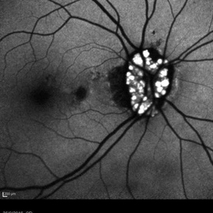
Female patient, 37 years old, Caucasian, with complaint of low lateral stroke in abos the eyes. In the retinal mapping examination and retinography, important alterations in the optic nerve head suggestive of DRUSAS DE PAPILA were observed. After being confirmed in the Autofluorescence examination, we observed Autohyperfluorescence compatible with deposits of calcified hyaline material, as well as another complementary exam such as USG and OCT.
Photographer: JEFFERSON R SOUSA - Study Center and Ophthalmological Research Dr. Andre M V Gomes, Institute Dr. Suel Abujamra, Clinic Marco Antonio Albhy Ophthalmology / São Paulo-Brazil
Imaging device: Heidelberg - HRA Angiograph, Autofluorescence com 30 degrees.
Condition/keywords: calcified drusen, drusen of optic disc
-
 Unilateral Acute Idiopathic Maculopathy Fundus
May 7 2019 by William Ensor
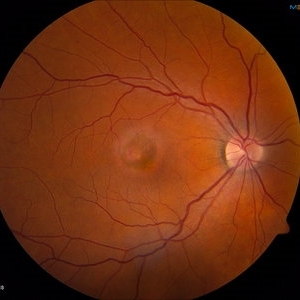
A 37-year-old female presented with a two-week history of vision loss in the right eye. She experienced a flu-like illness including rash on the hands, feet, and mouth 2 days prior to her vision change. Her 3-year-old son had a similar illness diagnosed as hand, foot, and mouth disease by his pediatrician one week prior. Her visual acuity was 20/150 of the right eye, and 20/20 of the left eye. On dilated fundus examination, the left eye was unremarkable; the right eye revealed a circular, variably pigmented lesion of the macula. OCT imaging showed areas of RPE loss and clumping, with overlying loss of the photoreceptor layer. Fluorescein angiography showed central and peripheral hyperfluorescence consistent with window defect, and blockage in area of RPE loss. No treatment was initiated at this time. The patient returned 10 days later; her visual acuity improved to 20/50 in the right eye. Dilated fundus exam showed increased pigmentation of the macular lesion. OCT of the right eye showed further RPE clumping without recovery of the photoreceptor layer, despite her improved visual acuity.
Condition/keywords: unilateral acute idiopathic maculopathy
-
---thumb.jpg/image-square;max$300,300.ImageHandler) Acute Posterior Multifocal Placoid Pigment Epitheliopathy
Acute Posterior Multifocal Placoid Pigment Epitheliopathy
Feb 27 2013 by Henry J. Kaplan, MD
APMPPE. F/A .Late hyperfluorescence and staining of the lesions apparent #3.
Condition/keywords: acute posterior multifocal placoid pigment epitheliopathy (APMPPE), white dot syndrome
-
 Circumscribed Choroidal Hemangioma
Oct 20 2012 by Hyung-Woo Kwak, MD
Fundus and OCT examination showed an oval mass at the posterior pole with indistinct margins that blend with surrounding choroid. FA early phase showed hyperfluorescence.
-
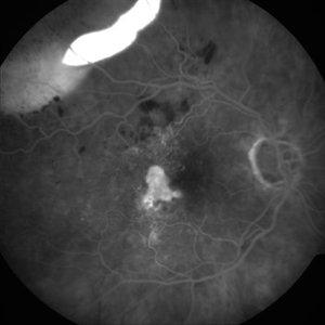
---thumb.jpg/image-square;max$300,300.ImageHandler) Coats Disease
Coats Disease
Oct 30 2012 by Lihteh Wu, MD
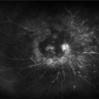
FA frame showing blocked fluorescence from the massive lipid exudation. There is also hyperfluorescence secondary to vascular leakage and hypofluorescence from the hyperplastic RPE. Superotemporal to the fovea there are areas of telangiectasia.
Condition/keywords: massive lipid exudation, retinal pigment epithelium, retinal telangiectasia
-
 Drusen
Mar 29 2018 by JEFFERSON R SOUSA, Tecg.º (Biomedical Systems Technology)
Male patient, 27-years-old, with complaint of low vision in both eyes. The fundoscopic evaluation was found the presence of drusen topography in the posterior pole with foveal. Fluorescein angiography shows the typical pattern of hyperfluorescence of drusen in the first minute of angiography.
Photographer: JEFFERSON R SOUSA - Study Center and Ophthalmological Research Dr. Andre M V Gomes, Institute Dr. Suel Abujamra São Paulo-Brazil
Imaging device: Topcon TRC-50 DX, Imaginet 5.0, angle de 20 graus. Flash 150.
Condition/keywords: colloidal drusen, drusen
-
 Extrafoveal PED with RPE rip FA3
Dec 23 2012 by Alex P. Hunyor, MD
80-year-old female with subfoveal occult CNV and large extrafoveal PED which underwent spontaneous RPE rip. FA shows intense hyperfluorescence in area of absent RPE, progressive filling of extrafoveal PED, and hyperfluorescence in macula from atrophy and occult CNV.
Condition/keywords: pigment epithelial detachment (PED), retinal pigment epithelium (RPE) tear
-
 Multiple Evanescent White Dot Syndrome (MEWDS)
Oct 20 2012 by Hyung-Woo Kwak, MD
Numerous small deep ill-defined, grey-white dot were seen at the posterior pole and mid-periphery. Some lesions showed mild hyperfluorescence in autofluorescence (AF) but were of limited diagnostic value. ICG showed more numerous hypofluorescent spots than are apparent clinically or on AF/FA
Condition/keywords: hypofluorescent spots, multiple evanescent white dot syndrome (MEWDS)
-
 Optic Pit FA
Jul 4 2012 by John T. Thompson, MD
Hyperfluorescence in optic pit due to fluorescein leakage
Imaging device: Zeiss FF4
Condition/keywords: fluorescein leakage, optic disc pit
-
---thumb.jpg/image-square;max$300,300.ImageHandler) Frosted Branch Angiitis
Frosted Branch Angiitis
Feb 26 2013 by Henry J. Kaplan, MD
Frosted branch angiitis patient. Left eye: F/A leakage seen as hyperfluorescence along involved vessels. #2
Condition/keywords: frosted branch angiitis
-
 "Smoke Stack" Hyperfluorescence in Central Serous Chorioretinopathy
Mar 2 2014 by Homayoun Tabandeh, MD, FASRS
"Smoke Stack" hyperfluorescence in central serous chorioretinopathy.
Condition/keywords: central serous chorioretinopathy (CSCR)
-
 Active Proliferative Diabetic Retinopathy
Jul 12 2024 by Korey Starkey
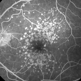
Fluorescein angiogram performed on 35 year old female with active proliferative diabetic retinopathy. Patient has peripapillary vascular loop and history of PRP treatment in both eyes. Patients left eye vision measured at Dcc20/200-1 at this visit.
Photographer: Korey Starkey
Imaging device: Optos
Condition/keywords: FLUORESCEIN ANGIOGRAPHY, hyperfluorescence, laser scarring, Optos, proliferative diabetic retinopathy (PDR), sea fan, ultra-wide field imaging, vascular loop
-
---thumb.jpg/image-square;max$300,300.ImageHandler) acute retinal necrosis
acute retinal necrosis
Feb 15 2013 by From the Collections of Thomas M. Aaberg, MD and Thomas M. Aaberg Jr., MD
early phase FA corresponding to slide 55, showing punctate hyperfluorescence consistent with microvascular damage and staining of areas of retinal necrosis
Condition/keywords: acute retinal necrosis
-
---thumb.jpg/image-square;max$300,300.ImageHandler) Adult Vitelliform Dystrophy
Adult Vitelliform Dystrophy
Aug 7 2013 by From the Collections of Thomas M. Aaberg, MD and Thomas M. Aaberg Jr., MD
Typical FA in adult vitelliform dystrophy shows central hypofluorescene with a ring of hyperfluorescence, left eye #2.
Condition/keywords: adult vitelliform dystrophy
-
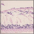 Bilateral Calcific Retina Arteriolar Occlusions in a Patient with Metastatic Ovarian Carcinoma
Dec 10 2020 by McGill University Health Centre
47-year-old female with cough and fever. Imaging showed a right pulmonary infiltrate. Transbronchial needle biopsy revealed lymphangitic spread of papillary adenocarcinoma with psammoma bodies (MRI of thyroid, CT of abdomen and pelvis were negative) gynecologic evaluation negative at that time . The patient had bilateral floaters, VA: 20/40 OD and 20/20 OS. Fundus examination showed retinal arteriolar sheathing and a flat choroidal lesion OS and vitritis OD. Fluorescein angiogram showed staining of left superior temporal retinal arterioles and bilateral midperipheral patchy hyperfluorescence at RPE. The patient vision in the OD deteriorated to 20/400, and in the OS 20/50. Four months later a new choroidal lesion was diagnosed OS. An abdominal mass consistent with a cystadenoma of the ovary was diagnosed. After a year patient developed systemic metastasis. Autopsy: Metastatic adenocarcinoma to the lung, both adrenals, para-aortic lymph nodes, left hip, right breast, occipital skin, serosal surface of liver, pituitary. In almost all metastatic lesions psammoma bodies were found. Presumptive diagnosis is a primary tumor of the ovary. Histopathologic examination of both eyes disclosed : Bilateral metastatic adenocarcinoma to the vitreous with partially calcified proliferation along internal limiting membrane, OS. Metastatic adenocarcinoma to choroid, OS. Bilateral optic atrophy secondary to retinal arteriolar occlusion with calcification.
Condition/keywords: bilateral, calcification, histopathology, metastatic adenocarcinoma, pathology, retinal arteriolar occlusion
 Loading…