Search results (372 results)
-
 Retinal capillary hemangiomas 3
Jan 11 2013 by Alex P. Hunyor, MD
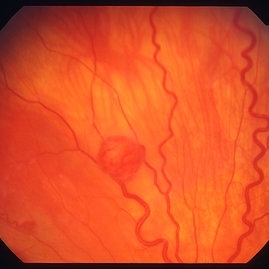
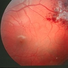
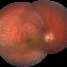
Retinal capillary haemangiomas, left superior periphery, in a 20 year old female with von Hippel-Lindau disease.
Condition/keywords: hemangioma, retinal capillary hemangioma, Von Hippel-Lindau
-
 Racemose Hemangioma
Feb 20 2013 by From the Collections of Thomas M. Aaberg, MD and Thomas M. Aaberg Jr., MD
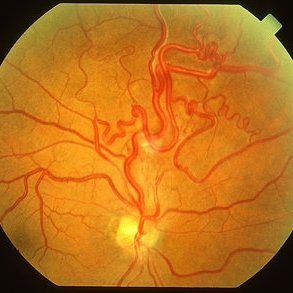
Dilated tortuous blood vessels.
Condition/keywords: racemose hemangioma
-
 Sturge-Weber Episcleral-Vessels
Apr 17 2014 by Susanna S. Park, MD, PhD
External photo of the right eye of this 8-year-old Hispanic boy with Sturge -Weber Syndrome and diffuse choroidal hemangioma showing dilated episcleral vessels.
Photographer: Ellen Redenbo, University of California Davis Eye Center
Condition/keywords: dilated episcleral vessels, Sturge-Weber syndrome
-
 Retinal capillary hemangioma 2
Jan 11 2013 by Alex P. Hunyor, MD
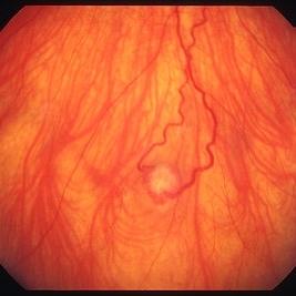
Retinal capillary haemangioma, right inferior periphery, in a 20-year-old female with von Hippel-Lindau disease.
Condition/keywords: hemangioma, retinal capillary hemangioma, Von Hippel-Lindau
-
 Retinal Angiomas In VHL
Dec 24 2012 by Roy D. Brod, MD
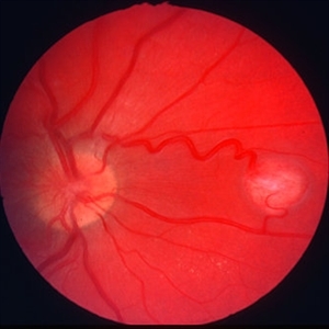
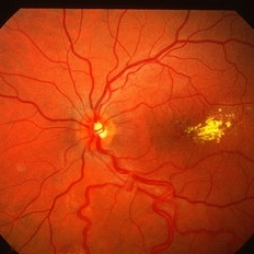
Fundus photograph of 16 year old male with recent diagnosis of Von Hippel-Lindau disease showing typical appearance of a retinal angioma in superior mid periphery OD. Note unrelated choroidal nevus above superior arcade.
Photographer: Julia Walker
Condition/keywords: hemangioma, Von Hippel-Lindau
-
---thumb.jpg/image-square;max$300,300.ImageHandler) Sturge-Weber Diffuse Hemangioma and Retinal Detachment on B-scan
Sturge-Weber Diffuse Hemangioma and Retinal Detachment on B-scan
Apr 18 2014 by Susanna S. Park, MD, PhD
B-scan ultrasonogram of the right eye of an 8 year old Hispanic boy with Sturge -Weber Syndrome showing diffuse choroidal thickening from diffuse choroidal hemangioma and associated total exudative retinal detachment.
Photographer: Ellen Redenbo, University of California Davis Eye Center
Condition/keywords: B scan ultrasound, diffuse choroidal hemangioma, Sturge-Weber syndrome
-
 Retinal capillary hemangioma
Jan 11 2013 by Alex P. Hunyor, MD
Retinal capillary haemangioma nasal to optic disc, right eye.
Condition/keywords: retinal capillary hemangioma, Von Hippel-Lindau
-
 Wyburn Mason Syndrome
May 2 2013 by Henry J. Kaplan, MD
Racemose angioma of the retina in Wyburn Mason syndrome.
Condition/keywords: racemose hemangioma
-
 Diffuse Choroidal Hemangioma
Nov 7 2012 by Rajiv Anand, MD, FRCS, FASRS
Fundus photo shows classic 'tomato-ketchup' red appearance of diffuse hemangioma. Due to chronic SRF , there is subretinal fibrosis.
Condition/keywords: subretinal fibrosis
-
---thumb.jpg/image-square;max$300,300.ImageHandler) Retinal capillary hemangioma 4 image 1
Retinal capillary hemangioma 4 image 1
Jan 11 2013 by Alex P. Hunyor, MD
Retinal capillary haemangioma, left eye, in a young female with von Hippel-Lindau disease. Color image 1 showing extensive lipid deposition in the macula.
Condition/keywords: retinal capillary hemangioma, Von Hippel-Lindau
-
 Wyburn-Mason (Raecemose Hemangioma)
Oct 2 2013 by Jerald A. Bovino, MD
The patieint has typical Wyburn Mason syndrome with dilated, extrement tortuous venules.
Condition/keywords: dilated and tortous veins, racemose hemangioma
-
 Retinal Angiomas In VHL
Dec 24 2012 by Roy D. Brod, MD
Fundus photograph of 16 year old male with recent diagnosis of Von Hippel-Lindau disease showing 2 retinal angiomas in inferior mid periphery OD.
Photographer: Julia Walker
Condition/keywords: hemangioma, Von Hippel-Lindau
-
 Cavernous Hem of Retina
Oct 9 2012 by Alan D. Letson, MD
12-year-old boy with cavernous hemangioma of the retina.
Photographer: Beverly Radcliffe
Condition/keywords: cavernous hemangioma of the retina, hamartoma, phakoma
-
 Retinal Cavernous Hemangioma
Oct 30 2012 by Roy D. Brod, MD
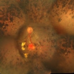
Color fundus photograph of right eye in a 32-year-old asymptomatic female showing typical "cluster of grapes" appearence. Normal MRI of brain. No skin lesions.
Photographer: Julia Walker
Condition/keywords: cavernous hemangioma of the retina, cluster of grapes
-
 Retinal Angiomas In VHL
Dec 24 2012 by Roy D. Brod, MD
Mid phase fluorescein angiogram of 16 year old male with recent diagnosis of Von Hippel-Lindau disease showing hyperfluorescent angioma in superior mid periphery OD.
Photographer: Julia Walker
Condition/keywords: hemangioma, Von Hippel-Lindau
-
 Choroidal Hemangioma
Oct 20 2012 by Hyung-Woo Kwak, MD
Fundus, ICG, and OCT examination showed a typical chorioretinal scar lying concentric to the optic disc. Typical choroidal rupture was performed after intravitreal gas injection under the diagnosis of submacular hemorrhage caused by trauma, after the absorption of submacular hemorrhage
Condition/keywords: chorioretinal scar, choroidal rupture, submacular hemorrhage
-
 Discrete Choroidal Hemangioma B-Scan
Jul 14 2014 by Susanna S. Park, MD, PhD
B-scan ultrasonography of a discrete choroidal hemangioma.
Photographer: Ellen Redenbo
Condition/keywords: B scan ultrasound, choroidal hemangioma
-
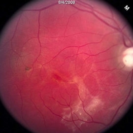
 Capillary Hemongima, Coat's Response
May 2 2013 by Henry J. Kaplan, MD
Coat's response as exudation in the macula in the same patient with retinal capillary hemangioma. Notice the dilated feeder vessles from the optic nerve infriorly; #2.
Condition/keywords: Coats' disease, retinal capillary hemangioma, Von Hippel-Lindau
-
 Cavernous Hemangioma of the Retina
Sep 20 2013 by Hector E. Ibanez, MD, FACS, FASRS, FABO, FAAO
Fundus photograph of an 8-year-old boy with a cavernous hemangioma of the retina.
Photographer: Hector E. Ibanez, MD, FACS
Imaging device: Topcon TRC 50
Condition/keywords: cavernous hemangioma of the retina, fundus photograph
-
---thumb.jpg/image-square;max$300,300.ImageHandler) Cavernous Hemangioma of the Retina
Cavernous Hemangioma of the Retina
Sep 20 2013 by Hector E. Ibanez, MD, FACS, FASRS, FABO, FAAO
Fundus photograph of an 8-year-old boy with a cavernous hemangioma of the retina.
Photographer: Hector E. Ibanez, MD, FACS
Imaging device: Topcon TRC 50
Condition/keywords: cavernous hemangioma of the retina
-
 Cavernous Hemangioma of the Retina
Sep 20 2013 by Hector E. Ibanez, MD, FACS, FASRS, FABO, FAAO
Fluorescein angiogram of an 8 year-old boy with a cavernous hemangioma of the retina.
Photographer: Hector E. Ibanez, MD, FACS
Imaging device: Topcon TRC 50
Condition/keywords: cavernous hemangioma of the retina
-
 Racemose Hemangioma
Feb 20 2013 by From the Collections of Thomas M. Aaberg, MD and Thomas M. Aaberg Jr., MD
Dilated tortuous blood vessels.
Condition/keywords: racemose hemangioma
-
 Retinal Angiomas In VHL
Dec 24 2012 by Roy D. Brod, MD
Mid phase fluorescein angiogram of 16 year old male with recent diagnosis of Von Hippel-Lindau disease showing hyperfluorescent angiomas in inferior mid periphery OD.
Photographer: Julia Walker
Condition/keywords: hemangioma, Von Hippel-Lindau
-
 Retinitis Pigmentosa With Hemangioma CF
Dec 15 2016 by Manish Nagpal, MD, FRCS (UK), FASRS
Fluorescein angiography OS of a patient having retinitis pigmentosa with a hemangioma inferiorly.
Condition/keywords: hemangioma, retinitis pigmentosa
-
 Diffuse Choroidal Hemangioma OD
Aug 24 2012 by John S. King, MD
Child c poor vision OD
Photographer: Kristin Konecki, OcuSight Eye Care Center, Rochester, NY
Condition/keywords: diffuse choroidal hemangioma
 Loading…