Search results (372 results)
-
 Capillary Heamngioma of the Optic Disc
Jun 30 2022 by Thirumalesh Mochi Basavaraj, MD
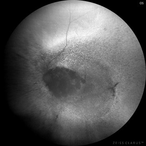
fundus image showing a large Capillary Hemangioma at the Optic Disc
Photographer: Thirumalesh Mochi Basavaraj
Imaging device: intraoperative picture
Condition/keywords: Hemangioma, Von Hippel-Lindau
-
 Capillary Hemangioma
Mar 27 2019 by Gary R. Cook, MD, FACS
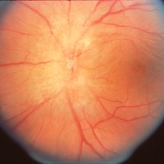
Left eye of a 35-year-old white female with a capillary hemangioma of the optic disc OS; V.A.= 20/40. This hemangioma showed significant preretinal gliosis, and was not associated with von Hippel-Lindau disease.
Condition/keywords: hemangioma
-
 Capillary Hemangioma
Apr 1 2019 by Gary R. Cook, MD, FACS
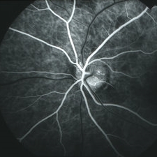
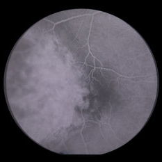
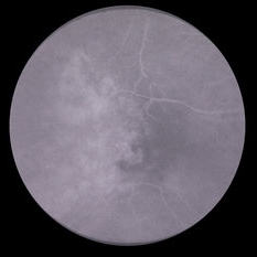
Mid-phase (laminar venous phase) fluorescein angiogram image of a capillary hemangioma of the optic disc OS showing delayed filling and relative hypofluorescence in the area of the hemangioma on the superior aspect in a 28-year-old white female
Imaging device: Topcon VT-50
Condition/keywords: FA mid phase, fluorescein angiogram (FA), hemangioma
-
 Capillary Hemangioma
Apr 1 2019 by Gary R. Cook, MD, FACS
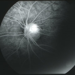
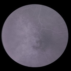
Late-phase (6 minutes) fluorescein angiogram image of a capillary hemangioma of the optic disc OS in a 28-year-old, asymptomatic white female showing late staining in the area of the hemangioma superiorly.
Imaging device: Topcon VT-50
Condition/keywords: FA late phase, fluorescein angiogram (FA), hemangioma, retinal capillary hemangioma
-
 Choroid hemangioma
Sep 7 2022 by JEFFERSON R SOUSA, Tecg.º (Biomedical Systems Technology)
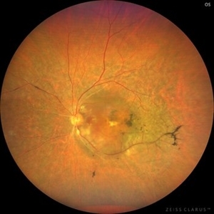
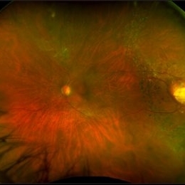
Patient 54 years old, Female, progressive loss of vision. In the multimodal evaluation of the retina showed important retinal alterations. A discreet opacity of the media impairs the quality of the images. In the Autofluorescent Background Image with a green filter, because it reaches a depth in the retinal tissue, it is able to show changes that affect the retinal pigment epithelium, it was better in this case than with the green filter. WF retinography shows an elevated, slightly reddish lesion, probable serous retinal detachment, mobilization of pigments and phantom vessels.
Photographer: JEFFERSON ROCHA DE SOUSA - Retinal Department at Instituto Dr. Suel Abujamra Sao Paulo-Brazil
Imaging device: Clarus 700 - Zeiss 135 degree images. Multimodal Evaluation
Condition/keywords: elevated retinal lesion, hemangioma, melanoma, serous retinal detachment
-
 Choroid hemangioma
Sep 7 2022 by JEFFERSON R SOUSA, Tecg.º (Biomedical Systems Technology)
Patient 54 years old, Female, progressive loss of vision. In the multimodal evaluation of the retina showed important retinal alterations. A discreet opacity of the media impairs the quality of the images. In the Autofluorescent Background Image with a green filter, because it reaches a depth in the retinal tissue, it is able to show changes that affect the retinal pigment epithelium, it was better in this case than with the green filter. WF retinography shows an elevated, slightly reddish lesion, probable serous retinal detachment, mobilization of pigments and phantom vessels.
Photographer: JEFFERSON ROCHA DE SOUSA - Retinal Department at Instituto Dr. Suel Abujamra Sao Paulo-Brazil
Imaging device: Clarus 700 - Zeiss 135 degree images. Multimodal Evaluation
Condition/keywords: elevated retinal lesion, hemangioma, melanoma, serous retinal detachment
-
 Choroidal Hemangioma
Mar 13 2025 by Virginia Gebhart
64 year old male referred for lesion in the STA with worsening SRF. Pt had been receiving injections for wetAMD q4weeks for 7 months. Reddish, elevated choroidal lesion, chronic SRF and pigment clumping consistent with hemangioma. FA/ICG/Bscan ultrasound also performed to confirm. Pt scheduled for PDT
Photographer: Virginia Gebhart, Retina Consultants of Carolina
Imaging device: Optos California
Condition/keywords: choroidal hemangioma, hemangioma, subretinal fluid
-
 Choroidal Hemangioma 4 Ways
Mar 13 2025 by Virginia Gebhart
Color fundus, FAF, late FA, late ICG of 64 year old male with choroidal hemangioma. Early hyperfluorescence with late leakage on FA, early hypercyanescence with late washout (25 min) on ICG.
Photographer: Virginia Gebhart, Retina Consultants of Carolina
Imaging device: Optos California
Condition/keywords: autofluorescence imaging, choroidal hemangioma, FA late phase, Fluorescein angiography, hemangioma, indocyanine green (ICG) angiography
-
 Choroidal Osteoma Or Hemangioma
-
 Hemangioma
Feb 9 2021 by Kim Barrett
66-year-old female with a history of thyroid and uterine cancer in her 30's. She has a family history of cancers also. Current VA 20/40-2 PH OS. Patient and doctor chose observation at this time with possible surgical intervention in the future. She also has a small Hemangioma temporally in the right eye. Von Hippel-Lindau is also suspected and genetic testing was suggested.
Photographer: Kim Barrett C.O.A. Retina Specialists of Michigan, Grand Rapids, MI
Imaging device: Optos California
Condition/keywords: cancer, genetic testing, Optos, retinal hemangioblastoma, Von Hippel-Lindau
-
 Hemangioma
Jan 7 2015 by H. Michael Lambert, MD
Fluorescein angiogram of cavernous hemangioma.
Condition/keywords: cavernous hemangioma of the retina
-
 Hemangioma
Jan 7 2015 by H. Michael Lambert, MD
Fluorescein angiogram of cavernous hemangioma.
Condition/keywords: hemangioma
-
 Hemangioma
Jan 7 2015 by H. Michael Lambert, MD
Fluorescein angiogram of cavernous hemangioma.
Condition/keywords: cavernous hemangioma of the retina
-
 Hemangioma
Oct 16 2012 by Anat Loewenstein, MD
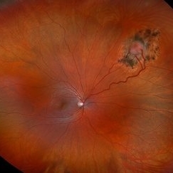
Fundus examination of a 68 year old lady with decreased vision in her left eye for several months. VA 20/30 in her RE and counting fingers in the left eye. In the left eye there was a large red mass protruding and covering almost the entire optic nerve. Diagnosed as retinal hemangioma. The patient underwent low fluence PDT.
Photographer: Galit Yair-Pur
Condition/keywords: hemangioma
-
 Hemangioma Capilar Retina
Apr 9 2023 by Gustavo Aguirre-Suarez
Fundus photograph composition of a Retinal Capilar Hemangioma
Photographer: Dr. Gustavo Aguirre-Suarez
Imaging device: Visucam 500
Condition/keywords: hemangioma, Von Hippel-Lindau
-
 Hemangioma of Retina
Mar 5 2025 by Virginia Gebhart
64 year old male with choroidal hemangioma in the macula and STA. Persistent IRF and new cuff of SRF compared to previous photos. BCVA CF@face. Pt has had PDT in the past with no significant improvement. Will observe closely
Photographer: Virginia Gebhart, Retina Consultants of Carolina
Imaging device: Optos California
Condition/keywords: hemangioma, inferior subretinal fluid
-
 Hemangioma of Retina
Sep 11 2018 by Carolyn Daley
50 degree OCT imaging of a 20-year-old with multiple bilateral hemangiomas. Patient was diagnosed with Von Hippel-Lindau Syndrome.
Photographer: Carolyn Daley, Retina Specialists of Michigan
Imaging device: Heidelberg Spectralis
Condition/keywords: 50 degrees, edema, hemangioma, optical coherence tomography (OCT), Von Hippel-Lindau
-
 Hemangioma of Retina
Sep 11 2018 by Carolyn Daley
Optos ultra wide field imaging of a 20-year-old with multiple bilateral hemangiomas. Patient was diagnosed with Von Hippel-Lindau Syndrome.
Photographer: Carolyn Daley, Retina Specialists of Michigan
Imaging device: Optos Ultra Wide Field
Condition/keywords: edema, hemangioma, Optos, Von Hippel-Lindau
-
 Hemangioma of Retina
Sep 11 2018 by Carolyn Daley
Optos ultra wide field auto fluorescence imaging of a 20-year-old with multiple bilateral hemangiomas. Patient was diagnosed with Von Hippel-Lindau Syndrome.
Photographer: Carolyn Daley, Retina Specialists of Michigan
Imaging device: Optos Ultra Wide Field
Condition/keywords: edema, hemangioma, Optos, Von Hippel-Lindau
-
 Hemangioma of Retina (FAF)
Mar 5 2025 by Virginia Gebhart
Fundus autofluorescence of 64 year old male with choroidal hemangioma in the macula and STA. Persistent IRF and new cuff of SRF compared to previous photos. BCVA CF@face. Pt has had PDT in the past with no significant improvement. Will observe closely
Photographer: Virginia Gebhart, Retina Consultants of Carolina
Imaging device: Optos California
Condition/keywords: autofluorescence imaging, hemangioma, inferior subretinal fluid
-
 Hemangioma Retina
Apr 15 2023 by Lulwa El Zein, MD
40 year old female with a incidental finding of retinal capillary hemangioma.
Photographer: Lulwa El Zei, Mayo clinic, Rochester MN
Condition/keywords: fluorescein angiogram (FA), hemangioma, retina
-
 Hemangioma-redfree
-
---thumb.jpg/image-square;max$300,300.ImageHandler) Liver Capillary Hemangioma in Same Patient With Optic Disc Capillary Hemangioblastoma(Von-Hippel Disease)
Liver Capillary Hemangioma in Same Patient With Optic Disc Capillary Hemangioblastoma(Von-Hippel Disease)
Mar 18 2014 by Arwa Azmeh, MD, PhD
CT scan of chest, abdomen, and pelvis was WNL except for a small hemangioma in the liver measuring 32 mm and this hemangioma was also noticed on abdomen ultrasound as a hyper-reflective area. Brain MRI was WNL.
Condition/keywords: hemangioma, Von Hippel-Lindau
-
 Optic Nerve/Retinal Capillary Hemangioma
Aug 12 2021 by Stefanie Palmer
Optic Nerve/Retinal Capillary Hemangioma of the Right eye.
Photographer: Stefanie Palmer, CRA
Condition/keywords: hemangioma, optic nerve
-
 Reactive Retinal Astrocytic Tumor
Dec 6 2024 by Virginia Gebhart
27 year old female self-referred for continued follow-up care of hemangioma of retina. Previous genetic testing negative for Von Hippel-Lindau. Pt recently diagnosed with Ehlers-Danlos Arthroclasia, most likely reactive retinal astrocytic tumor. Tumor is stable and surrounded by good laser barricade, will continue to observe.
Photographer: Virginia Gebhart, Retina Consultants of Carolina
Imaging device: Optos California
Condition/keywords: feeder vessel, hemangioma, RRAT
 Loading…