Search results (372 results)
-
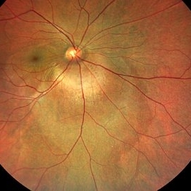
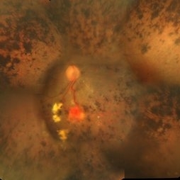 Retinitis Pigmentosa With Hemangioma CF
Dec 15 2016 by Manish Nagpal, MD, FRCS (UK), FASRS
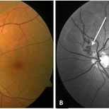
Fluorescein angiography OS of a patient having retinitis pigmentosa with a hemangioma inferiorly.
Condition/keywords: hemangioma, retinitis pigmentosa
-
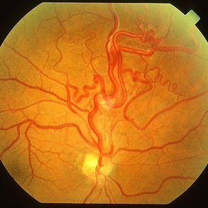
 Retinal Arteriovenous Malformations (Racemose Hemangiomatosis)
Mar 30 2018 by Rameez N Hussain, MD
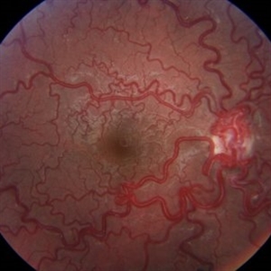
A 7-years-old Portuguese girl with unilateral retinal arteriovenous malformations composed of dilated, tortuous vessels with normal vision.
Photographer: Thambi Durai. Consultant Optometrist, Orbit Health Care - Dr Agarwal's Eye Hospital, Maputo, Mozambique
Imaging device: TOPCON
Condition/keywords: racemose hemangioma, retinal arteriovenous malformations, Wyburn-Mason
-
 Wyburn-Mason Syndrome (Racemose Hemangiomatosis)
Mar 30 2018 by Rameez N Hussain, MD
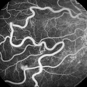
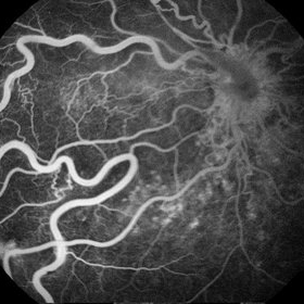
A 7-year-old Portuguese girl with unilateral retinal arteriovenous malformations composed of dilated, tortuous vessels with normal vision.
Photographer: Thambi Durai
Imaging device: TOPCON
Condition/keywords: arteriovenous malformation, racemose hemangioma, Wyburn-Mason
-
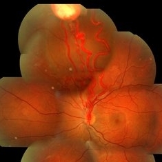
 Hemangioma Capilar Retina
Apr 9 2023 by Gustavo Aguirre-Suarez
Fundus photograph composition of a Retinal Capilar Hemangioma
Photographer: Dr. Gustavo Aguirre-Suarez
Imaging device: Visucam 500
Condition/keywords: hemangioma, Von Hippel-Lindau
-
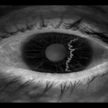 Iris Racemose Hemangioma
Jan 1 2023 by Maxwell J Wingelaar, MD
Fluorescein Angiogram of 66 year old female presented with an iris racemose hemangioma
Photographer: Ken Huff
Condition/keywords: Racemose hemangioma
-
 VHL With Capillary Hemangioma Pre-Rx
Dec 29 2016 by Manish Nagpal, MD, FRCS (UK), FASRS
VHL with hemangioma with feeder vessels.
Photographer: rakesh juneja
Condition/keywords: cryotherapy, hemangioma, laser, Von Hippel-Lindau
-
 Wyburn Mason Syndrome
May 2 2013 by Henry J. Kaplan, MD
Racemose angioma of the retina in Wyburn Mason syndrome.
Condition/keywords: racemose hemangioma
-
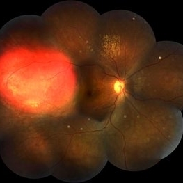
 Capillary Hemangioma
Dec 14 2016 by Young Hee Yoon, MD, PhD
Wide fundus photo of a 35-year-old man with huge capillary hemagioma in the right eye. He is diagnosed with Von Hippel-Lindau disease. His best-corrected visual acuity was 20/50.
Photographer: Yu Jin Jang and Hun Eui Hong, Asan Medical Center
Imaging device: Wide fundus camera
Condition/keywords: retinal capillary hemangioma, Von Hippel-Lindau
-
---thumb.jpg/image-square;max$300,300.ImageHandler) Cavernous Hemangioma of the Retina
Cavernous Hemangioma of the Retina
Sep 20 2013 by Hector E. Ibanez, MD, FACS, FASRS, FABO, FAAO
Fundus photograph of an 8-year-old boy with a cavernous hemangioma of the retina.
Photographer: Hector E. Ibanez, MD, FACS
Imaging device: Topcon TRC 50
Condition/keywords: cavernous hemangioma of the retina
-
 Circumscribed Choroidal Hemangioma with Serous Macular Retinal Detachment
Oct 2 2023 by Aditya S Kelkar, MS, FRCS, FASRS,FRCOphth
Fundus photograph of a 43-year-old male with a circumscribed choroidal hemangioma with serous macular retinal detachment associated with diminision of vision.
Photographer: Dr. Harsh Jain, National Institute of Ophthalmology
Imaging device: Clarus 500
Condition/keywords: choroidal hemangioma
-
 Focal Chroidal Hemangioma
Sep 18 2018 by Somnath Chakraborty, MD
Right eye fundus photo montage of a 17-year-old boy showing a focal choridal hemangioma temporally.
Photographer: Saptarshi Mehta, Retina Institute of Bengal
Condition/keywords: choroidal hemangioma
-
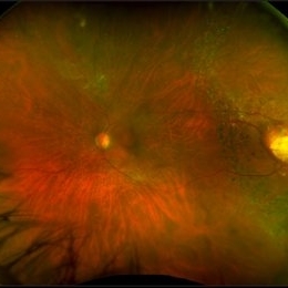 Hemangioma
Feb 9 2021 by Kim Barrett
66-year-old female with a history of thyroid and uterine cancer in her 30's. She has a family history of cancers also. Current VA 20/40-2 PH OS. Patient and doctor chose observation at this time with possible surgical intervention in the future. She also has a small Hemangioma temporally in the right eye. Von Hippel-Lindau is also suspected and genetic testing was suggested.
Photographer: Kim Barrett C.O.A. Retina Specialists of Michigan, Grand Rapids, MI
Imaging device: Optos California
Condition/keywords: cancer, genetic testing, Optos, retinal hemangioblastoma, Von Hippel-Lindau
-
 Hemangioma of Retina
Sep 11 2018 by Carolyn Daley
50 degree OCT imaging of a 20-year-old with multiple bilateral hemangiomas. Patient was diagnosed with Von Hippel-Lindau Syndrome.
Photographer: Carolyn Daley, Retina Specialists of Michigan
Imaging device: Heidelberg Spectralis
Condition/keywords: 50 degrees, edema, hemangioma, optical coherence tomography (OCT), Von Hippel-Lindau
-
 Hemangioma of Retina
Mar 5 2025 by Virginia Gebhart
64 year old male with choroidal hemangioma in the macula and STA. Persistent IRF and new cuff of SRF compared to previous photos. BCVA CF@face. Pt has had PDT in the past with no significant improvement. Will observe closely
Photographer: Virginia Gebhart, Retina Consultants of Carolina
Imaging device: Optos California
Condition/keywords: hemangioma, inferior subretinal fluid
-
 Hemangioma of Retina (FAF)
Mar 5 2025 by Virginia Gebhart
Fundus autofluorescence of 64 year old male with choroidal hemangioma in the macula and STA. Persistent IRF and new cuff of SRF compared to previous photos. BCVA CF@face. Pt has had PDT in the past with no significant improvement. Will observe closely
Photographer: Virginia Gebhart, Retina Consultants of Carolina
Imaging device: Optos California
Condition/keywords: autofluorescence imaging, hemangioma, inferior subretinal fluid
-
 Indocyanine Green (ICG) of Circumscribed Choroidal Hemangioma (CCH)
Feb 6 2025 by Jack B Margines, MD, MHCI
Peripheral patchy hyperfluorescence is seen on this early image of ICG-A on a 53-year-old asymptomatic with an extramacular circumscribed choroidal hemangioma.
Photographer: W Ryan Miliam, CRA, OCT-C, University of California, Irvine Gavin Herbert Eye Institute
Imaging device: Optos
Condition/keywords: choroidal hemangioma, indocyanine green (ICG) angiography
-
 Multiple Cavernous Hemangioma
Jan 20 2020 by Sarah Oelrich
Multiple Cavernous Hemangioma
Photographer: Sarah Oelrich CRA COT OCT-C Southeastern Retina Associates
Condition/keywords: cavernous hemangioma of the retina
-
 Optic Nerve/Retinal Capillary Hemangioma
Aug 12 2021 by Stefanie Palmer
Optic Nerve/Retinal Capillary Hemangioma of the Right eye.
Photographer: Stefanie Palmer, CRA
Condition/keywords: hemangioma, optic nerve
-
 Peri-papillary Vascular Loop
Jun 2 2020 by Dhaivat Shah
Peri-papillary vascular loops (PVL) are rare congenital vascular malformations, which are usually detected as accidental finding during routine fundus examination. They can often be confused with tributary vein occlusion or racemose hemangioma. Although benign and asymptomatic, they can be rarely associated with vitreous hemorrhage and arterial occlusion. We herein present a case of a 60-year-old hypertensive male, who was diagnosed elsewhere to have a tributary vein occlusion and was referred to us. FFA was advised to rule out neovascularization, surrounding capillary non perfusion and mass lesion (hemangioma). On FFA, the arterial loop showed a slightly delayed filling (3-5 seconds) as compared to the other arterial vessels and the original vessel appeared to be a branch arising from central retinal artery. The choroidal filling was delayed in the area supplied by the loop. A cilioretinal artery was also noted. The patient was diagnosed to have a Peri-papillary vascular arterial loop (PVL), likely to be congenital in origin. The patient was reassured and was advised yearly follow up. These loops are usually accidental findings discovered during routine fundus examination. Since these vessels are looped and tortuous, they exhibit a slower and laminar blood flow, which make them more prone for arterial occlusions. The vitreous in this area tends to be adherently attached, so during PVD induction, it is likely to cause a tear and hemorrhage leading to vitreous hemorrhage. Until and unless there is a break, this hemorrhage tends to resolve on its own and does not warrant treatment. If there is an evident break, it can be dealt with laser barrage.
Photographer: Choithram Netralaya
Condition/keywords: congenital prepapillary vascular loop
-
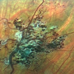
 Racemose Hemangioma
Feb 20 2013 by From the Collections of Thomas M. Aaberg, MD and Thomas M. Aaberg Jr., MD
Dilated tortuous blood vessels.
Condition/keywords: racemose hemangioma
-
---thumb.jpg/image-square;max$300,300.ImageHandler) Racemose Hemangioma
Racemose Hemangioma
Feb 20 2013 by From the Collections of Thomas M. Aaberg, MD and Thomas M. Aaberg Jr., MD
dilated vessels
Condition/keywords: racemose hemangioma
-
 Racemose Hemangioma
Mar 14 2021 by Luiz A Zago, PhD
Racemose hemangioma in a 30-year-old woman with ambliopia in this eye.
Photographer: Luiz Alberto Zago Filho
Imaging device: Topcon 50IX
Condition/keywords: racemose hemangioma
-
 Racemose Hemangioma and Retinal Vein Occlusion
Mar 5 2013 by Eduardo Torres-Porras, MD
A 13-year-old woman had a history of decreased vision for 2 years. Visual acuity was 20/400.
Photographer: Camelia Rosales Lara
Condition/keywords: retinal angiomatous proliferation (RAP)
-
 Racemose Hemangioma and Retinal Vein Occlusion
Mar 5 2013 by Eduardo Torres-Porras, MD
A 13-year-old woman had a history of decreased vision for 2 years. Visual acuity was 20/400.
Photographer: Camelia Rosales Lara
Condition/keywords: retinal angiomatous proliferation (RAP)
-
 Racemose Hemangiomatosis
May 27 2020 by Jamin S. Brown, MD
Fundus photo of 25-year-old female with racemose hemangiomatosis OS.
Photographer: Stefanie Palmer CRA, Retina-Vitreous Surgeons of CNY
Condition/keywords: racemose hemangioma
 Loading…