Search results (372 results)
-
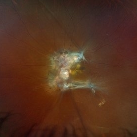
 Choroidal Hemangioma (AF)
Jul 5 2025 by Gustavo Uriel Fonseca Aguirre
This wide-field fundus autofluorescence image demonstrates a mushroom-shaped choroidal melanoma adjacent to the optic nerve head, exhibiting hypo-autofluorescence (melanin). Vitreous pigment dispersion (tobacco dust sign) is evident, indicating tumor activity.
Photographer: Gustavo U. Fonseca Aguirre, Hospital Conde de Valenciana, Ciudad de México
Condition/keywords: choroidal melanoma
-
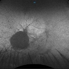
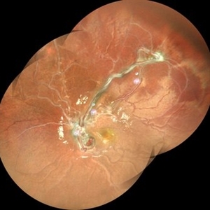
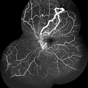
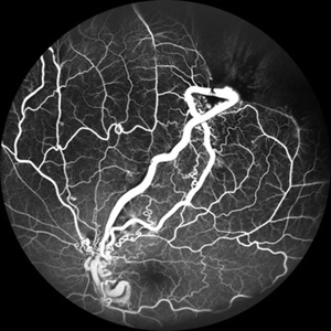
 From Artery to Vein, No Detour: Meet the AV Maverick: Racemose Hemangioma
Jul 1 2025 by rohan jain
A case of 10 year old girl with defective vision in LE (6/60) who presented us with this condition.
Photographer: Dr. ROHAN JAIN
Imaging device: mirante
Condition/keywords: arteriovenous malformation, FFA in a case of Racemose angioma, racemose hemangioma
-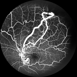
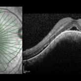 Choroidal Hemangioma
Jun 18 2025 by Moazzam Parvez
An OCT image of a 42 year old man presenting with a vision of 20/80 and complaining of distortion. OCT reveals serous retinal detachment with RPE alteration and disruption of outer retinal layers.
Photographer: Moazzam Parvez , Netralayam , Kolkata
Imaging device: Heidelberg Spectralis
Condition/keywords: Choroidal Hemangioma, Sub retinal fluid, tumor
-
 Choroidal Hemangioma
Jun 18 2025 by Moazzam Parvez
Multicolor and infrared reflectance image of a 42 year old gentleman with a Choroidal hemangioma lesion temporal to the fovea complaining of distortion in his right eye . Fundus imaging revealed a well-circumscribed ,elevated, reddish orange lesion within the choroid involving the posterior pole temporally .
Photographer: Moazzam Parvez, Netralayam , Kolkata
Imaging device: Heidelberg Spectralis
Condition/keywords: Choroidal Hemangioma, tumor
-
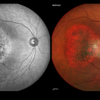
 Choroidal Hemangioma 4 Ways
Mar 13 2025 by Virginia Gebhart
Color fundus, FAF, late FA, late ICG of 64 year old male with choroidal hemangioma. Early hyperfluorescence with late leakage on FA, early hypercyanescence with late washout (25 min) on ICG.
Photographer: Virginia Gebhart, Retina Consultants of Carolina
Imaging device: Optos California
Condition/keywords: autofluorescence imaging, choroidal hemangioma, FA late phase, Fluorescein angiography, hemangioma, indocyanine green (ICG) angiography
-
 Choroidal Hemangioma
Mar 13 2025 by Virginia Gebhart
64 year old male referred for lesion in the STA with worsening SRF. Pt had been receiving injections for wetAMD q4weeks for 7 months. Reddish, elevated choroidal lesion, chronic SRF and pigment clumping consistent with hemangioma. FA/ICG/Bscan ultrasound also performed to confirm. Pt scheduled for PDT
Photographer: Virginia Gebhart, Retina Consultants of Carolina
Imaging device: Optos California
Condition/keywords: choroidal hemangioma, hemangioma, subretinal fluid
-
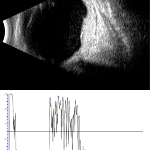 Choroidal Hemangioma, Ecography
Mar 11 2025 by Gustavo Uriel Fonseca Aguirre
45-year-old female with choroidal hemangioma in the macular area. A B-mode ultrasound study is presented showing a well-circumscribed, elevated lesion at the level of the choroid, involving the posterior pole, with intralesional fluid. The standardized A-mode ultrasound shows a lesion with medium-high reflectivity characteristic of choroidal hemangioma.
Photographer: Gustavo U. Fonseca Aguirre, Hospital Conde de Valenciana, Ciudad de México
Condition/keywords: Choroidal Hemangioma, ecography
-
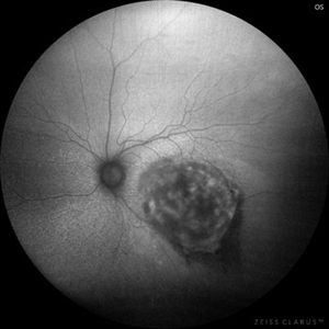 Choroidal Hemangioma, Autofluorescence
Mar 11 2025 by Gustavo Uriel Fonseca Aguirre
45-year-old female with choroidal hemangioma in the macular area. An autofluorescence image of the lesion is presented, with heterogeneous hypoautofluorescent characteristics.
Photographer: Gustavo U. Fonseca Aguirre, Hospital Conde de Valenciana, Ciudad de México
Condition/keywords: Choroidal Hemangioma
-
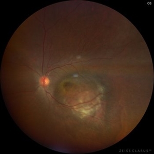 Choroidal Hemangioma
Mar 11 2025 by Gustavo Uriel Fonseca Aguirre
45-year-old female with choroidal hemangioma in the macular area.
Photographer: Gustavo U. Fonseca Aguirre, Hospital Conde de Valenciana, Ciudad de México
Condition/keywords: Choroidal Hemangioma
-
 Hemangioma of Retina (FAF)
Mar 5 2025 by Virginia Gebhart
Fundus autofluorescence of 64 year old male with choroidal hemangioma in the macula and STA. Persistent IRF and new cuff of SRF compared to previous photos. BCVA CF@face. Pt has had PDT in the past with no significant improvement. Will observe closely
Photographer: Virginia Gebhart, Retina Consultants of Carolina
Imaging device: Optos California
Condition/keywords: autofluorescence imaging, hemangioma, inferior subretinal fluid
-
 Hemangioma of Retina
Mar 5 2025 by Virginia Gebhart
64 year old male with choroidal hemangioma in the macula and STA. Persistent IRF and new cuff of SRF compared to previous photos. BCVA CF@face. Pt has had PDT in the past with no significant improvement. Will observe closely
Photographer: Virginia Gebhart, Retina Consultants of Carolina
Imaging device: Optos California
Condition/keywords: hemangioma, inferior subretinal fluid
-
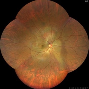
 FAF-G Circumscribed Choroidal Hemangioma
Mar 1 2025 by Vishal Agrawal, MD, FRCS,FACS,FASRS
A 37-year-old male presented with decreased vision in the right eye. This is the fundus autofluorescence (FAF-G) of the right eye showing hypo auto fluorescent lesion with surrounding hyper auto fluorescence extending inferiorly corresponding to the fluid tract.
Photographer: Dr Ayushi Gupta
Imaging device: Clarus 700
Condition/keywords: Circumscribed Choroidal Hemangioma, fundus autofluorescence (FAF)
-
 VHL Syndrome with Capillary Hemangioblastomas
Feb 26 2025 by Virginia Gebhart
39 year old female with choroidal hemangioma with capillary hemangioblastomas. Positive genetic testing for Von Hippel-Lindau Syndrome. Hemangioblastomas are stable compared to initial imaging in 2021. Pt started Welireg in Dec 2024, CNS tumors have started shrinking. No lesions in OD
Photographer: Virginia Gebhart, Retina Consultants of Carolina
Imaging device: Optos California
Condition/keywords: retinal capillary hemangioblastoma, Von Hippel-Lindau
-
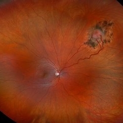
 Vortex-pattern Exudative Retinal Detachment
Feb 22 2025 by CUI YUELING
Patient: Male, 40 years old. Chief Complaint: Blurred vision and metamorphopsia in the left eye for more than 10 days. Past Medical History Hypertension for 4 years, with a highest recorded blood pressure of 160/80 mmHg. Currently controlled with oral "Nifedipine Sustained-Release Tablets, 2 tablets daily." Long-term history of heavy alcohol consumption and smoking. Ophthalmic Examination: Visual Acuity: Right eye (OD): 0.4 (uncorrected, no improvement with correction). Left eye (OS): 0.5 (-1.5DS = 1.0). Intraocular Pressure (IOP): OD: 15 mmHg. OS: 17 mmHg. Anterior Segment:Unremarkable. Fundus Examination: Right eye: Optic disc margins are clear, with a slightly reddish hue. Cup-to-disc ratio (C/D) = 0.2. A scalloped, orange-red elevated lesion is observed superior to the optic disc, with anterior displacement of the focal point. This is accompanied by a secondary, turbine-like exudative retinal detachment centered around the optic disc, involving the macula. The macular region shows scattered punctate yellow-white exudates. Diagnosis: Choroidal hemangioma with secondary exudative retinal detachment(OD).
Photographer: Cui yueling The First People's Hospital of Zunyi, Guizhou, Zunyi, China
Imaging device: Zeiss Clarus 500
Condition/keywords: choroidal hemangioma, exudative retinal detachment
-
 Indocyanine Green (ICG) of Circumscribed Choroidal Hemangioma (CCH)
Feb 6 2025 by Jack B Margines, MD, MHCI
Peripheral patchy hyperfluorescence is seen on this early image of ICG-A on a 53-year-old asymptomatic with an extramacular circumscribed choroidal hemangioma.
Photographer: W Ryan Miliam, CRA, OCT-C, University of California, Irvine Gavin Herbert Eye Institute
Imaging device: Optos
Condition/keywords: choroidal hemangioma, indocyanine green (ICG) angiography
-
 CCH
CCH
Feb 6 2025 by Jack B Margines, MD, MHCI
SD-OCT of an extramacular Circumscribed Choroidal Hemangioma in an asymptomatic 53 year-old female
Photographer: Ryan Milam, University of California, Irvine Gavin Herbert Eye Institute
Imaging device: Zeiss Cirrus
Condition/keywords: Circumscribed Choroidal Hemangioma, OCT
-
 Reactive Retinal Astrocytic Tumor
Dec 6 2024 by Virginia Gebhart
27 year old female self-referred for continued follow-up care of hemangioma of retina. Previous genetic testing negative for Von Hippel-Lindau. Pt recently diagnosed with Ehlers-Danlos Arthroclasia, most likely reactive retinal astrocytic tumor. Tumor is stable and surrounded by good laser barricade, will continue to observe.
Photographer: Virginia Gebhart, Retina Consultants of Carolina
Imaging device: Optos California
Condition/keywords: feeder vessel, hemangioma, RRAT
-
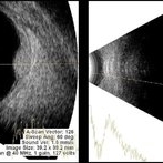 USG in Case of Choroidal Hemangioma With Associated Exudative RD
Nov 29 2024 by Anand Temkar
USG of right eye of a 42 year old male, showing us the BRIGHT choroidal hemangioma. This is because they are a mass of relatively large and well-formed blood vessels. Each blood vessel reflects sound waves producing characteristically intense reflections or moderately “MODERATELY HIGH INTERNAL REFLECTIVITY” from within the hemangioma tumor. Moderately high internal reflectivity is an important diagnostic characteristic.
Photographer: Dr.Anand Temkar- Retina Foundation, Ahmedabad
Imaging device: Mirante
Condition/keywords: B scan ultrasound, Choroidal Hemangioma
-
 Fluorescein and Indocyanine Green Angiography in Right Eye in Case of Choroidal Hemangioma
Nov 29 2024 by Anand Temkar
Right eye Fluorescein and Indocyanine green angiography of a 42 year old male in case of Choroidal hemangioma. Choroidal hemangioma have a unique pattern of circulation where the large blood vessels produce a “COARSE VASCULAR PATTERN.” Fluorescein angiography of circumscribed choroidal hemangiomas typically reveals very early hyperfluorescence of larger-caliber choroidal blood vessels either before or simultaneously with the initial filling of the retinal arterioles. Indocyanine green angiography typically shows filling of the intralesional vascular channels, intense hypercyanescence of the lesion by the intermediate frames (peaks around 3-4 minutes) and late washout of the central portion of the lesion.
Photographer: Dr.Anand Temkar- Retina Foundation, Ahmedabad
Imaging device: Mirante
Condition/keywords: Choroidal Hemangioma, FLUORESCEIN ANGIOGRAPHY, indocyanine green (ICG) angiography
-
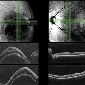 Both Eyes OCT in Case of Right Eye Choroidal Hemangioma
Nov 29 2024 by Anand Temkar
BE OCT of a 42 year old male, showing the elevation of the right eye retina along with the cystic spaces and subretinal fluid.
Photographer: Dr.Anand Temkar- Retina Foundation, Ahmedabad
Imaging device: Mirante
Condition/keywords: OCT
-
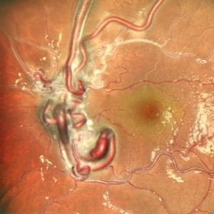
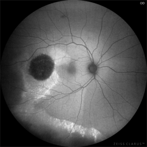
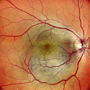 Right Eye Color Photo in Case of Choroidal Hemangioma
Nov 29 2024 by Anand Temkar
Right eye color photo of a 42 year old male in case of choroidal hemangioma. We can see the reddish-orange, round to oval choroidal tumor located completely in the posterior half of the fundus.
Photographer: Dr.Anand Temkar- Retina Foundation, Ahmedabad
Imaging device: Mirante
Condition/keywords: Choroidal Hemangioma
 Loading…