Search results (65 results)
-
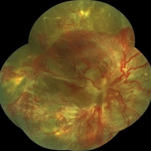
 Total Rhegmatogenous Retinal Detachment With Severe PVR
May 27 2015 by Darin R. Goldman, MD
63-year-old pseudophakic male with hand motion vision in the left eye due to a total retinal detachment with severe proliferative vitreoretinopathy.
Condition/keywords: proliferative vitreoretinopathy (PVR), retinal tear
-
000---thumb.jpg/image-square;max$300,300.ImageHandler) Fundus Panorama Finding of Tractional Retinal Detachment Due to Proliferative Diabetic Retinopathy
Fundus Panorama Finding of Tractional Retinal Detachment Due to Proliferative Diabetic Retinopathy
Dec 25 2013 by Dong Yoon Kim, MD
47-year-old woman visited our clinic for decreased visual acuity on her right eye. Her visual acuity of right eye was hand motion. Fundus examination showed traction retinal detachment.
Condition/keywords: fundus photograph, tractional retinal detachment
-
 Retinitis pigmentosa AR slide 1
Oct 22 2012 by Ronald C. Gentile, MD
17 year-old girl, born of consanguineous parents, presented with nyctalopia and poor vision in both eyes. Vision was hand motions in both eyes. Electroretinogram was extinguished for both photopic and scotopic responses.
Photographer: The New York Eye & Ear Infirmary Department of Medical Imaging
Condition/keywords: retinitis pigmentosa
-
---thumb.JPG/image-square;max$300,300.ImageHandler) Retinal Detachment With Dislocated IOL Lens
Retinal Detachment With Dislocated IOL Lens
Jun 30 2013 by Jason S. Calhoun
47-year-old male who had trauma to the right eye. Patient had retinal detachment surgery in the past (scleral buckle), to the right eye. Patient came in with another retinal detachment with dislocated PC IOL lens. Notice the haptics tearing the retina. Patient underwent vitrectomy with gas exchange. VA was hand motion 1 day post-op.
Photographer: Jason S. Calhoun, Mayo Clinic Jacksonville, Florida
Condition/keywords: dislocated posterior chamber intraocular lens (PCIOL), retinal tear
-
---thumb.jpg/image-square;max$300,300.ImageHandler) Terson's Syndrome
Terson's Syndrome
Oct 8 2013 by Maurice F. Rabb
39 year female with a long history of chronic back pain treated by a sequence of epidural injections. Following her last injection, she complained of a moderately severe protracted headache and had several attempts at placement of an epidural blood patch without success. Under general anesthesia, she underwent injection of a larger volume of saline in an attempt to stem a presumed CSF leak producing "spinal headache". In the left eye she demonstrated multiple superficial and deep intraretinal hemorrhages associated with mild disc swelling and a central scotoma. In the right eye she showed a posterior subhyaloid and sub-internal limiting membrane hemorrhage with buffy coat layering superiorly. The visual acuity measured hand motions OD, 20/200 OS. The patient underwent a surgical evacuation of the sub-ILM hemorrhage.
Condition/keywords: Terson's Syndrome
-
 Choroidal Melanoma With Radiation Retinopathy
Jul 8 2013 by Jason S. Calhoun
Patient came with follow up on choroidal melanoma. Right eye that was treated back in June of 2009 with a radioactive implant. Vein occlusion is also present with VA - hand motion. Hemorrhages visible with hard exudates from the radiation retinopathy.
Photographer: Jason S. Calhoun, Department of Ophthalmology, Mayo Clinic Jacksonville, Florida
Condition/keywords: radiation retinopathy
-
---thumb.jpg/image-square;max$300,300.ImageHandler) Terson's Syndrome
Terson's Syndrome
Oct 8 2013 by Maurice F. Rabb
39 year female with a long history of chronic back pain treated by a sequence of epidural injections. Following her last injection, she complained of a moderately severe protracted headache and had several attempts at placement of an epidural blood patch without success. Under general anesthesia, she underwent injection of a larger volume of saline in an attempt to stem a presumed CSF leak producing "spinal headache". In the left eye she demonstrated multiple superficial and deep intraretinal hemorrhages associated with mild disc swelling and a central scotoma. In the right eye she showed a posterior subhyaloid and sub-internal limiting membrane hemorrhage with buffy coat layering superiorly. The visual acuity measured hand motions OD, 20/200 OS. The patient underwent a surgical evacuation of the sub-ILM hemorrhage.
Condition/keywords: Terson's Syndrome
-
---thumb.jpg/image-square;max$300,300.ImageHandler) Fundus Panorama: Post Operative Findings of Tractional Retinal Detachment
Fundus Panorama: Post Operative Findings of Tractional Retinal Detachment
Dec 25 2013 by Dong Yoon Kim, MD
47-year-old woman underwent vitrectomy and silicone oil tampoande for tractional retinal detachment due to proliferative diabetic retinopathy. 3 years after silicone oil removal and cataract surgery, her visual acuity was improved from hand motion to 20/40.
Condition/keywords: silicone oil, tractional retinal detachment
-
 Retinal Detachment With Dislocated IOL Lens
Jun 30 2013 by Jason S. Calhoun
47-year-old male who had trauma to the right eye. Patient had retinal detachment surgery in the past (scleral buckle), to the right eye. Patient came in with another retinal detachment with dislocated PC IOL lens. Notice the haptics tearing the retina. Patient underwent vitrectomy with gas exchange. VA was hand motion 1 day post-op.
Photographer: Jason S. Calhoun, Mayo Clinic Jacksonville, Florida
Condition/keywords: dislocated posterior chamber intraocular lens (PCIOL), retinal tear
-
 Valsalva Retinopathy
May 30 2014 by Mitzy E Torres Soriano, MD
A 45-year-old woman presented sudden loss of vision (hand motion) in the right eye. Fundus examination revealed multiple deep retinal hemorrhages and a large pre macular subhyaloid hemorrhage. Spontaneous resorption occurred at one month and visual acuity improved to 20/25.
Photographer: Mitzy E Torres Soriano. Hospital Central de Maracay. Venezuela
Condition/keywords: macular hemorrhage, subhyaloid hemorrhage, valsalva retinopathy
-
 Pseudoexfoliation With Partial Subluxation
Jul 12 2013 by Jason S. Calhoun
81-year-old female woke up with loss of vision in the left eye. Patient was hand motion in the left eye. Slit lamp examination shows dislocated lens with pseudoexfoliation material around the iris rim. Proceeded with cataract surgery.
Photographer: Jason S. Calhoun, Department of Ophthalmology, Mayo Clinic Jacksonville, Florida
Condition/keywords: pseudoexfoliation of lens capsule, subluxation of lens
-
 Lupus Retinopathy
Mar 27 2014 by Jason S. Calhoun
Female patient in for evaluation on lupus retinopathy. Has poor vision in the right eye. VA is hand motion in the right eye. Fundus photos show fibrosis along the temporal arcades and narrowing of the arteries. No macular edema found.
Photographer: Jason S. Calhoun, Mayo Clinic Jacksonville, Department of Ophthalmology
Imaging device: TOPCON TRC 50-EX
Condition/keywords: lupus, retinopathy
-
---thumb.JPG/image-square;max$300,300.ImageHandler) Retinal Detachment With Dislocated IOL Lens
Retinal Detachment With Dislocated IOL Lens
Jun 30 2013 by Jason S. Calhoun
47-year-old male who had trauma to the right eye. Patient had retinal detachment surgery in the past (scleral buckle), to the right eye. Patient came in with another retinal detachment with dislocated PC IOL lens. Notice the haptics tearing the retina. Patient underwent vitrectomy with gas exchange. VA was hand motion 1 day post-op.
Photographer: Jason S. Calhoun, Mayo Clinic Jacksonville, Florida
Condition/keywords: dislocated posterior chamber intraocular lens (PCIOL), retinal tear
-
---thumb.JPG/image-square;max$300,300.ImageHandler) CRAO With Embolis
CRAO With Embolis
Jul 16 2013 by Jason S. Calhoun
Patient with CRAO in the right eye. VA was hand motion. Fundus photo shows white veil over the retina with 2-emboli in a branch artery temporal in the right eye. She will be evaluated for emboli and followed up in a month.
Photographer: Jason S. Calhoun, Department of Ophthalmology, Mayo Clinic Jacksonville, Florida
Imaging device: TOPCON TRC 50-EX
Condition/keywords: central retinal artery occlusion (CRAO), retinal microembolism
-
 Lupus Retinopathy
Mar 27 2014 by Jason S. Calhoun
Female patient in for evaluation on lupus retinopathy. Has poor vision in the right eye. VA is hand motion in the right eye. Fundus photos show fibrosis along the temporal arcades and narrowing of the arteries. No macular edema found.
Photographer: Jason S. Calhoun, Mayo Clinic Jacksonville, Department of Ophthalmology
Imaging device: TOPCON TRC 50-EX
Condition/keywords: lupus, retinopathy
-
---thumb.jpg/image-square;max$300,300.ImageHandler) Terson's Syndrome
Terson's Syndrome
Oct 8 2013 by Maurice F. Rabb
39 year female with a long history of chronic back pain treated by a sequence of epidural injections. Following her last injection, she complained of a moderately severe protracted headache and had several attempts at placement of an epidural blood patch without success. Under general anesthesia, she underwent injection of a larger volume of saline in an attempt to stem a presumed CSF leak producing "spinal headache". In the left eye she demonstrated multiple superficial and deep intraretinal hemorrhages associated with mild disc swelling and a central scotoma. In the right eye she showed a posterior subhyaloid and sub-internal limiting membrane hemorrhage with buffy coat layering superiorly. The visual acuity measured hand motions OD, 20/200 OS. The patient underwent a surgical evacuation of the sub-ILM hemorrhage.
Condition/keywords: Terson's Syndrome
-
 Tractional Retinal Detachment
Jul 29 2017 by FELIPE PEREIRA
Fundus photograph of an 40-year-old woman with diabetes mellitus diagnosed 20 years ago in insulin use. This image is from her right eye and it was diagnosed with severe total tractional retinal detachment. The best correct visual acuity was hand motion in this eye.
Photographer: Felipe Pereira, Federal University of Sao Paulo, Sao Paulo, Brazil
Imaging device: VISUCAM 524 Fundus Imaging
Condition/keywords: diabetes, tractional retinal detachment
-
 Pseudoexfoliation With Partial Subluxation
Jul 12 2013 by Jason S. Calhoun
81-year-old female who woke up with loss of vision in the left eye. Patient was hand motion in the left eye. Slit lamp examination shows dislocated lens with pseudoexfoliation material around the iris rim. Proceeded with cataract surgery.
Photographer: Jason S. Calhoun, Department of Ophthalmology, Mayo Clinic Jacksonville, Florida
Condition/keywords: pseudoexfoliation of lens capsule, subluxation of lens
-
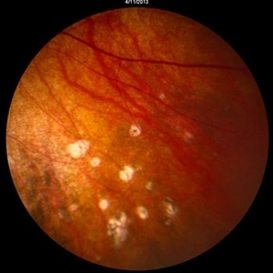
---thumb.jpg/image-square;max$300,300.ImageHandler) Resolution of retinal lesions associated with Retinitis
Resolution of retinal lesions associated with Retinitis
Feb 15 2013 by From the Collections of Thomas M. Aaberg, MD and Thomas M. Aaberg Jr., MD
Montage of color fundus photographs eye showing resolution of retinal lesions associated with retinitis. There is optic atrophy, vessel whitening and attentuation, macular pigmentary changes, and diffuse chorioretinal atrophy. Visual acuity was hand motions.
Condition/keywords: retinitis
-
 Chronical Submacular Hemorrhage in the Setting of Neovascular AMD
Mar 23 2015 by Rita Couceiro, MD, MS
An 80-year-old male, with a history of hypertension and high cholesterol, complained of acute and painless vision loss in his left eye (OS) in the previous 5 months. On observation best corrected visual acuity in OS was hand motion. A dense vitreous opacity in OS precluded fundus examination. Ocular ultrasound revealed vitreous hemorrhage and thickening of the macular area. The patient was submitted to pars plana vitrectomy, which disclosed a large submacular hemorrhage with chronical features and disciform scarring in the setting of neovascular AMD.
Imaging device: Intraoperative fundus photograph
Condition/keywords: neovascular age-related macular degeneration (AMD), submacular hemorrhage, wet age-related macular degeneration (wet AMD)
-
 Welder's Maculopathy
Dec 14 2016 by Young Hee Yoon, MD, PhD
Fundus photograph of a 53-year-old man with welder's maculopathy in the right eye. He complained sudden visual loss in the right eye at thirty minutes after welding arc work. His best-corrected visual acuity was hand motion.
Photographer: Kyung Wun Kim, Asan Medical Center
Condition/keywords: central retinal artery occlusion (CRAO), Welder's maculopathy
-
 VKH2
VKH2
May 14 2013 by David Callanan, MD
Photos of both eyes of a 66-year-old male with a 4 year history of VKH treated with oral prednisone and mycophenolate. His presenting acuity was hand motions OD and counting fingers at 4 feet OS. Now 20/20-1 OU. Areas of previous inflammatory activity are seen as atrophic scars now.
-
 Pseudoexfoliation With Partial Subluxation
Jul 12 2013 by Jason S. Calhoun
81-year-old female who woke up with loss of vision in the left eye. Patient was hand motion in the left eye. Slit lamp examination shows dislocated lens with pseudoexfoliation material around the iris rim. Proceeded with cataract surgery.
Photographer: Jason S. Calhoun, Department of Ophthalmology, Mayo Clinic Jacksonville, Florida
Condition/keywords: pseudoexfoliation of lens capsule, subluxation of lens
-
 Welder's Maculopathy
Dec 14 2016 by Young Hee Yoon, MD, PhD
Early phase fluorescein angiography of a 53-year-old man with welder's maculopathy in the right eye. He complained sudden visual loss in the right eye at thirty minutes after welding arc work. His best-corrected visual acuity was hand motion.
Photographer: Kyung Wun Kim, Asan Medical Center
Condition/keywords: central retinal artery occlusion (CRAO), Welder's maculopathy
 Loading…