Search results (2824 results)
-
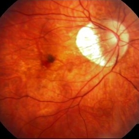
---thumb.JPG/image-square;max$300,300.ImageHandler) Weiss Ring (Floater)
Weiss Ring (Floater)
Jul 10 2013 by Jason S. Calhoun
Patient comes in complaining of a floater towards the nasal aspect of her vision. Fundus photograph with anterior shot, shows a weiss ring pulled off from the optic nerve.
Photographer: Jason S. Calhoun, Department of Ophthalmology, Mayo Clinic Jacksonville, Florida
Condition/keywords: floaters, Weiss ring
-
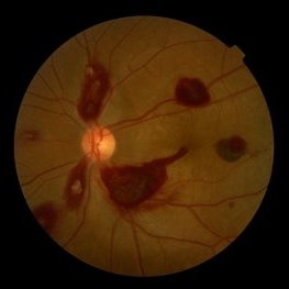
 Bilateral Retinoschisis Retinal Detachment
Sep 15 2012 by Barbara Parolini, MD
Fundus photograph of a case of bilateral retinoschisis and retinal detachment. The border of the external layer breaks and the border of the schisis have been treated with argon laser. An epiretinal membrane formed after the formation of retinal detachment.
Photographer: Dr Rino Frisina, Istituto Clinico S.Anna, Brescia, Italy
Imaging device: optos
Condition/keywords: epiretinal membrane formation, retinoschisis
-
---thumb.jpg/image-square;max$300,300.ImageHandler) Normal Fundus Photo
Normal Fundus Photo
Feb 13 2013 by From the Collections of Thomas M. Aaberg, MD and Thomas M. Aaberg Jr., MD
Normal fundus photo.
Condition/keywords: fundus photograph, normal eye
-
 Lacquer Cracks in Pathologic Myopia
Apr 19 2013 by Theodore Leng, MD, MS, FASRS
Color fundus photograph of a 48-year-old woman with lacquer cracks in the setting of pathologic myopia.
Condition/keywords: lacquer cracks, pathologic myopia
-
---thumb.JPG/image-square;max$300,300.ImageHandler) Weiss Ring (Floater)
Weiss Ring (Floater)
Jul 10 2013 by Jason S. Calhoun
Patient comes in complaining of a floater towards the nasal aspect of her vision. Fundus photograph with anterior shot, shows a weiss ring pulled off from the optic nerve.
Photographer: Jason S. Calhoun, Department of Ophthalmology, Mayo Clinic Jacksonville, Florida
Condition/keywords: floaters, Weiss ring
-
 Proliferative Diabetic Retinopathy
Sep 17 2012 by Michael P. Kelly, FOPS
Retinal fundus photograph of a patient with PDR and NVD.
Photographer: Michael P. Kelly, FOPS Director, Duke Eye Labs, Duke University Hospital, Duke Eye Center
Imaging device: Topcon
Condition/keywords: neovascularization of the disc (NVD)
-
 Venous Loop & Venous Beading
May 31 2014 by Hamid Ahmadieh, MD
Color fundus photograph of the left eye of a diabetic patient with NVD, NVE, venous loop and venous beading.
Photographer: Elham Salehi, Negah Eye Center, Tehran
Condition/keywords: color fundus photograph, neovascularization elsewhere (NVE), neovascularization of the disc (NVD), proliferative diabetic retinopathy (PDR), venous beading, venous loop
-
 Ocular Manifestation of Acute Leukemia
Sep 8 2012 by Hamid Ahmadieh, MD
Color fundus photograph of a 26-year-old man with acute leukemia.
Photographer: Hamid Ahmadieh, MD, Ophthalmic Research Center, Labbafinejad Medical Center, Shahid Beheshti University of Medical Sciences , Tehran
Imaging device: Topcon Fundus Camera
Condition/keywords: acute leukemia, white centered retinal hemorrhage (Roth Spot)
-
 Asteroid Hyalosis
Aug 27 2015 by René Hernán Parada Vásquez
Fundus photograph of 60-year-old male with an asteroid hyalosis, showing a multiple-yellow-white, round, particles composed of calcium.
Photographer: Parada René, ESO, Guatemala.
Condition/keywords: asteroid hyalosis, floaters
-
 "Boat-Shaped" Preretinal Hemorrhage
Feb 21 2019 by Mitzy E Torres Soriano, MD
Color fundus photograph showing preretinal (subhyaloid) hemorrhage in a diabetic patient with proliferative diabetic retinopathy.
Photographer: Andrea Vitale, MD
Condition/keywords: preretinal hemorrhage, proliferative diabetic retinopathy (PDR), subhyaloid hemorrhage
-
---thumb.jpg/image-square;max$300,300.ImageHandler) Presumed Ocular Histoplasmosis Syndrome
Presumed Ocular Histoplasmosis Syndrome
Feb 26 2013 by Henry J. Kaplan, MD
Color fundus photograph of the right eye of a patient with POHS shows typical punched out scars and peripapillary atrophy.
Condition/keywords: presumed ocular histoplasmosis syndrome (POHS)
-
 Coloboma, In Stereo
Oct 1 2012 by Michael P. Kelly, FOPS
This is a stereo retinal fundus photograph of a coloboma, with the optic nerve centered, using a Zeiss FF3C retinal fundus camera.
Photographer: Michael P. Kelly, FOPS Director, Duke Eye Labs, Duke University Hospital, Duke Eye Center, Durham, NC
Condition/keywords: coloboma, fundus photograph, stereo pair
-
 Snail Track Peripheral Retinal Degeneration
Apr 29 2022 by Otakar Dušek, M.D. Ph.D.
Colour fundus photograph of 22-year-old woman with incidentally found snail track retinal degeneration in the superior temporal periphery of the retina of the right eye.
Photographer: Otakar Dušek, Charles University, Prague
Imaging device: Zeiss Clarus
Condition/keywords: peripheral retinal degeneration
-
---thumb.jpg/image-square;max$300,300.ImageHandler) C3F8 gas bubble after retinal detachment surgery
C3F8 gas bubble after retinal detachment surgery
Feb 1 2013 by Sharon Fekrat, MD FACS FASRS
63 year old man s/p encircling scleral buckle and 23g pars plana vitrectomy for a macula off phakic rhegmatogenous retinal detachment. This fundus photograph shows the effect of the encircling buckle and the residual C3F8 intravitreal gas bubble in the right eye.
Photographer: Tiffanie Keaton, Duke Eye Imaging, Duke University Eye Center, Durham, NC
Imaging device: Optos
Condition/keywords: intravitreal gas bubble, vitrectomy
-
 Normal Fundus image
Aug 30 2018 by Timothy Adeyemo
Fundus photograph of a healthy 31-year-old lady being examined for glaucoma.
Photographer: Adeyemo Timothy, National Eye Centre, Kaduna, Nigeria.
Imaging device: Topcon's TRC- NW8 plus
Condition/keywords: normal retina
-
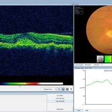 Choroidal Osteoma Plus CNV
Sep 2 2012 by Hamid Ahmadieh, MD
Color fundus photograph and OCT imaging of a 47-year-old man with a juxtafoveal CNV superimposed on a choroidal osteoma.
Photographer: Hamid Ahmadieh, Ophthalmic Research Center, Labbafinejad Medical Center
Imaging device: Topcon
Condition/keywords: choroidal neovascularization (CNV), choroidal osteoma, optical coherence tomography (OCT)
-
 Retinoschisi and Retinal Detachment
Sep 15 2012 by Barbara Parolini, MD
Fundus photograph of an eye with retinoschisis and retinal detachment. The other eye has a retinoschisis and retinal detachment with epiretinal membrane.
Photographer: Dr Rino Frisina, Istituto Clinico S.Anna, Brescia, Italy
Imaging device: Optos ultra wide-field retinographer
Condition/keywords: epiretinal membrane formation, retinoschisis
-
---thumb.jpg/image-square;max$300,300.ImageHandler) Bergmeister's Papilla
Bergmeister's Papilla
Mar 22 2014 by Hamid Ahmadieh, MD
Color fundus photograph of the right eye of a 50-year-old man with Bergmeister's papilla.
Photographer: Naghmeh Nozhat, Negah Eye Center, Tehran
Imaging device: Topcon Fundus Camera
Condition/keywords: Bergmeister's Papillae, color photo
-
 Vortex Vein In A Patient With A Blond Fundus
Oct 2 2013 by Jerald A. Bovino, MD
The vortex vein and vortex vein ampulla are visible in this patient with a blonde (lightly pigmented) fundus.
Condition/keywords: choroidal circulation, fundus photograph, vortex vein
-
 Kearns-Sayre Syndrome
Sep 18 2012 by Michael P. Kelly, FOPS
Retinal fundus photograph of a Kearns-Sayre Syndrome patient.
Photographer: Michael P. Kelly, FOPS Director, Duke Eye Labs, Duke University Hospital, Duke Eye Center
Imaging device: Canon 60UV
Condition/keywords: bilateral pigmentary retinopathy, cardiac conduction abnormalities, chronic progressive ophthalmoplegia, heart-block, Kearns-Sayre Syndrome, ptosis
-
 Geographic Atrophy, Fundus photograph
Aug 23 2012 by Gerardo Garcia-Aguirre, MD
Fundus photograph of an 85-year-old patient with age related macular degeneration and geographic atrophy. A large area with well-defined borders is observed, in which the choroidal vasculature is visualized.
Photographer: Noemí Hernández, Asociación para Evitar la Ceguera en México
Imaging device: Zeiss FF4
Condition/keywords: geographic atrophy
-
 Myelinated Nerve Fibers
Sep 17 2012 by Michael P. Kelly, FOPS
Retinal fundus photograph of a macular hole.
Photographer: Michael P. Kelly, FOPS Director, Duke Eye Labs, Duke University Hospital, Duke Eye Center
Imaging device: Topcon
Condition/keywords: macular hole, myelinated nerve fibers
-
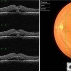 Chronic Active Central Serous Chorioretinopathy (CSCR)
Sep 11 2012 by Hamid Ahmadieh, MD
Color fundus photograph and OCT image of a 30-year-old man with chronic active CSCR.
Photographer: Hamid Ahmadieh, MD, Ophthalmic Research Center, Labbafinejad Medical Center, Shahid Beheshti University of Medical Sciences
Imaging device: Topcon
Condition/keywords: central serous chorioretinopathy (CSCR), optical coherence tomography (OCT)
-
 Branch Retinal Artery Occlusion
Sep 21 2012 by Allen Chiang, MD, FASRS
Fundus photograph of a 27-year old male with a branch retinal artery occlusion. Systemic medical evaluation identified anti-phospholipid antibody syndrome.
Imaging device: Topcon
Condition/keywords: antiphospholipid antibody syndrome, branch retinal artery occlusion (BRAO)
-
 Myopic Choroidal Neovascular Membrane
Mar 25 2013 by Ratimir Lazic, MD, PhD
Color fundus photography of a 33-year-old female. In macular area subretinal hemorrhage can be seen. Area of atrophy temporal from PNO. Myopic changes of posterior pole and mid periphery can be noticed. The patient has been treated with 2 consecutive ranibizumab intravitreal injections. BCVA at baseline was 0,05 (Snellen lines) and 0,3 (Snellen lines) 2 months after.
Photographer: Marko Lukic, MD
Imaging device: Zeis Visucam Lite 2
Condition/keywords: high myopia, myopic choroidal neovascularization (CNV), ranibizumab
 Loading…