Search results (113 results)
-
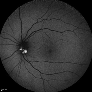
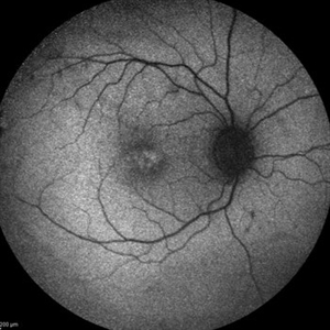
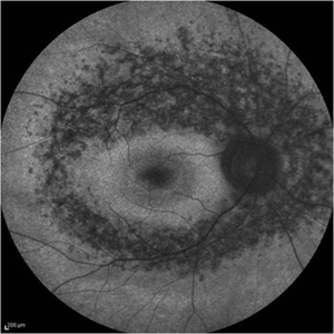 Retinitis Pigmentosa - Fundus Autofluorescence
Sep 20 2014 by Rameez N Hussain, MD
Fundus autofluorescence of retinitis pigmentosa showing hyperautofluorescent rings or foveal hyperautofluorescence.
Photographer: Dr.Rameez N Hussain, MD, Central Imaging Center, Vitreo Retinal Services, Giridhar Eye Institute, Cochin, India
Imaging device: Heidelberg Blue Peak Autofluorescence imaging.
Condition/keywords: bone spicule, cystoid macular edema (CME), fundus autofluorescence (FAF), retinitis pigmentosa
-
 Retinal Angiomatous Proliferation in Age-Related Macular Degeneration with Subretinal Neovascularization
Sep 24 2012 by James B. Soque, CRA, OCT-C, COA, FOPS
75-year-old white male with classic SRN with RAP. Lesion OD is active, and patient is receiving anti-VEGF treatment. Mid phase FA, 50 Deg, Mag 2x.
Photographer: James Soque, CRA, COA, Island Retina, Shirley, NY, USA
Imaging device: Topcon TRC 50 DX, OIS 5.0 MP Color, FA Camera, OIS Software
Condition/keywords: age-related macular degeneration (AMD), fundus autofluorescence (FAF), leakage, retinal angiomatous proliferation (RAP), subretinal neovascularization (SRNV)
-
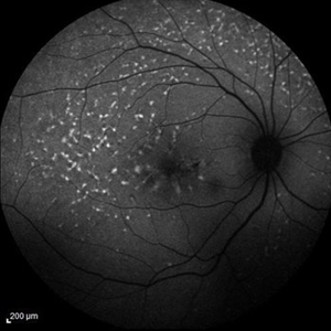
 Stargardts Disease in FAF
Sep 14 2012 by Michael P. Kelly, FOPS
This is a scanning laser ophthalmoscopic FAF image of a patient with Stargardts Disease captured with a Heidelberg Spectralis imaging unit. Note, besides the obvious hyper-autofluorescent areas centrally, the much smaller, and in greater number, pinpoints of hyper-autofluorescence extending from the vascular arcades into the mid-periphery.
Photographer: Michael P. Kelly, FOPS, Director, Duke Eye Center Labs, Duke Universtiy Hospital
Imaging device: Heidelberg Spectralis
Condition/keywords: fundus autofluorescence (FAF), Stargardt disease
-
 Macular Hole, Autofluorescence
Sep 14 2012 by Michael P. Kelly, FOPS
Fundus autofluorescence (FAF) of a macular hole captured using a Heidelberg Spectralis.
Photographer: Michael P. Kelly, FOPS, Director, Duke Eye Cneter Labs, Duke Universty Hospital
Imaging device: Heidelberg Spectralis
Condition/keywords: fundus autofluorescence (FAF), macular hole
-
Angioid Streaks & CNV (Fig 1)
Aug 25 2012 by Hamid Ahmadieh, MD
Fundus autofluorescence (FAF) of a 53-year-old woman with a juxtafoveal CNV secondary to angioid streaks.
Photographer: Hamid Ahmadieh, Ophthalmic Research Center, Labbafinejad Medical Center
Imaging device: Heidelberg Spectralis
Condition/keywords: angioid streaks, choroidal neovascularization (CNV), fundus autofluorescence (FAF)
-
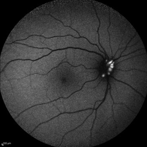 Optic Disc Drusen
Jul 10 2013 by Hamid Ahmadieh, MD
Fundus autofluorescence image of the right eye of a 24-year-old woman with optic disc drusen and VA 20/20.
Photographer: Solmaz Shahmohammadi, Negah Eye Center, Tehran
Imaging device: Heidelberg Spectralis
Condition/keywords: fundus autofluorescence (FAF), optic disc drusen
-
 FFA - PDR
Mar 30 2018 by Lanin Chen
Fundus fluorescein angiography photo of the left eye of a 62-year-old woman with history of Type 2 diabetes mellitus since 20 years showing proliferative diabetic retinopathy.
Photographer: Lanin Chen
Condition/keywords: fundus autofluorescence (FAF), proliferative diabetic retinopathy (PDR)
-
 Tamoxifen Retinopathy- FAF
Aug 30 2012 by Young Hee Yoon, MD, PhD
Fundus autofluorescence (FAF) of an 58-year-old woman with a bilateral tamoxifen maculopathy. She had taken tamoxifen for 24 months due to breast cancer. In spite of discontinuation 2 years ago, her macula remained unchanged. Her best-corrected visual acuity was 20/50 in the right and 20/100 in the left.
Photographer: Soo Hyun Cho, Asan Medical Center
Imaging device: Heidelberg HRA II
Condition/keywords: drug toxicity, toxic maculopathy
-
Stargardts Disease in Fundus Autofluorescence
Sep 12 2012 by Michael P. Kelly, FOPS
Fundus autofluorescence of a patient with Stargardts disease. Note the central area of hypo-autofluorescence indicating atrophy surrounded by smaller areas of hyper-autofluorescence. Note also the much smaller, and in greater number, pinpoints of hyper-autofluorescence extending from the vascular arcades into the mid-periphery.
Photographer: Michael P. Kelly, FOPS, Director, Duke Eye Labs, Duke University Hospital, Duke Eye Center
Imaging device: Heidelberg Spectralis
Condition/keywords: fundus autofluorescence (FAF), Stargardt disease
-
 Cystoid Macular Edema (CME)
Sep 11 2012 by Hamid Ahmadieh, MD
Fundus autofluorescence (FAF) of the right eye a 17-year-old boy with chronic intermediate uveitis showing CME.
Photographer: Hamid Ahmadieh, MD, Ophthalmic Research Center, Labbafinejad Medical Center, Shahid Beheshti University of Medical Sciences
Imaging device: Heidelberg Spectralis
Condition/keywords: cystoid macular edema (CME), fundus autofluorescence (FAF), intermediate uveitis
-
 Optic Disc Drusen
Jul 10 2013 by Hamid Ahmadieh, MD
Fundus autofluorescence image of the left eye of a 24-year-old woman with optic disc drusen and VA 20/20.
Photographer: Solmaz Shahmohammad, Negah Eye Center, Tehran
Imaging device: Heidelberg Spectralis
Condition/keywords: fundus autofluorescence (FAF), optic disc drusen
-
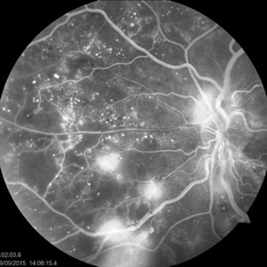
 Proliferative Diabetic Retinopathy
Sep 15 2012 by Hamid Ahmadieh, MD
Fundus autofluorescence image of a 30-year-old woman with the history of scatter laser photocoagulation and a preretinal hemorrhage due to active PDR .
Photographer: Hamid Ahmadieh, MD, Ophthalmic Research Center, Labbafinejad Medical Center, Shahid Beheshti University of Medical Sciences
Imaging device: Heidelberg HRA
Condition/keywords: fundus autofluorescence (FAF), preretinal hemorrhage
-
---thumb.jpg/image-square;max$300,300.ImageHandler) Tamoxifen Retinopathy- FAF
Tamoxifen Retinopathy- FAF
Aug 30 2012 by Young Hee Yoon, MD, PhD
Fundus autofluorescence (FAF) of an 58-year-old woman with a bilateral tamoxifen maculopathy. She had taken tamoxifen for 24 months due to breast cancer. In spite of discontinuation 2 years ago, her macula remained unchanged. Her best-corrected visual acuity was 20/50 in the right and 20/100 in the left.
Photographer: Kyoung Woon Kim, Asan Medical Center
Imaging device: Heidelberg
Condition/keywords: drug toxicity, toxic maculopathy
-
---thumb.jpg/image-square;max$300,300.ImageHandler) Polypoidal Choroidal Vasculopathy: Case 1 - Image 2 of 7
Polypoidal Choroidal Vasculopathy: Case 1 - Image 2 of 7
Oct 4 2012 by Gregg T. Kokame, MD, MMM, FASRS
Bluepeak Autofluorescence image of a 57-year-old woman with treatment-naive polypoidal choroidal vasculopathy. Series of images provides an comparative view of the same condition while utilizing a variet of different imaging procedures.
Photographer: Andrew Yuen, Retina Consultants of Hawaii
Imaging device: Heidelberg Spectralis
Condition/keywords: autofluorescence imaging, fundus autofluorescence (FAF), polypoidal choroidal vasculopathy (PCV)
-
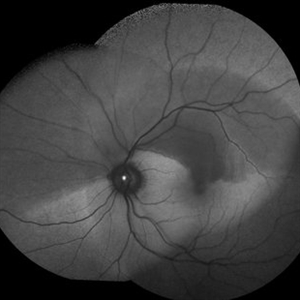
 Fundus autofluorescence of geographic atrophy
Apr 30 2015 by Mitzy E Torres Soriano, MD
Fundus autofluorescence of geographic atrophy (AMD).
Photographer: Mitzy E. Torres Soriano, MD; Centro medico Cagua-Estado Aragua. Venezuela
Imaging device: TOPCON
Condition/keywords: dry age-related macular degeneration (dry AMD), fundus autofluorescence (FAF), geographic atrophy
-
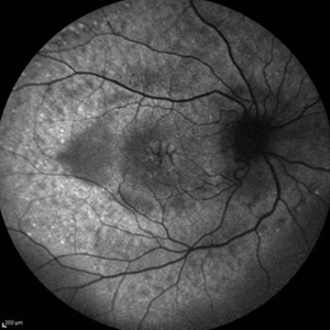
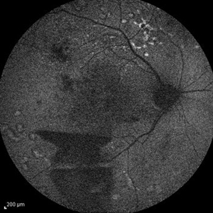
 Geographic Atrophy
Mar 27 2013 by Michael P. Kelly, FOPS
This is a combined FAF/SD-OCT in EDI mode of a patient with geographic atrophy and foveal sparing.
Photographer: Michael P. Kelly, FOPS. Director, Duke Eye Labs, Duke University Eye Center
Imaging device: Heidelberg Spectralis
Condition/keywords: enhanced depth imaging, foveal sparing, fundus autofluorescence (FAF), geographic atrophy, optical coherence tomography (OCT)
-
 Autofluorescence of Choroidal Melanoma
Oct 22 2017 by Daniel Rojas Abatte
Female patient, 53-years-old, diagnosis of choroidal melanoma, already operated in 2009 with brachytherapy.
Photographer: Daniel Rojas
Imaging device: Topcon TRC 50 DX
Condition/keywords: fundus autofluorescence (FAF)
-
 Fundus Flavimaculatus and CNV
Nov 14 2013 by Hamid Ahmadieh, MD
FAF image of the right eye of a 35-year-old woman with subfoveal CNV secondary to fundus flavimaculatus .
Photographer: Nayereh Hadipour, Negah Eye Center, Tehran
Condition/keywords: choroidal neovascularization (CNV), fundus autofluorescence (FAF), fundus flavimaculatus, retinal flecks
-
 Congenital Retinal Pigment Epithelial Hypertrophy (CHRPE) Associated with Gardner's Syndrome
Mar 13 2018 by Olivia Rainey
Ultra-wide field fundus autofluorescence images of a 14-year-old patient with congenital retinal pigment epithelial hypertrophy affecting both eyes as a manifestation of Gardner's Syndrome.
Photographer: Olivia Rainey
Imaging device: Optos
Condition/keywords: bilateral, familial adenomatous polyposis, fundus autofluorescence (FAF), Gardner Syndrome, hypofluorescent lesions, Optos, ultra-wide field imaging
-
---thumb.JPG/image-square;max$300,300.ImageHandler) Optic Pit Maculopathy - Fundus Autofluorescence
Optic Pit Maculopathy - Fundus Autofluorescence
Oct 14 2013 by Cagri G Besirli, MD, PhD, FASRS
10-year-old girl with congenital optic pit and recent vision loss secondary to optic pit maculopathy.
Imaging device: Optos
Condition/keywords: fundus autofluorescence (FAF), maculopathy
-
 X-Linked Retinoschisis
Jan 31 2015 by Hamid Ahmadieh, MD
FAF of the right eye of a 35-year-old man with x-linked retinoschisis. Please notice the foveal schisis.
Photographer: Solmaz Shahmohammad, Negah Eye Center, Tehran, Iran
Imaging device: Heidelberg
Condition/keywords: foveal schisis, fundus autofluorescence (FAF), x-linked retinoschisis (XLRS)
-
---thumb.jpg/image-square;max$300,300.ImageHandler) Primary Subhyaloid Hemorrhage Due to Valsalva Retinopathy
Primary Subhyaloid Hemorrhage Due to Valsalva Retinopathy
Nov 13 2013 by Hamid Ahmadieh, MD
FAF image of the left eye of a 25-year-old man with primary subhyaloid hemorrhage due to Valsalva retinopathy.
Photographer: Nayereh Hadipour, Negah Eye Center, Tehran
Condition/keywords: fundus autofluorescence (FAF), subhyaloid hemorrhage, valsalva retinopathy
-
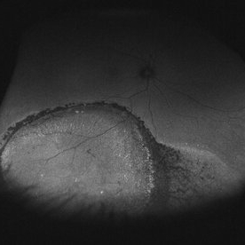 Asymptomatic Chronic Retinal Detachment With Demarcation Line
Jun 11 2016 by Philip J. Polkinghorne, MD
A 65-year-old emmetrope with asymptomatic chronic retinal detachment with demarcation line.
Photographer: Alex Fraser, Greenlane Clinical Center, Auckland, New Zealand
Condition/keywords: chronic retinal detachment, fundus autofluorescence (FAF)
-
 Color Fundus Photographs of Optic Disc Drusen
Apr 26 2018 by Ahmad B. Tarabishy, MD
Fundus photographs and autofluorescence of a 75-year-old man with an epiretinal membrane in the left eye. Incidentally, he had a history of optic disc drusen, which show a striking hyperautofluorescence on FAF imaging.
Photographer: Michelle Howarth, Lakeland Eye Clinic
Imaging device: Zeiss Visucam
Condition/keywords: fundus autofluorescence (FAF), optic disc drusen
-
 Branch Retinal Artery Occlusion With Calcium Embolus at the Disc - Fundus Autofluorescence Imaging (FAF)
Apr 7 2018 by Rameez N Hussain, MD
Acute branch retinal artery occlusion with a calcium embolus at the disc which is hyper autofluorescent in fundus autofluorescence Imaging (FAF) -resembles an LED light source ('LED sign').
Photographer: DR RAMEEZ N HUSSAIN
Imaging device: Heidelberg Spectralis
Condition/keywords: branch retinal artery occlusion (BRAO), embolus, fundus autofluorescence (FAF), retinal edema
 Loading…