Search results (173 results)
-
 Fundus Flavimaculatus
May 2 2013 by Henry J. Kaplan, MD
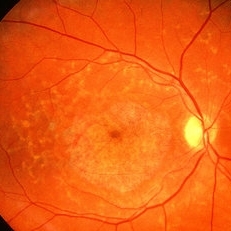
Yellow pisciform flecks at the level of RPE accompanied by Bull's eye, Right eye; #1.
Condition/keywords: bull's eye maculopathy, fundus flavimaculatus, Stargardt disease
-
 Stargardt macular dystrophy slide 1
Oct 22 2012 by Ronald C. Gentile, MD
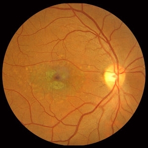
16-year-boy with difficulty in school seeing the black board. The macula area of the right eye had areas with a beaten bronze appearance and atrophy. Small pisci-form flecks can be seen surrounding the fovea.
Photographer: The New York Eye & Ear Infirmary Department of Medical Imaging
Condition/keywords: small pisci-form flecks, Stargardt disease
-
 Stargardt Disease
Feb 28 2013 by Theodore Leng, MD, MS, FASRS
Fundus photograph of a 63-year-old man with Stargardt disease. Multiple pisciform flecks are visible.
Imaging device: Zeiss FF450
Condition/keywords: flecks, Stargardt disease
-
 Stargardt macular dystrophy slide 1
Oct 22 2012 by Ronald C. Gentile, MD
45-year-old man with progressive central vision loss since the age of 10. Small flecks can be seen surrounding the central area of atrophy.
Photographer: The New York Eye & Ear Infirmary Department of Medical Imaging
Condition/keywords: Stargardt disease, vision loss
-
 Fundus Flavimaculatus
May 2 2013 by Henry J. Kaplan, MD
Widespread pisciform flecks in the fundus accompanied by macular beaten bronze lesion; Left eye; #2.
Condition/keywords: fundus flavimaculatus, Stargardt disease
-
 Fundus Flavimaculatus and CNV
Nov 14 2013 by Hamid Ahmadieh, MD
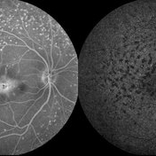
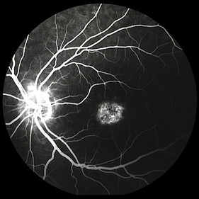
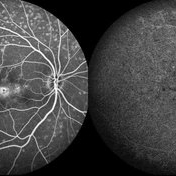
Late phase FA and ICG angiography images of the right eye of a 35-year-old woman with subfoveal CNV secondary to fundus flavimaculatus .
Photographer: Nayereh Hadipour, Negah Eye Center, Tehran
Condition/keywords: choroidal neovascularization (CNV), fundus flavimaculatus, indocyanine green (ICG) angiography, retinal flecks
-
 Stargardt macular dystrophy slide 2
Oct 22 2012 by Ronald C. Gentile, MD
Fundus examination of the left eye had similar findings with centrally atrophic macula area with surrounding flecks.
Photographer: The New York Eye & Ear Infirmary Department of Medical Imaging
Condition/keywords: Stargardt disease
-
 Pattern Dystrophy slide 1
Oct 22 2012 by Ronald C. Gentile, MD
Asymptomatic middle-age man with normal vision and a multifocal pattern dystrophy. The pattern dystrophy simulates Stargardt disease/fundus flavimaculatus with irregular yellow-white flecks scattered throughout the posterior pole. Some lesions extend beyond the retinal vascular arcades.
Photographer: The New York Eye & Ear Infirmary Department of Medical Imaging
Condition/keywords: pattern macular dystrophy
-
 Senile Retinoschisis
Nov 9 2012 by Norman Byer
This 48-year-old woman has senile retinoschisis involving the most common location, the lower temporal quadrant. The lesion shown here illustrates one of the two clinical features which are most often responsible for attracting the attention of the examiner to such lesions, namely the multitude of yellow flecks lying on the inner surface of the inner layer. The nature of these flecks is not known, but it seems clear that they do not originate in the schisis cavity for they do not represent remnants of ruptured Miller’s fibers. In this photograph you can easily detect the fluid space which separates the inner and outer retinal layers.
Condition/keywords: lower temporal quadrant, senile retinoschisis, yellow flecks
-
 Pattern Dystrophy slide 2
Oct 22 2012 by Ronald C. Gentile, MD
The irregular yellow-white flecks are noted in both eyes and are symmetric.
Photographer: The New York Eye & Ear Infirmary Department of Medical Imaging
Condition/keywords: pattern macular dystrophy
-
 Stargardt disease case 3 FA LE
Jan 11 2013 by Alex P. Hunyor, MD
Stargardt disease - left eye. Atrophic maculopathy without flecks. Fluorescein angiogram - note silent choroid.
Condition/keywords: Stargardt disease
-
 Fundus Flavimaculatus and CNV
Nov 14 2013 by Hamid Ahmadieh, MD
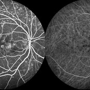
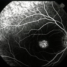
FA and ICG angiography images of the right eye of a 35-year-old woman with subfoveal CNV secondary to fundus flavimaculatus .
Photographer: Nayereh Hadipour, Negah Eye Center, Tehran
Condition/keywords: choroidal neovascularization (CNV), fundus flavimaculatus, indocyanine green (ICG) angiography, retinal flecks
-
 Fundus Flavimaculatus and CNV
Nov 14 2013 by Hamid Ahmadieh, MD
FAF image of the right eye of a 35-year-old woman with subfoveal CNV secondary to fundus flavimaculatus .
Photographer: Nayereh Hadipour, Negah Eye Center, Tehran
Condition/keywords: choroidal neovascularization (CNV), fundus autofluorescence (FAF), fundus flavimaculatus, retinal flecks
-
 Stargardt Disease
Feb 28 2013 by Theodore Leng, MD, MS, FASRS
Fundus photograph of a 63-year-old man with Stargardt disease. Multiple pisciform flecks are visible.
Imaging device: Zeiss FF450
Condition/keywords: flecks, Stargardt disease
-
 Fundus Flavimaculatus and CNV
Nov 14 2013 by Hamid Ahmadieh, MD
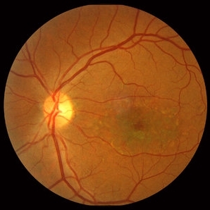
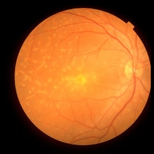
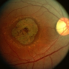
Color fundus photograph of the right eye of a 35-year-old woman with subfoveal CNV secondary to fundus flavimaculatus .
Photographer: Nayereh Hadipour, Negah Eye Center, Tehran
Condition/keywords: choroidal neovascularization (CNV), fundus flavimaculatus, fundus photograph, retinal flecks
-
 Fundus Flavimaculatus and CNV
Nov 14 2013 by Hamid Ahmadieh, MD
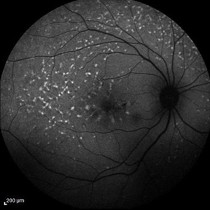
Infrared image of the right eye of a 35-year-old woman with subfoveal CNV secondary to fundus flavimaculatus .
Photographer: Nayereh Hadipour, Negah Eye Center, Tehran
Condition/keywords: choroidal neovascularization (CNV), fundus flavimaculatus, infrared image, retinal flecks
-
 Macular Dystrophy With Flecks
Jul 31 2013 by From the Collections of Thomas M. Aaberg, MD and Thomas M. Aaberg Jr., MD
Macular dystrophy with flecks.
Condition/keywords: flecks, macular dystrophy
-
 Flecks Retinopathy
Jul 31 2013 by From the Collections of Thomas M. Aaberg, MD and Thomas M. Aaberg Jr., MD
Flecks retinopathy.
Condition/keywords: fleck retinopathy
-
 Stargardt's disease
May 2 2013 by Henry J. Kaplan, MD
Bull's eye and surrounding retinal flecks in the right eye; #1.
Condition/keywords: bull's eye maculopathy, Stargardt disease
-
 Stargardt's Disease
Oct 4 2018 by Aditya S Kelkar, MS, FRCS, FASRS,FRCOphth
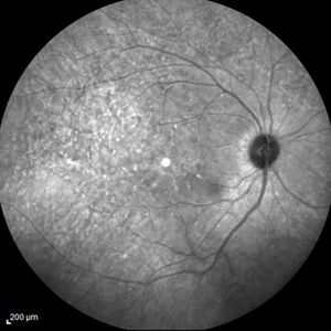
Auto-fluorescence image of a 19-year-old male showing flecks of both increased and decreased autofluorescence and reduced central macular autofluorescence surrounded by an increased signal.
Photographer: Dr. Aanchal Agarwal
Condition/keywords: Stargardt disease
-
 Macular Dystrophy With Flecks
Jul 31 2013 by From the Collections of Thomas M. Aaberg, MD and Thomas M. Aaberg Jr., MD
Macular dystrophy with flecks.
Condition/keywords: flecks, macular dystrophy
-
 Fundus Flavimaculatus and CNV
Nov 14 2013 by Hamid Ahmadieh, MD
Late phase FA and ICG angiography images of the right eye of a 35-year-old woman with subfoveal CNV secondary to fundus flavimaculatus .
Photographer: Nayereh Hadipour, Negah Eye Center, Tehran
Condition/keywords: choroidal neovascularization (CNV), fundus flavimaculatus, indocyanine green (ICG) angiography, retinal flecks
-
 Stargardt disease case 3 FA RE
Jan 11 2013 by Alex P. Hunyor, MD
Stargardt disease - right eye. Atrophic maculopathy without flecks. Fluorescein angiogram - note silent choroid.
Condition/keywords: Stargardt disease
-
 Flecks Retinopathy
Jul 31 2013 by From the Collections of Thomas M. Aaberg, MD and Thomas M. Aaberg Jr., MD
Flecks retinopathy.
Condition/keywords: fleck retinopathy
-
 Stargardt Disease
Feb 28 2013 by Theodore Leng, MD, MS, FASRS
Fluorescein angiogram of a 62-year-old woman with Stargardt disease. Multiple pisciform flecks that stain are visible.
Imaging device: Zeiss FF450
Condition/keywords: flecks, Stargardt disease
 Loading…