Search results (172 results)
-
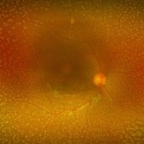 Benign Familial Fleck Retina
Nov 7 2018 by Vedang Shah
Flecks over the entire retinal mid-periphery and periphery of a 12-year-old male with no visual complaints.
Photographer: Dr. Vedang Shah
Imaging device: OPTOS IMAGING SYSTEM
Condition/keywords: fleck retinopathy
-
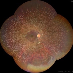 Benign Familial Fleck Retina
Dec 21 2023 by Vishal Agrawal, MD, FRCS,FACS,FASRS
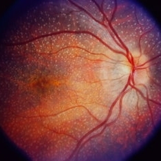
10-year male with high myopia on examination revealed diffuse flecks distributed all over fundus in both eyes sparing macula. Inferior lattice with WWOP areas were also noted in right eye.
Photographer: Dr Ayushi
Imaging device: Clarus 700
Condition/keywords: fleck retinopathy, myopia
-
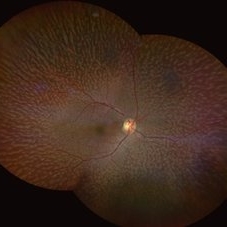 Fundus Albipunctatus
Aug 25 2022 by Aditya S Kelkar, MS, FRCS, FASRS,FRCOphth
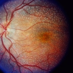
65 year old female, presented for cataract evaluation. Fundus examination showed whitish-yellow flecks in the retina.
Imaging device: Clarus 500
Condition/keywords: fundus albipunctatus, fundus photograph
-
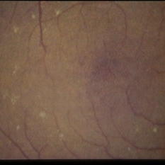
 Stargardt's Disease
Oct 4 2018 by Aditya S Kelkar, MS, FRCS, FASRS,FRCOphth
Auto-fluorescence image of a 19-year-old male showing flecks of both increased and decreased autofluorescence and reduced central macular autofluorescence surrounded by an increased signal.
Photographer: Dr. Aanchal Agarwal
Condition/keywords: Stargardt disease
-
 Stargardt's Disease
Oct 23 2024 by Virginia Gebhart
62 year old female with bullseye RPE changes and flecks, mottled FAF, and silent choroid on FA consistent with late onset Stargardt's Disease. Pt is asymptomatic with 20/20 vision OU at this time
Photographer: Virginia Gebhart, Retina Consultants of Carolina
Imaging device: Optos California
Condition/keywords: Stargardt disease, Stargardts Disease
-
 Tamoxifen
Jan 7 2015 by H. Michael Lambert, MD
High mag right macula with yellow flecks from tamoxifen retinopathy.
Condition/keywords: tamoxifen retinopathy
-
 Stargardt Disease
Feb 28 2013 by Theodore Leng, MD, MS, FASRS
Fundus photograph of a 63-year-old man with Stargardt disease. Multiple pisciform flecks are visible.
Imaging device: Zeiss FF450
Condition/keywords: flecks, Stargardt disease
-
 Stargardt Disease
Feb 28 2013 by Theodore Leng, MD, MS, FASRS
Fundus photograph of a 63-year-old man with Stargardt disease. Multiple pisciform flecks are visible.
Imaging device: Zeiss FF450
Condition/keywords: flecks, Stargardt disease
-
 Stargardt Disease
Feb 28 2013 by Theodore Leng, MD, MS, FASRS
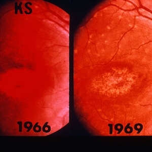
Fluorescein angiogram of a 62-year-old woman with Stargardt disease. Multiple pisciform flecks that stain are visible.
Imaging device: Zeiss FF450
Condition/keywords: flecks, Stargardt disease
-
 Benign Familial Fleck Retina
Dec 2 2024 by KANWALJEET HARJOT MADAN, M.S. (Ophthalmology), FAICO (Vitreous - Retina)
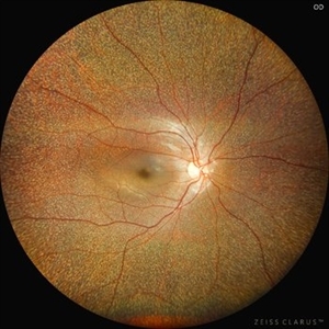
This is fundus picture of a 21 year old female patient who had come for refractive surgery consultation. Her best corrected vision in both eyes was 20/20. She had myopic astigmatism in both eyes. Fundus exam revealed presence of multiple yellowish white flecks spread throughout retina sparing macular area in both eyes. Her color vision was normal. Electroretinogram and electrooculogram were normal. She gave no history of night blindness. A diagnosis of Benign Familial Fleck Retina was made. She was also advised ocular exam of her parents and elder brother which was normal.
Photographer: Dr. Kanwaljeet Harjot Madan, M.S. (Ophthalmologist) Fellow in Vitrous & Retina. Thind Eye Hospital, Jalandhar City. Punjab. India
Imaging device: Zeiss Clarus
Condition/keywords: Benign familial fleck retina, Night Blindness
-
 Benign Familial Fleck Retina
Jul 13 2024 by Tejaswita Verma
Fundus photograph of the LE of a 46 y/o female with bilateral diffuse yellow-white fleck like lesions with 6/6 vision bilaterally.
Photographer: DR. TEJASWITA VERMA
Imaging device: MIRANTE
Condition/keywords: Benign familial fleck retina, flecks
-
 Benign Familial Fleck Retina
Jul 13 2024 by Tejaswita Verma
Fundus photograph of the RE of a 46 y/o female with bilateral diffuse yellow-white fleck like lesions with 6/6 vision bilaterally.
Photographer: DR. TEJASWITA VERMA
Imaging device: MIRANTE
Condition/keywords: Benign familial fleck retina, flecks
-
 Benign Fleck Retina
Sep 21 2023 by Ben Serar
Fundus photograph of LE showing golden-yellow flecks at the posterior pole in a case of Benign Fleck Retina.
Condition/keywords: Benign Fleck Retina
-
 Benign Fleck Retina
Sep 21 2023 by Ben Serar
Fundus photograph of RE showing golden-yellow flecks at the posterior pole in a case of Benign Fleck Retina.
Condition/keywords: Benign Fleck Retina
-
 Best Disease
Nov 7 2024 by Virginia Gebhart
Fluorescein angiogram of 49 year female with Best Disease. Genetic testing done in 2000 confirms Best Disease and also possible Stargardts mutation. Characteristic bullseye maculopathy with surrounding yellowish flecks are present in both eyes.
Photographer: Virginia Gebhart, Retina Consultants of Carolina
Imaging device: Optos California
Condition/keywords: Best disease, fluorescein angiogram (FA)
-
 Bull's Eye Maculopathy With Flecks
Jul 31 2013 by From the Collections of Thomas M. Aaberg, MD and Thomas M. Aaberg Jr., MD
Bull's eye maculopathy with flecks.
Condition/keywords: bull's eye maculopathy, flecks
-
 Bull's Eye Maculopathy With Flecks
Jul 31 2013 by From the Collections of Thomas M. Aaberg, MD and Thomas M. Aaberg Jr., MD
Bull's eye maculopathy with flecks.
Condition/keywords: bull's eye maculopathy, flecks
-
 Central Areolar Choroidal Dystrophy
Oct 30 2020 by Mihir Trivedi
Fundus photo of a 43-year-old female with gradual onset diminution of vision in both eyes since 2-3 years. BCVA in OU was 3/60. She was diagnosed to have central areolar choroidal dystrophy(CACD). Central areolar choroidal dystrophy (CACD) is a rare inherited disease, which causes progressive profound loss of vision in patients during their fourth decade. It is characterized by atrophy of retinal pigment epithelium, photoreceptors and choriocapillaris. IT is a progressive macular dystrophy characterized by subtle, mottled depigmentation in the posterior pole in the early stages. The depigmentation area gradually enlarges until an oval or round surface of atrophy of the retinal pigmentary epithelium and choriocapillaris is formed. Drusen or flecks are absent in a typical presentation.
Photographer: Mr Ganesh Naidu
Imaging device: TOPCON DRI Triton
Condition/keywords: central areolar choroidal dystrophy (CACD)
-
 Central atrophic pigment changes
Apr 4 2013 by Jerald A. Bovino, MD
No history, seems to have flecks, probably hereditary macular degeneration
Condition/keywords: pigment changes
-
 Choroideremia With Periperal Pigment Changes, Drusenoid Flecks, Patchy Atrophy
Aug 1 2013 by From the Collections of Thomas M. Aaberg, MD and Thomas M. Aaberg Jr., MD
Choroideremia with periperal pigment changes, drusenoid flecks, patchy atrophy.
Condition/keywords: choroideremia, drusenoid flecks, periperal pigment changes
-
 Choroideremia With Periperal Pigment Changes, Drusenoid Flecks, Patchy Atrophy
Aug 1 2013 by From the Collections of Thomas M. Aaberg, MD and Thomas M. Aaberg Jr., MD
Choroideremia with periperal pigment changes, drusenoid flecks, patchy atrophy.
Condition/keywords: atrophy, choroideremia, drusenoid flecks
-
 Choroideremia With Periperal Pigment Changes, Drusenoid Flecks, Patchy Atrophy
Aug 1 2013 by From the Collections of Thomas M. Aaberg, MD and Thomas M. Aaberg Jr., MD
Choroideremia with periperal pigment changes, drusenoid flecks, patchy atrophy.
Condition/keywords: choroideremia, drusenoid flecks
 Loading…