Search results (173 results)
-
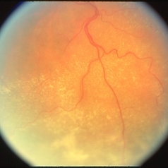
 Starstruck by Stargardt
Nov 17 2025 by SHRADDHA RAJ SHRIVASTAVA
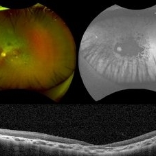
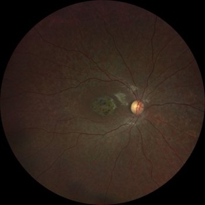
Left eye G-FAF image of a 26 year old patient diagnosed with Stargardt Disease, showing hyperautofluorescent flecks of increased lipofuscin accumulation and dark areas of hypoautofluorescence representing retinal pigment epithelium (RPE) atrophy.
Photographer: Dr. Shraddha Raj Shrivastava
Imaging device: Nidek Mirante SLO/OCT (Confocal scanning/Spectral domain OCT)
Condition/keywords: fleck dystrophy, fundus autofluorescence (FAF), hereditary macular dystrophy, heredomacular degeneration, lipofuscin, Stargardt Disease
-
 LCA type 10
Apr 10 2025 by Joshua Friedman
LCA type 10 due to mutations in CEP290. 36-year-old male with best corrected visual acuity of light perception in both eyes since childhood. On color fundus imaging, there is a mix of polymorphous white flecks and pigmentary changes. On autofluorescence imaging, there is almost complete loss of macular RPE. On OCT, there is complete loss of inner and outer retinal layers, the greatest losses occurring centrally.
Photographer: Stephen Tsang, MD, PhD
Condition/keywords: Leber Congenital Amaurosis
-
 Benign Familial Fleck Retina
Dec 2 2024 by KANWALJEET HARJOT MADAN, M.S. (Ophthalmology); FAICO (Vitreous - Retina)
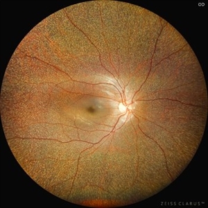
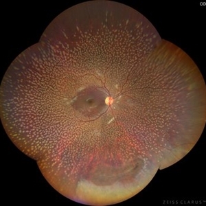
This is fundus picture of a 21 year old female patient who had come for refractive surgery consultation. Her best corrected vision in both eyes was 20/20. She had myopic astigmatism in both eyes. Fundus exam revealed presence of multiple yellowish white flecks spread throughout retina sparing macular area in both eyes. Her color vision was normal. Electroretinogram and electrooculogram were normal. She gave no history of night blindness. A diagnosis of Benign Familial Fleck Retina was made. She was also advised ocular exam of her parents and elder brother which was normal.
Photographer: Dr. Kanwaljeet Harjot Madan, M.S. (Ophthalmologist) Fellow in Vitrous & Retina. Thind Eye Hospital, Jalandhar City. Punjab. India
Imaging device: Zeiss Clarus
Condition/keywords: Benign familial fleck retina, Night Blindness
-
 MIDD (Maternally Inherited Diabetes and Deafness) - Left FP
Nov 30 2024 by John S. King, MD
Both the right and left Eye have fairly symmetrical, extrafoveal drusenoid-like flecks and focal and faint areas of RPE hyperplasia (in addition to mild NPDR and PPA) 57 yo WF referred for AMD vs Pattern Dystrophy that was diagnosed 10 years ago. Reported some slow progressive vision loss in both eyes for distance and near. Denies nyctalopia or hemeralopia. Background medical history includes HTN, CVD, and DM. No family history of eye problems. Denied pentosan use. Anterior segment showed moderate cataracts (OD>OS). Posterior segment exam showed macular changes and mild NPDR. The macular appearance showed a symmetrical, paramacular ring of fleck-like drusenoid material with some faint focal areas of RPE hyperplasia. Fundus Photos, AF, OCT were performed as well as a gene test. Further questioning showed revealed that her mother and maternal grandmother had both diabetes mellitus and sensorineural hearing loss. The patient developed diabetes in her teens, and some high frequency hearing loss in her early twenties. She had not had a previous genetic test or diagnosis of MIDD. Gene testing is pending for the mitochondrial component. Invitae's retinal panel, which does not include mitochondrial disorders, only showed a variant of uncertain significance, HMCN1. I discussed this case with Dr. Freund, and it is similar to a the case report : Inoue M, Kiss S, Freund KB. MACULAR PIGMENT RINGS AS THE PRESENTING FINDING OF MITOCHONDRIAL MYOPATHY, ENCEPHALOPATHY, LACTIC ACIDOSIS, AND STROKELIKE EPISODES. Retin Cases Brief Rep. 2015 Fall;9(4):260-4. doi: 10.1097/ICB.0000000000000182. PMID: 26200388.
Photographer: Grace Melton and Carley Gunn
Imaging device: Clarus
Condition/keywords: Macular Dystrophy, Maternally Inherited Diabetes and Deafness, MIDD, Mitochondrial Disorder
-
 MIDD (Maternally Inherited Diabetes and Deafness) - Right FP
Nov 30 2024 by John S. King, MD
Both the right and left Eye have fairly symmetrical, extrafoveal drusenoid-like flecks and focal and faint areas of RPE hyperplasia (in addition to mild NPDR and PPA) 57 yo WF referred for AMD vs Pattern Dystrophy that was diagnosed 10 years ago. Reported some slow progressive vision loss in both eyes for distance and near. Denies nyctalopia or hemeralopia. Background medical history includes HTN, CVD, and DM. No family history of eye problems. Denied pentosan use. Anterior segment showed moderate cataracts (OD>OS). Posterior segment exam showed macular changes and mild NPDR. The macular appearance showed a symmetrical, paramacular ring of fleck-like drusenoid material with some faint focal areas of RPE hyperplasia. Fundus Photos, AF, OCT were performed as well as a gene test. Further questioning showed revealed that her mother and maternal grandmother had both diabetes mellitus and sensorineural hearing loss. The patient developed diabetes in her teens, and some high frequency hearing loss in her early twenties. She had not had a previous genetic test or diagnosis of MIDD. Gene testing is pending for the mitochondrial component. Invitae's retinal panel, which does not include mitochondrial disorders, only showed a variant of uncertain significance, HMCN1. I discussed this case with Dr. Freund, and it is similar to a the case report : Inoue M, Kiss S, Freund KB. MACULAR PIGMENT RINGS AS THE PRESENTING FINDING OF MITOCHONDRIAL MYOPATHY, ENCEPHALOPATHY, LACTIC ACIDOSIS, AND STROKELIKE EPISODES. Retin Cases Brief Rep. 2015 Fall;9(4):260-4. doi: 10.1097/ICB.0000000000000182. PMID: 26200388.
Photographer: Grace Melton and Carley Gunn
Imaging device: Clarus
Condition/keywords: Macular Dystrophy, Maternally Inherited Diabetes and Deafness, MIDD, Mitochondrial Disorder
-
 Best Disease
Nov 7 2024 by Virginia Gebhart
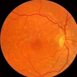
Fluorescein angiogram of 49 year female with Best Disease. Genetic testing done in 2000 confirms Best Disease and also possible Stargardts mutation. Characteristic bullseye maculopathy with surrounding yellowish flecks are present in both eyes.
Photographer: Virginia Gebhart, Retina Consultants of Carolina
Imaging device: Optos California
Condition/keywords: Best disease, fluorescein angiogram (FA)
-
 Stargardt's Disease
Oct 23 2024 by Virginia Gebhart
62 year old female with bullseye RPE changes and flecks, mottled FAF, and silent choroid on FA consistent with late onset Stargardt's Disease. Pt is asymptomatic with 20/20 vision OU at this time
Photographer: Virginia Gebhart, Retina Consultants of Carolina
Imaging device: Optos California
Condition/keywords: Stargardt disease, Stargardts Disease
-
 Pattern dystrophies – Asymptomatic middle-aged man with normal vision and a multifocal PD
Sep 17 2024 by Nicolas A Yannuzzi, MD
The PD simulates Stargardt disease/fundus flavimaculatus with irregular yellow-white flecks scattered throughout the posterior pole. Some lesions extend beyond the retinal vascular arcades.
Condition/keywords: inherited retinal disease, pattern dystrophy
-
 Fundus Flavimaculatus
Aug 12 2024 by Virginia Gebhart
77 year old female with symmetrical retinal flecks consistent with hereditary dystrophy. Unable to complete genetic testing today, will consider in the future. Pt Asymptomatic, Dcc 20/20 OU
Photographer: Virginia Gebhart
Imaging device: Optos California
Condition/keywords: fundus flavimaculatus
-
 Benign Familial Fleck Retina
Jul 13 2024 by Tejaswita Verma
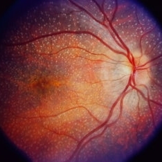
Fundus photograph of the RE of a 46 y/o female with bilateral diffuse yellow-white fleck like lesions with 6/6 vision bilaterally.
Photographer: DR. TEJASWITA VERMA
Imaging device: MIRANTE
Condition/keywords: Benign familial fleck retina, flecks
-
 Benign Familial Fleck Retina
Jul 13 2024 by Tejaswita Verma
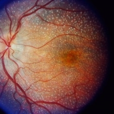
Fundus photograph of the LE of a 46 y/o female with bilateral diffuse yellow-white fleck like lesions with 6/6 vision bilaterally.
Photographer: DR. TEJASWITA VERMA
Imaging device: MIRANTE
Condition/keywords: Benign familial fleck retina, flecks
-
 Stargardt Disease
Apr 8 2024 by T. P . VIGNESH, MBBS,MS
Fundus photo of left eye of 20 year old woman revealing beaten bronze foveal atrophy and wide-spread flecks radiating from the posterior pole to the periphery .
Photographer: Bharathi
Imaging device: ZEISS CLARUS
Condition/keywords: fundus flavimaculatus, Stargardt disease, Stargardts Disease
-
 Stargardt Disease
Apr 8 2024 by T. P . VIGNESH, MBBS,MS
Fundus photo of right eye of 20 year old woman revealing beaten bronze foveal atrophy and wide-spread flecks radiating from the posterior pole to the periphery .
Photographer: Bharathi
Imaging device: ZEISS CLARUS
Condition/keywords: Stargardt disease
-
 Benign Familial Fleck Retina
Dec 21 2023 by Vishal Agrawal, MD, FRCS,FACS,FASRS
10-year male with high myopia on examination revealed diffuse flecks distributed all over fundus in both eyes sparing macula. Inferior lattice with WWOP areas were also noted in right eye.
Photographer: Dr Ayushi
Imaging device: Clarus 700
Condition/keywords: fleck retinopathy, myopia
-
 Benign Fleck Retina
Sep 21 2023 by Ben Serar
Fundus photograph of RE showing golden-yellow flecks at the posterior pole in a case of Benign Fleck Retina.
Condition/keywords: Benign Fleck Retina
-
 Benign Fleck Retina
Sep 21 2023 by Ben Serar
Fundus photograph of LE showing golden-yellow flecks at the posterior pole in a case of Benign Fleck Retina.
Condition/keywords: Benign Fleck Retina
-
 Stargardt’s Disease
Sep 21 2023 by Ben Serar
Fundus photograph of RE showing golden-yellow flecks at the macula in a case of Stargardt’s disease.
Condition/keywords: Stargardts Disease
-
 Stargardt’s Disease
Sep 12 2023 by Ben Serar
Fundus Photograph of LE showing golden yellow flecks at the posterior pole in a case of Stargardt’s Disease.
Condition/keywords: fundus flavimaculatus, Stargardts Disease
-
 Fundus Albipunctatus
Aug 25 2022 by Aditya S Kelkar, MS, FRCS, FASRS,FRCOphth
65 year old female, presented for cataract evaluation. Fundus examination showed whitish-yellow flecks in the retina.
Imaging device: Clarus 500
Condition/keywords: fundus albipunctatus, fundus photograph
-
 Fundus Albipunctatus
Apr 27 2021 by Priya Rasipuram Chandrasekaran, MBBS, DO, DNB, FRCS
This is the fundus photo montage of a 23-year-old male demonstrating whitish-yellow spots all over the fundus sparing the fovea at the level of retinal pigment epithelium. This belongs to the group of congenital stationary night blindness with flecks in the retina.
Condition/keywords: fleck retinopathy
-
 Central Areolar Choroidal Dystrophy
Oct 30 2020 by Mihir Trivedi
Fundus photo of a 43-year-old female with gradual onset diminution of vision in both eyes since 2-3 years. BCVA in OU was 3/60. She was diagnosed to have central areolar choroidal dystrophy(CACD). Central areolar choroidal dystrophy (CACD) is a rare inherited disease, which causes progressive profound loss of vision in patients during their fourth decade. It is characterized by atrophy of retinal pigment epithelium, photoreceptors and choriocapillaris. IT is a progressive macular dystrophy characterized by subtle, mottled depigmentation in the posterior pole in the early stages. The depigmentation area gradually enlarges until an oval or round surface of atrophy of the retinal pigmentary epithelium and choriocapillaris is formed. Drusen or flecks are absent in a typical presentation.
Photographer: Mr Ganesh Naidu
Imaging device: TOPCON DRI Triton
Condition/keywords: central areolar choroidal dystrophy (CACD)
-
 Fundus Flavimaculatus
Apr 2 2019 by Gary R. Cook, MD, FACS
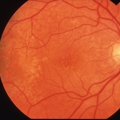
53-year-old white female with asymmetric fundus flavimaculatus demonstrating multiple yellow-white flecks of variable size and shape throughout the posterior pole OS also but with significant macular atrophy; V.A. = 20/200
Imaging device: Topcon VT-50
Condition/keywords: fundus flavimaculatus, Stargardt disease
-
 Fundus Flavimaculatus
Apr 2 2019 by Gary R. Cook, MD, FACS
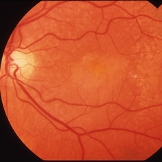
53-year-old white female with asymmetric fundus flavimaculatus demonstrating multiple yellow-white flecks of variable size and shape throughout the posterior pole OD but no macular atrophy; V.A. = 20/20-2
Imaging device: Topcon VT-50
Condition/keywords: fundus flavimaculatus, Stargardt disease
-
 Whitish Flecks in Bullous Retinoschisis
Apr 2 2019 by Gary R. Cook, MD, FACS
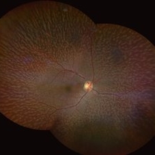
Image demonstrating numerous whitish surface flecks in an area of bullous retinoschisis OS.
Condition/keywords: bullous retinoschisis, retinoschisis
-
 Stargardt's Disease
Dec 17 2018 by Deepak Bhojwani, MS
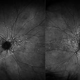
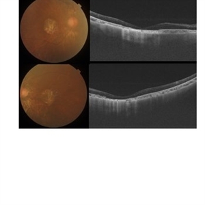
Fundus image and autofluorescence image of left eye a 26- year-old male with large area of RPE atrophy over macular region surrounded by pisciform flecks. Note the flecks are better appreciated on autofluorescence images (classic hypofluorescent pisciform lesions).
Photographer: Dr. Deepak Bhojwani
Imaging device: AUTO FLOROSCENCE IMAGING
Condition/keywords: autofluorescence imaging, macular dystrophy, Stargardt disease
 Loading…