Search results (885 results)
-
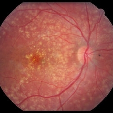
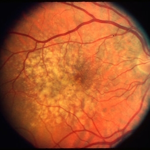
 Familial Dominant Drusen
Nov 22 2015 by Mallika Goyal, MD
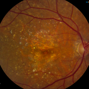
Bilateral drusen over the entire retinal mid-periphery and periphery of a 29-year-old male with no visual complaints. Macular centre is normal though there are some drusen in the temporal macula.
Photographer: Mallika Goyal, MD, Apollo Health City, Jubilee Hills, Hyderabad, India
Condition/keywords: familial drusen
-
 Optic Disc Drusen
Jul 31 2016 by Mitzy E Torres Soriano, MD
Optic Disc Drusen (Right eye)
Photographer: Mitzy E. Torres Soriano. Retina Department. Hospital Provincial del Centenario. Rosario, Argentina
Imaging device: TOPCON
Condition/keywords: optic disc drusen, optic nerve drusen
-
 Familial Dominant Drusen
Nov 22 2015 by Mallika Goyal, MD
Bilateral drusen over the entire retinal mid-periphery and periphery of a 29-year-old male with no visual complaints. Macular centre is normal though there are some drusen in the temporal macula.
Photographer: Mallika Goyal, MD, Apollo Health City, Jubilee Hills, Hyderabad, India
Condition/keywords: familial drusen
-
 Spontaneous Flattening of Drusenoid PED
Jul 1 2014 by John S. King, MD
Consult to r/o ExAMD; observed; scans about a year apart.
Photographer: Wayne A Ladlee Jr
Imaging device: Cirrus
Condition/keywords: drusenoid PED, macular drusenoid lesion, pigment epithelial detachment (PED)
-
 Reticular Drusen
-
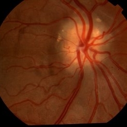
 Optic Nerve Head Drusen With Idiopathic CNV
Feb 17 2017 by Kristen Wagner
22-year-old female fundus photograph of a right eye with Optic Nerve Drusen with Idiopathic CNV.
Photographer: Kristen Wagner, COT, OSC Ophthalmic Photographer, Tennessee Retina, Nashville TN
Condition/keywords: choroidal neovascularization (CNV), drusen of optic disc, optic disc drusen
-
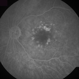
 Dry Age-Related Macular Degeneration, Fluorescein Angiogram
Aug 23 2012 by Gerardo Garcia-Aguirre, MD
Fluroescein angiogram of a 66 year-old patient with several hyperfluorescent spots corresponding to drusen.
Photographer: Noemí Hernández, Asociación para Evitar la Ceguera en México
Imaging device: FF4
Condition/keywords: age-related macular degeneration (AMD), dry age-related macular degeneration (dry AMD)
-
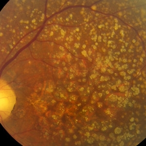
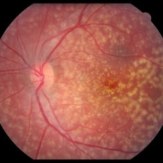 Familial drusen LE
Dec 29 2012 by Barbara Parolini, MD
Fundus photograph of a 25-year-old woman with familial drusen observed over time for growth. BCVA is 20\20. Electrooculogram is abnormal. Electroretinogram is normal.
Photographer: Fausto Lorenzi, MD
Condition/keywords: familial drusen
-
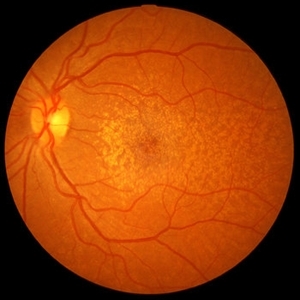
 Dry AMD
Jun 4 2014 by Henry J. Kaplan, MD
Multiple drusen with RPE changes in the macula #2.
Condition/keywords: age-related macular degeneration (AMD), dry age-related macular degeneration (dry AMD)
-
 Wet Age Related Macular Degeneration (WET AMD)
Sep 8 2012 by Ratimir Lazic, MD, PhD
Color fundus image of a 65 - year- old male. Drusen with suspect CNV
Photographer: Ratimir Lazic, PhD MD
Imaging device: Zeis Visucam Lite 2
Condition/keywords: fundus photograph
-
 Yellow Globular Lesion
Nov 9 2012 by Norman Byer
This glistening yellow globular lesion is a so-called pearl of the ora serrata in a 45-year-old man. Notice location in the tooth of the ora, which is a characteristic of this lesion. Histologically pearls are drusen-like structures which form on the inner side of Bruch’s membrane beneath the pigment epithelium. They are seen in about 20% of eyes and are often bilaterally symmetrical. They have no clinical significance but are valuable as landmarks.
Condition/keywords: Bruch's membrane, drusen-like, lesion, ora serrata
-
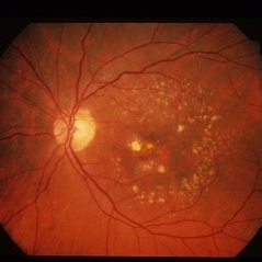
 AMD with Calcific Drusen and Geographic Atrophy
Apr 19 2013 by Brandon G. Busbee, MD
AMD with calcific drusen and geographic atrophy.
Photographer: Alecia Camp, CRA - Tennessee Retina - Nashville, TN
Imaging device: Topcon TRC 50-EX
Condition/keywords: geographic atrophy
-
---thumb.jpg/image-square;max$300,300.ImageHandler) Optic Disc Drusen
Optic Disc Drusen
Jul 10 2013 by Hamid Ahmadieh, MD
SD-OCT image of the left eye of a 24-year-old woman with optic disc drusen and VA 20/20.
Photographer: Solmaz Shahmohammadi, Negah Eye Center, Tehran
Imaging device: Heidelberg Spectralis
Condition/keywords: optic disc drusen, optical coherence tomography (OCT)
-
 Reticular Drusen, Doyne's Honeycomb Retinal Dystrophy, Malattia Leventinese, Familial Dominant Drusen
Feb 22 2018 by Nichole Lewis
Reticular Drusen, Doyne's Honeycomb Retinal Dystrophy, Malattia Leventinese, Familial Dominant Drusen
Photographer: Nichole Lewis
Condition/keywords: Doyne's Honeycomb, Familial Dominant Drusen, Malattia Leventinese, reticular drusen
-
 Calcified drusen, fundus photograph
Aug 23 2012 by Gerardo Garcia-Aguirre, MD
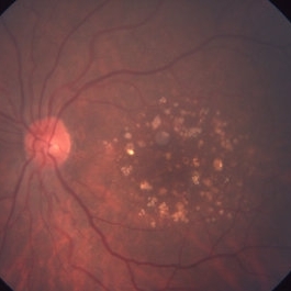
Fundus photograph showing multiple white-yellowhish lesions corresponding to calcified drusen.
Photographer: Noemí Hernández, Asociación para Evitar la Ceguera en México
Imaging device: Zeiss FF4
Condition/keywords: calcified drusen, dry age-related macular degeneration (dry AMD)
-
 Choroidal Nevus
May 2 2013 by Henry J. Kaplan, MD
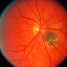
Juxtapapillary choroidal nevus with overlying drusen.
Condition/keywords: choroidal nevus
-
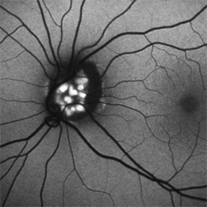
 Optic Disc Drusen
Jul 10 2013 by Hamid Ahmadieh, MD
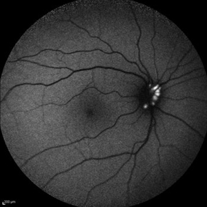
Fundus autofluorescence image of the right eye of a 24-year-old woman with optic disc drusen and VA 20/20.
Photographer: Solmaz Shahmohammadi, Negah Eye Center, Tehran
Imaging device: Heidelberg Spectralis
Condition/keywords: fundus autofluorescence (FAF), optic disc drusen
-
 Familial drusen RE
Dec 29 2012 by Barbara Parolini, MD
Fundus photograph of a 25-year-old woman with familial drusen observed over time for growth. BCVA is 20\20. Electrooculogram is abnormal. Electroretinogram is normal.
Photographer: Fausto Lorenzi, MD
Condition/keywords: familial drusen
-
 Calcified Drusen
Mar 1 2014 by Homayoun Tabandeh, MD, FASRS
Calcified drusen in a patient with dry age-related macular degeneration.
Condition/keywords: age-related macular degeneration (AMD), calcified drusen
-
 Choroidal Neovascularization
Oct 20 2012 by Hyung-Woo Kwak, MD
This 35-year-old young female patient has drusen-like lesions under the macular in both eyes. Such patients have a considerable risk of developing choroidal neovascular lesions.
Condition/keywords: choroidal neovascularization (CNV)
-
 Calcified Drusen, Fluorescein Angiogram
Aug 23 2012 by Gerardo Garcia-Aguirre, MD
Fluorescein angiogram of a 72 year-old patient with dry age-related macular degeneration and calcified drusen.
Photographer: Noemí Hernández, Asociación para Evitar la Ceguera en México
Imaging device: Zeiss FF4
Condition/keywords: age-related macular degeneration (AMD), calcified drusen, dry age-related macular degeneration (dry AMD)
-
 Optic Disc Drusen Autofluorescence
Apr 2 2016 by David Callanan, MD
30-year-old Caucasian male with visual field defect OD > OS.
Condition/keywords: optic disc drusen
-
 Optic Disc Drusen
Sep 21 2012 by Suber S. Huang, MD, MBA, FASRS
Fundus photograph of a 50-year-old woman with optic disc drusen complicated by anterior ischemic optic neuropathy
Condition/keywords: optic disc drusen
-
 Bulls eye retinopathy
Nov 20 2012 by Roy Schwartz, MD
75-YEAR-OLD FEMALE PRESENTS WITH BILATERAL GRADUAL VISUAL LOSS 6/30 Dx BE PSEUDOPHAKIA + PCO BE BULLS EYE MACULOPATHY PER FA VA IMPROVES TO 6/10 S/P YAG CAPSULOTOMY OCT - BE MACULAR SUBRETINAL FLUID NO HISTORY OF CHLOROQUINE THERAPY NO DRUSEN OR SIGNS OF AMD WORKING DIAGNOSIS - BE CHRONIC CSCR
Imaging device: Heidelberg spectralis
Condition/keywords: bull's eye maculopathy, optical coherence tomography (OCT)
-
 Dry Age-Related Macular Degeneration
Mar 29 2013 by Henry J. Kaplan, MD
Dry AMD with multiple confluent soft drusens
Condition/keywords: age-related macular degeneration (AMD), dry age-related macular degeneration (dry AMD)
 Loading…