Search results (885 results)
-
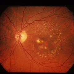
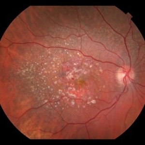
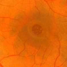
 AMD with Calcific Drusen and Geographic Atrophy
Apr 19 2013 by Brandon G. Busbee, MD
AMD with calcific drusen and geographic atrophy.
Photographer: Alecia Camp, CRA - Tennessee Retina - Nashville, TN
Imaging device: Topcon TRC 50-EX
Condition/keywords: geographic atrophy
-
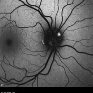
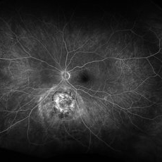
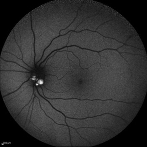
 Optic Nerve Head Drusen With Idiopathic CNV
Feb 17 2017 by Kristen Wagner
22-year-old female fundus photograph of a right eye with Optic Nerve Drusen with Idiopathic CNV.
Photographer: Kristen Wagner, COT, OSC Ophthalmic Photographer, Tennessee Retina, Nashville TN
Condition/keywords: choroidal neovascularization (CNV), drusen of optic disc, optic disc drusen
-
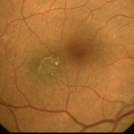
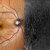
 Cuticular and soft drusen
Jun 14 2021 by Gerardo Garcia-Aguirre, MD
Fundus photograph (left) and Retro mode infrared image (right) of an eye with soft and cuticular drusen. Drusen are highlighted and better visualized with retro mode imaging.
Photographer: Gerardo Garcia-Aguirre
Imaging device: Nidek Mirante
Condition/keywords: drusen, dry age-related macular degeneration (dry AMD)
-
 Cuticular Drusen
Jan 17 2024 by John Lee
Heidelberg SD-OCT of a 65-year-old woman with age-related macular degeneration demonstrating classic sawtooth appearance of cuticular drusen.
Photographer: Natasha Vinson
Imaging device: Heidelberg Spectralis
Condition/keywords: age-related macular degeneration (AMD), cuticular drusen
-
 Dry AMD
Jun 4 2014 by Henry J. Kaplan, MD
Multiple drusen with RPE changes in the macula #2.
Condition/keywords: age-related macular degeneration (AMD), dry age-related macular degeneration (dry AMD)
-
 Familial drusen LE
Dec 29 2012 by Barbara Parolini, MD
Fundus photograph of a 25-year-old woman with familial drusen observed over time for growth. BCVA is 20\20. Electrooculogram is abnormal. Electroretinogram is normal.
Photographer: Fausto Lorenzi, MD
Condition/keywords: familial drusen
-
 Adult Onset Foveomacular Vitelliform Dystrophy
Jan 5 2015 by H. Michael Lambert, MD
Central, creamy elevation of pigment epithelium without drusen.
Condition/keywords: adult vitelliform dystrophy
-
 AMD With Calcified Drusen and Small, Deep IRH
Jul 22 2018 by John S. King, MD
AMD with calcified drusen and small, deep IRH
Photographer: Stacey
Imaging device: Topcon
Condition/keywords: calcified drusen
-
 Blue autofluroscence of Right eye optic nerve head showing auto fluorescence of the drusen
Aug 5 2022 by Kavitha Duraipandi, MD DNB FICO FRCS
A 20 year old patient referred to the clinic with blurred disc margins to rule out papilledema.
Photographer: Natalie Fox- Bussell
Condition/keywords: Blue autofluroscence, Heidelburg Spectralis
-
 Choroidal Melanoma FA
Nov 14 2023 by Virginia Gebhart
Fluorescein angiogram of 69 year old male with small lesion consistent with choroidal melanoma. Small pigmented elevated choroidal lesion just below ON with drusen, RPE changes and trace questionable OP present in the left eye. Extensive imaging and ultrasound was performed for further evaluation and documentation.
Photographer: Virginia Gebhart
Imaging device: Optos
Condition/keywords: fluorescein angiogram (FA), Fluorescein angiography
-
 Choroidal Nevus Associated with Drusen
Jan 11 2021 by Gabriel Costa Andrade, PhD
Fundus photograph of an 53-year-old man with a macular melanotic nevus.
Photographer: Gabriel Andrade
Condition/keywords: choroidal nevus
-
 Color Photo and Retro Mode of Cuticular Drusens
Aug 30 2020 by Sham Talati, DOMS
A patient With cuticular drusens in both eyes.
Photographer: Dr. Sham Talati,Retina Foundation,Ahmedabad
Imaging device: Nidek Mirante
Condition/keywords: cuticular drusen
-
 Craters on the Moon
Apr 21 2025 by rohan jain
Retro image of a 44 year-old woman with Familial Dominant Drusen.
Photographer: Dr. ROHAN JAIN
Condition/keywords: drusen, FAMILIAL DOMINANT DRUSEN, retro mode
-
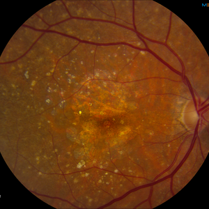
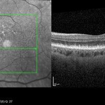
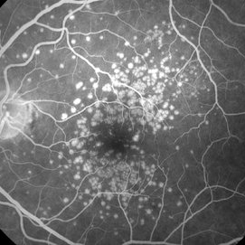 Drusen
Mar 29 2018 by JEFFERSON R SOUSA, Tecg.º (Biomedical Systems Technology)
Male patient 27-years-old, with complaint of low vision in both eyes. The fundoscopic evaluation found the presence of drusen topography in the posterior pole with foveal. Fluorescein angiography shows the typical pattern of hyperfluorescence of drusen in the first minute of angiography.
Photographer: JEFFERSON R SOUSA - Study Center and Ophthalmological Research Dr. Andre M V Gomes, Institute Dr. Suel Abujamra São Paulo-Brazil
Imaging device: Topcon TRC-50 DX, Imaginet 5.0, angle de 50 graus. Flash 150.
Condition/keywords: colloidal drusen, drusen
-
 Dry Macular Degeneration
May 8 2013 by Jerald A. Bovino, MD
Fundus photograph showing large drusen in dry AMD.
Condition/keywords: macular degeneration
-
 Example of AREDS Category 1 (Small Drusen But Not Considered AMD)
Feb 11 2013 by Neil M. Bressler, MD
Person in AREDS Category 1 were essentially free of age-related macular abnormalities, with a total drusen area less than 5 small drusen (<63 microns) within 3,000 microns of the center of the macula, and visual acuity of 20/32 or better in both eyes1. These are fundus photographs of a 53-year-old man, with visual acuity 20/20 OD and 20/32 OS presenting for evaluation of any diabetic retinopathy. Reference: 1 Age-Related Eye Disease Study Research Group. A randomized, placebo controlled clinical trial of high-dose supplementation with vitamins C and E, beta carotene, and zinc for age-related macular degeneration and vision loss: AREDS report No. 8. Arch Ophthalmol. 2001;119(10):1417-1436.
Condition/keywords: age-related macular degeneration (AMD)
-
 Familial Dominant Drusen
Nov 22 2015 by Mallika Goyal, MD
Bilateral drusen over the entire retinal mid-periphery and periphery of a 29-year-old male with no visual complaints. Macular centre is normal though there are some drusen in the temporal macula.
Photographer: Mallika Goyal, MD, Apollo Health City, Jubilee Hills, Hyderabad, India
Condition/keywords: familial drusen
-
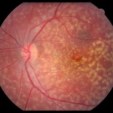
 Macular Degeneration with Significant Drusen
Jul 10 2018 by Karen Panzegrau
Zoomed-in ultra-wide field images of a 77-year-old female with macular degeneration with significant drusen.
Photographer: Karen Panzegrau
Imaging device: Optos
Condition/keywords: age-related macular degeneration (AMD), bilateral, drusen, fundus photograph, pseudocolor
-
 Macular Hole
Mar 29 2013 by Henry J. Kaplan, MD
Chronic macular hole with drusen like deposits and surrounding cuffing of subretinal fluid.
Condition/keywords: macular hole
-
 MIDD (Maternally Inherited Diabetes and Deafness) - Left AF
Nov 30 2024 by John S. King, MD
Both right and left eyes have symmetrical ring of mottled hypo/hyper AF around the fovea and disc. The HyperAF areas correspond to RPE deposits on OCT as well as areas of blockage on FA, and drusenoid deposits seen on fundus photos 57 yo WF referred for AMD vs Pattern Dystrophy that was diagnosed 10 years ago. Reported some slow progressive vision loss in both eyes for distance and near. Denies nyctalopia or hemeralopia. Background medical history includes HTN, CVD, and DM. No family history of eye problems. Denied pentosan use. Anterior segment showed moderate cataracts (OD>OS). Posterior segment exam showed macular changes and mild NPDR. The macular appearance showed a symmetrical, paramacular ring of fleck-like drusenoid material with some faint focal areas of RPE hyperplasia. Fundus Photos, AF, OCT were performed as well as a gene test. Further questioning showed revealed that her mother and maternal grandmother had both diabetes mellitus and sensorineural hearing loss. The patient developed diabetes in her teens, and some high frequency hearing loss in her early twenties. She had not had a previous genetic test or diagnosis of MIDD. Gene testing is pending for the mitochondrial component. Invitae's retinal panel, which does not include mitochondrial disorders, only showed a variant of uncertain significance, HMCN1. I discussed this case with Dr. Freund, and it is similar to a the case report : Inoue M, Kiss S, Freund KB. MACULAR PIGMENT RINGS AS THE PRESENTING FINDING OF MITOCHONDRIAL MYOPATHY, ENCEPHALOPATHY, LACTIC ACIDOSIS, AND STROKELIKE EPISODES. Retin Cases Brief Rep. 2015 Fall;9(4):260-4. doi: 10.1097/ICB.0000000000000182. PMID: 26200388.
Photographer: Grace Melton and Carley Gunn
Imaging device: Clarus
Condition/keywords: Macular Dystrophy, Maternally Inherited Diabetes and Deafness, MIDD, Mitochondrial Disorder
-
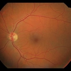
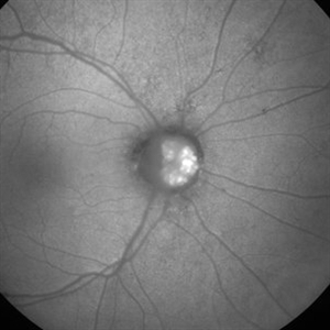
 Optic Disc Drusen
Jul 10 2013 by Hamid Ahmadieh, MD
Fundus autofluorescence image of the left eye of a 24-year-old woman with optic disc drusen and VA 20/20.
Photographer: Solmaz Shahmohammad, Negah Eye Center, Tehran
Imaging device: Heidelberg Spectralis
Condition/keywords: fundus autofluorescence (FAF), optic disc drusen
-
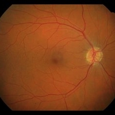
 Optic disc drusen
Dec 25 2012 by Alex P. Hunyor, MD
Autofluorescent image of the right optic disc showing autofluorescence of optic disc drusen.
Condition/keywords: optic disc drusen
-
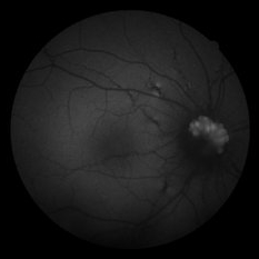
 Optic Disc Drusen
Jun 29 2022 by Mohamed Awadalla, MD, FRCSEd
Autofluorescence in optic disc drusen Red free fundus photo
Condition/keywords: Autofluorescence, optic disc drusen
-
 Optic Disc Drusen and Angioid Streaks
Jun 3 2020 by Mirko Ratkovic, MD
Optic disc drusen and angioid streaks.
Condition/keywords: angioid streaks, drusen of optic disc, fundus autofluorescence (FAF), fundus photograph
 Loading…