Search results (134 results)
-
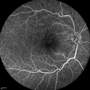 Optociliary Shunt Vessels in Old CRVO
Sep 8 2012 by Hamid Ahmadieh, MD
FA image of a 60-year-old woman with the history of central retinal vein occlusion.
Photographer: Hamid Ahmadieh, MD, Ophthalmic Research Center, Labbafinejad Medical Center, Shahid Beheshti University of Medical Sciences
Imaging device: Heidelberg Spectralis
Condition/keywords: central retinal vein occlusion (CRVO), shunts vessels
-
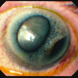
 Ciliary Body Melanoma With Partial Ring Configuration and Diffuse Sentinel Vessels
Feb 26 2014 by Susanna S. Park, MD, PhD
Slit lamp photo of a 57-year-old man with new vision loss from cataract formation. Large ciliary body mass with diffuse sentinel vessels is noted. The eye was removed and the tumor was noted to have a partial ring configuration with predominantly epithelioid cells and early vitreous seeding.
Photographer: Ellen Redenbo, University of California Davis Eye Center
Condition/keywords: ciliary body melanoma, melanoma
-
 Persistent Fetal Vasculature
Oct 10 2012 by Audina M. Berrocal, MD FASRS
Elongated ciliary body processes and zonules
Photographer: Ditte Hess CRA, BPEI
Imaging device: Ret Cam
Condition/keywords: persistent fetal vasculature (PFV)
-
 Persistent Fetal Vasculature
Oct 10 2012 by Audina M. Berrocal, MD FASRS
Elongated ciliary body processes Posterior capsule opacification with posterior tunica vasculosa
Photographer: Ditte Hess CRA, BPEI
Condition/keywords: persistent fetal vasculature (PFV)
-
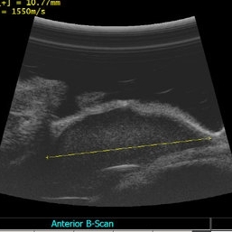
 Ciliary Body Melanoma B-Scan Ultrasound
Feb 27 2014 by Susanna S. Park, MD, PhD
Large ciliary body melanoma in a 57-year-old man.
Photographer: Ellen Redenbo, University of California Davis Eye Center
Condition/keywords: B scan ultrasound, ciliary body melanoma
-
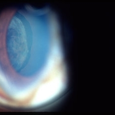
 Ciliary Body Melanoma
Jul 12 2013 by Jason S. Calhoun
71-year-old male who was recently diagnosed with a large ciliary body melanoma that is pushing into the anterior chamber of the left eye. Patient is going to proceed with proton therapy.
Photographer: Jason S. Calhoun, Department of Ophthalmology, Mayo Clinic Jacksonville, Florida
Condition/keywords: ciliary body melanoma
-
 Ciliary Body Ocular Melanoma
May 9 2016 by Nichole Lewis
Ciliary body ocular melanoma.
Photographer: Nichole Lewis
Condition/keywords: ciliary body melanoma
-
 Ciliary Body Cyst
Jul 14 2013 by Jason S. Calhoun
UBM image of ciliary body cyst.
Photographer: Jason S. Calhoun, Department of Ophthalmology, Mayo Clinic Jacksonville, Florida
Imaging device: Ultrasound
Condition/keywords: ciliary body cyst
-
 Ciliary Body Melanoma-UBM
Feb 27 2014 by Susanna S. Park, MD, PhD
UBM of a large ciliary body melanoma in a 57-year-old man. Histopathology after eye removal showed a diffuse ring component that also involved the anterior choroid.
Photographer: Ellen Redenbo, University of California Davis Eye Center
Condition/keywords: ciliary body melanoma, ultrasound
-
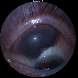
 Ciliary body melanoma
Jan 11 2013 by Alex P. Hunyor, MD
Left inferotemporal ciliary body melanoma with displacement of pupil, cataract, and large dilated episcleral vessels.
-
---thumb.jpg/image-square;max$300,300.ImageHandler) Intermediate Uveitis, Ciliary Body Cyst
Intermediate Uveitis, Ciliary Body Cyst
Feb 26 2013 by Henry J. Kaplan, MD
Intermediate uveitis, ciliary body cyst.
Condition/keywords: ciliary body cyst, intermediate uveitis
-
 Disseminated Chorioretinitis With Unknown Etiology
Apr 5 2018 by Kim Barrett
Ultra-wide field fluorescein angiogram of a 31-year-old female with intermittent pain in her left eye. Her condition has been managed in Liberia until recently when she moved to the United States. She suffers from multiple modalities including central retinal artery occlusion, posterior synechiae of the iris, interstitial keratitis, disseminated chorioretinitis, as well as HIV. An infectious cause is high on the differential in light of her HIV status. DDx: hypertensive crisis, an embolism (? IV drug use), coagulopathy, trauma, infectious. Blood work was normal. Her current vision is 20/30 right eye and 20/400 left eye.
Photographer: Kim Barrett, COA
Imaging device: Optos
Condition/keywords: central retinal artery occlusion (CRAO), chorioretinal scar, ciliary artery sparring, disseminated chorioretinitis, HIV, left eye, optic atrophy, staining
-
 Subluxated Sulcus IOL With Small AC Bubble
Aug 21 2018 by Russell Pokroy, MD
Anterior segment photograph of 71-year-old woman with 1-piece soft IOL decentered nasally with the haptics in the ciliary sulcus. A small area of the capsule remnant is evident suprotemporally. Small gas bubble in the AC is evident two weeks after PPV. The vitreous gas bubble decentered this IOL, with some spontaneous improvement of the IOL centering after gas dissipation.
Photographer: Russell Pokroy, Assaf Harofe Medical Center, Israel
Condition/keywords: intraocular lens (IOL)
-
 Ciliary body melanoma
May 2 2013 by Henry J. Kaplan, MD
Ciliary body melanoma visible through the pupil.
Condition/keywords: ciliary body melanoma
-
 Aniridic Fibrosis Syndrome - #5 of 7
Jan 24 2013 by Christopher D. Riemann, MD
6-year-old pseudophakic girl with aniridic fibrosis syndrome. Inferior view with HD endoscope. Note: severe inferior fibrosis fibrosis clearly extending across the ciliary body and obliterating the inferior ciliary processes and migrating onto the anterior pars plana .
Photographer: Christopher Riemann MD, Cincinnati Eye Institute, University of Cincinnati
Imaging device: Endoscope
Condition/keywords: aniridia, epiciliary membrane
-
 Central Retinal Vein Occlusion - After 3 Consecutive Ranibizumab Injections
Jan 26 2013 by Ratimir Lazic, MD, PhD
Color fundus photography of a 67-year-old female. Intraretinal hemorrhages in posterior pole, tortuous and dilated veins with optociliary shunts visible on optic nerve head. No macular edema can be noticed.
Photographer: Marko Lukic, MD
Imaging device: Zeis Visucam Lite 2
Condition/keywords: central retinal vein occlusion (CRVO)
-
 Ciliary mass ultrasound
Nov 7 2012 by Rajiv Anand, MD, FRCS, FASRS
Immersion ultrasound of mass shows cystic lesion 11mm in diameter
Photographer: Janae Sierocki
Condition/keywords: ciliary body mass, cystic lesion, immersion ultrasound
-
 Tumor in ciliary body.
Apr 9 2013 by Jerald A. Bovino, MD
No history. Associated with melanoma slides. Whole mount section of eye.
Condition/keywords: tumor
-
 Ciliary Body Melanoma
Jul 12 2013 by Jason S. Calhoun
71-year-old male who was recently diagnosed with a large ciliary body melanoma that is pushing into the anterior chamber of the left eye. Patient is going to proceed with proton therapy.
Photographer: Jason S. Calhoun, Department of Ophthalmology, Mayo Clinic Jacksonville, Florida
Condition/keywords: ciliary body melanoma
-
 Aniridic Fibrosis Syndrome - #1 of 7
Jan 24 2013 by Christopher D. Riemann, MD
6-year-old pseudophakic girl with aniridic fibrosis syndrome. Superior view with HD endoscope. Note: complete absence of fibrosis, a normal ciliary body, normal pars plana and normal anterior retina.
Photographer: Christopher Riemann MD, Cincinnati Eye Institute, University of Cincinnati
Imaging device: Endoscope
Condition/keywords: aniridia, epiciliary membrane
-
 Aniridic Fibrosis Syndrome #6 of 7
Jan 24 2013 by Christopher D. Riemann, MD
6-year-old girl with Aniridic Fibrosis Syndrome. Note: endoscopic view of 20 gauge vitreous cutter engaging epiciliary membrane.
Photographer: Christopher Riemann MD, Cincinnati Eye Institute, University of Cincinnati
Imaging device: Endoscope
Condition/keywords: aniridia, epiciliary membrane
-
 UBM of Ciliary Process
Sep 17 2015 by Jason S. Calhoun
UBM of a transverse section of the ciliary process.
Photographer: Jason Calhoun, Mayo Clinic, Department of Ophthalmology
Imaging device: Quantal Medical Aviso
Condition/keywords: glaucoma, ultrasound
-
 Ciliary mass
Nov 7 2012 by Rajiv Anand, MD, FRCS, FASRS
Patient presented with mass hidden by upper lid, 20/30 vision
Photographer: Mike Mackens
Condition/keywords: ciliary body mass
-
 Aniridic Fibrosis Syndrome - #3 of 7
Jan 24 2013 by Christopher D. Riemann, MD
6-year-old pseudophakic girl with aniridic fibrosis syndrome. Nasal view with HD endoscope. Note: increasing fibrosis fibrosis clearly extending onto the ciliary body and enveloping ciliary processes.
Photographer: Christopher Riemann MD, Cincinnati Eye Institute, University of Cincinnati
Imaging device: Endoscope
Condition/keywords: aniridia, epiciliary membrane
-
 Ciliary Body Melanoma
Jul 12 2013 by Jason S. Calhoun
71-year-old male who was recently diagnosed with a large ciliary body melanoma that is pushing into the anterior chamber of the left eye. Patient is going to proceed with proton therapy.
Photographer: Jason S. Calhoun, Department of Ophthalmology, Mayo Clinic Jacksonville, Florida
Condition/keywords: ciliary body melanoma
 Loading…