Search results (134 results)
-
 Slide 1-11
Feb 19 2019 by Lancaster Course in Ophthalmology
Cross-sections of two cilia implanted in the anterior chamber by trauma. Giant cells and scar surround each one. (H&E stain)
Condition/keywords: anterior chamber, cilia, giant cell, scar, trauma
-
 Central Retinal Artery Occlusion
May 26 2025 by yao zhang
Fundus photograph of an 84-year-old man with CRAO
Photographer: Yao Zhang,TongUniversity, Shanghai East Hospital, Department of ophthalmology
Condition/keywords: Ciliary artery sparing central retinal artery occlusion (CRAO)
-
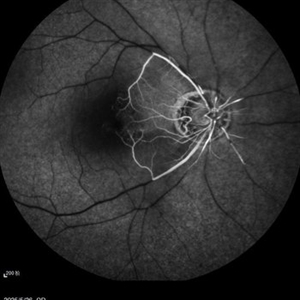
 Central Vein And Ciliar Oclusion FA
Sep 25 2013 by Alexandre Durao Alves Pereira, MD
Central vein and ciliar oclusion FA.
Condition/keywords: ciliar occlusion
-
 Choroidal and Ciliary Body Melanoma
Apr 11 2018 by Jason Griffith
15-year-old male patient referred for suspicious mass.
Photographer: Jason Griffith, Tennessee Retina, Nashville, TN
Imaging device: Topcon TRC 50EX
Condition/keywords: ciliary body melanoma
-
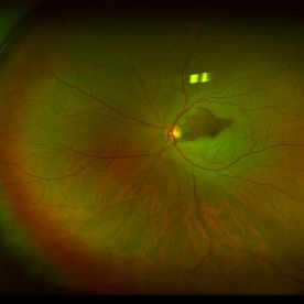 Ciliary Artery Sparing Central Retinal Artery Occlusion (CRAO)
Oct 27 2024 by César Adrián Gómez Valdivia, MD
Ciliary artery sparing central retinal artery occlusion (CRAO) is a condition where the central retinal artery is blocked, but the cilioretinal artery is patent, allowing for the preservation of central vision.
Photographer: @eyemissu2
Imaging device: California ICG OPTOS
Condition/keywords: Ciliary artery sparing central retinal artery occlusion (CRAO), ciliary artery sparring, oclussion
-
 Ciliary Body Choroidal Melanoma
Jan 29 2015 by H. Michael Lambert, MD
Invivo presentation of ciliary body melanoma.
Condition/keywords: ciliary body melanoma
-
 Ciliary Body Choroidal Melanoma
Jan 29 2015 by H. Michael Lambert, MD
Large mass hanging posterior to the lens superiorly in a dilated eye.
Condition/keywords: ciliary body melanoma
-
 Ciliary Body Cyst
Jul 14 2013 by Jason S. Calhoun
UBM image of ciliary body cyst.
Photographer: Jason S. Calhoun, Department of Ophthalmology, Mayo Clinic Jacksonville, Florida
Imaging device: Ultrasound
Condition/keywords: ciliary body cyst
-
Ciliary Body Detachment in Uveal Effusion Syndrome
Apr 11 2025 by Siri Uppuluri
Ultrasound biomicroscopy of a phakic left eye in an 82-year-old man demonstrating ciliary body detachment in the setting of uveal effusion syndrome. Patient also presented with 360 choroidal effusions and underwent sclerectomy and drainage of choroidal effusions with resolution after surgical intervention.
Photographer: Siri Uppuluri, MD; Rutgers New Jersey Medical School
Condition/keywords: uveal effusion syndrome
-
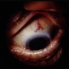
 Ciliary Body Melanoma
Nov 2 2024 by Virginia Gebhart
53 year old male with a large mass behind the lens as well as prominent scleral vessels. Clinical exam and ultrasound findings consistent with melanoma. Pt will be scheduled for enucleation pending CT scan results. Edit: Sadly patient has canceled all appointments and has requested no further contact
Photographer: Virginia Gebhart, Retina Consultants of Carolina
Imaging device: Optos California
Condition/keywords: ciliary body mass, ciliary body melanoma, ciliary body tumor
-
 Ciliary Body Melanoma
Feb 12 2025 by Virginia Gebhart
91 year old female with large collar button tumor emanating from the ciliary body with resolving vitreous hemorrhage. Melanoma cells in the AV as well as studded on the entire retina surface. Pt scheduled for enucleation. CT scans of chest and abdomen showed no evidence of metastatic disease.
Photographer: Virginia Gebhart, Retina Consultants of Carolina
Imaging device: Optos California
Condition/keywords: asteroid hyalosis, ciliary body mass, ciliary body melanoma, vitreous hemorrhage
-
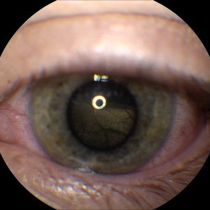
 Ciliary body melanoma
Jul 4 2021 by Gerardo Rivera Arroyo
Clinical image taken in a slit lamp with a gonioscopy of a 39-year-old female patient with ciliary body melanoma before enucleation and pathological study.
Condition/keywords: ciliary body melanoma, gonioscopy
-
 Ciliary Body Melanoma
Jul 4 2021 by Gerardo Rivera Arroyo
Clinical image taken in a slit lamp with a gonioscopy of a 39-year-old female patient with ciliary body melanoma before enucleation and pathological study.
Condition/keywords: ciliary body melanoma, gonioscopy
-
 Ciliary Body Melanoma
May 18 2020 by McGill University Health Centre
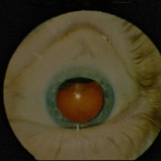
Uveal melanoma is the most common primary eye malignancy in adulthood, occurring mainly after age 60. The uveal tract — composed of the iris, ciliary body, and choroid — can be affected by uveal melanoma. Despite advances in treatment of the primary tumor, metastatic disease occurs in almost half of patients, generally affecting the liver and lungs via hematogenous dissemination of the primary tumor. Tumors have different levels of pigmentation, and some are amelanocytic (nonpigmented). The differential diagnosis for amelanotic choroidal melanoma is metastatic disease. Large tumors displace the lens. Of the 3 locations in the uveal tract, tumors of the ciliary body have the worst prognosis. The enucleation specimen in (A) shows a firm, dome-shaped, deeply pigmented tumor arising from the ciliary body (arrow). The lens has been removed, and a diffuse retinal detachment artifact is present.
Condition/keywords: enucleation, melanoma
-
 Ciliary Body Melanoma
May 18 2020 by McGill University Health Centre
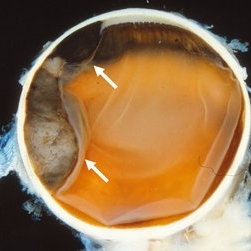
Uveal melanoma is the most common primary eye malignancy in adulthood, occurring mainly after age 60. The uveal tract — composed of the iris, ciliary body, and choroid — can be affected by uveal melanoma. Despite advances in treatment of the primary tumor, metastatic disease occurs in almost half of patients, generally affecting the liver and lungs via hematogenous dissemination of the primary tumor. Tumors have different levels of pigmentation, and some are amelanocytic (nonpigmented). The differential diagnosis for amelanotic choroidal melanoma is metastatic disease. Large tumors displace the lens. Of the 3 locations in the uveal tract, tumors of the ciliary body have the worst prognosis The enucleation specimen in (B) shows a large, dome-shaped, mixed melanotic and amelanotic choroidal melanoma. The anterior chamber is closed, and the angle is infiltrated (arrow). Total secondary retinal detachment with subretinal serous fluid and some subretinal hemorrhages are present (arrowhead). The lens is cataractous.
Condition/keywords: enucleation, melanoma
-
 Ciliary Body Melanoma
May 18 2020 by McGill University Health Centre
Uveal melanoma is the most common primary eye malignancy in adulthood, occurring mainly after age 60. The uveal tract — composed of the iris, ciliary body, and choroid — can be affected by uveal melanoma. Despite advances in treatment of the primary tumor, metastatic disease occurs in almost half of patients, generally affecting the liver and lungs via hematogenous dissemination of the primary tumor. Tumors have different levels of pigmentation, and some are amelanocytic (nonpigmented). The differential diagnosis for amelanotic choroidal melanoma is metastatic disease. Large tumors displace the lens. Of the 3 locations in the uveal tract, tumors of the ciliary body have the worst prognosis. This enucleation specimen shows a pigmented, nodular-shaped ciliary body melanoma (arrow) with extensive necrosis (*). A retinal detachment is present with subretinal fluid (arrowhead), and the retina is folded (•).
Condition/keywords: enucleation, melanoma
-
 Ciliary Body Melanoma
May 18 2020 by McGill University Health Centre
Large tumors displace the lens. Of the 3 locations in the uveal tract, tumors of the ciliary body have the worst prognosis. This enucleation specimen shows a pigmented, bilobed, dome-shaped tumor arising from the ciliary body (arrow). The lens has been removed, and a diffuse, flat retinal detachment artifact is present.
Condition/keywords: melanoma
-
 Ciliary body melanoma
May 2 2013 by Henry J. Kaplan, MD
Ciliary body melanoma visible through the pupil.
Condition/keywords: ciliary body melanoma
-
 Ciliary Body Melanoma
Apr 1 2019 by Gary R. Cook, MD, FACS
White male with a ciliary body melanoma OD seen as a dark, dome-shaped mass through a dilated pupil; VA=20/30-2.
Imaging device: Topcon VT-50
Condition/keywords: ciliary body mass, melanocytic lesion, melanoma
-
 Ciliary body melanoma
Jan 11 2013 by Alex P. Hunyor, MD
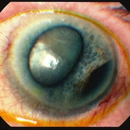
Left inferotemporal ciliary body melanoma with displacement of pupil, cataract, and large dilated episcleral vessels.
-
 Ciliary Body Melanoma
Jul 12 2013 by Jason S. Calhoun
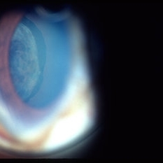
71-year-old male who was recently diagnosed with a large ciliary body melanoma that is pushing into the anterior chamber of the left eye. Patient is going to proceed with proton therapy.
Photographer: Jason S. Calhoun, Department of Ophthalmology, Mayo Clinic Jacksonville, Florida
Condition/keywords: ciliary body melanoma
-
 Ciliary Body Melanoma
Jul 12 2013 by Jason S. Calhoun
71-year-old male who was recently diagnosed with a large ciliary body melanoma that is pushing into the anterior chamber of the left eye. Patient is going to proceed with proton therapy.
Photographer: Jason S. Calhoun, Department of Ophthalmology, Mayo Clinic Jacksonville, Florida
Condition/keywords: ciliary body melanoma
 Loading…