Search results (134 results)
-
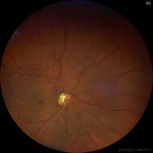
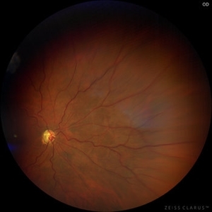
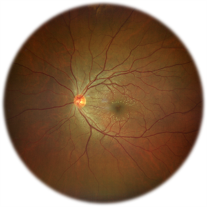 Commotio Retinae
Jun 10 2025 by CUI YUELING
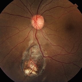
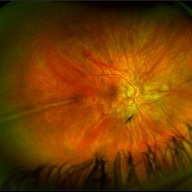
The patient presented 2 hours after sustaining a left eye injury caused by a stick. Visual acuity in the left eye was 0.2 without improvement upon correction, and intraocular pressure measured 15 mmHg. Examination of the anterior segment revealed ciliary conjunctival injection accompanied by patchy subconjunctival hemorrhage. The corneal surface remained smooth, and the anterior chamber was deep with hyphema characterized by blood-tinged aqueous humor predominantly settled inferiorly. The pupil was slightly irregular, approximately 3 mm in diameter, with a superotemporal notch; pupillary light reflex was intact. The lens appeared clear. Fundus examination showed well-defined optic disc margins with normal coloration and a cup-to-disc ratio of 0.2. Retinal arteries and veins were normally distributed with an artery-to-vein ratio of 2:3. At the posterior pole, the foveal reflex exhibited concentric ripple-like changes centered on the fovea, accompanied by localized pigment attenuation and reduced reflex intensity. Irregular reflectivity was noted in the superotemporal and inferotemporal nerve fiber layers.
Photographer: Yueling Cui
Imaging device: Zeiss Clarus 500
Condition/keywords: commotio retinae
-
 Central Retinal Artery Occlusion
May 26 2025 by yao zhang
Fundus photograph of an 84-year-old man with CRAO
Photographer: Yao Zhang,TongUniversity, Shanghai East Hospital, Department of ophthalmology
Condition/keywords: Ciliary artery sparing central retinal artery occlusion (CRAO)
-
 Cyclic Membrane
Apr 23 2025 by Gustavo Uriel Fonseca Aguirre
This UBM scan reveals pars planitis with characteristic findings: an inflammatory pupillary membrane, a cataractous lens, and cyclitic membrane causing ciliary body detachment and traction. The lens demonstrates spherical deformation due to zonular laxity from ciliary body traction.
Photographer: Gustavo U. Fonseca Aguirre, Hospital Conde de Valenciana, Ciudad de México
Condition/keywords: cyclic membrane, pars planitis
-
Ciliary Body Detachment in Uveal Effusion Syndrome
Apr 11 2025 by Siri Uppuluri
Ultrasound biomicroscopy of a phakic left eye in an 82-year-old man demonstrating ciliary body detachment in the setting of uveal effusion syndrome. Patient also presented with 360 choroidal effusions and underwent sclerectomy and drainage of choroidal effusions with resolution after surgical intervention.
Photographer: Siri Uppuluri, MD; Rutgers New Jersey Medical School
Condition/keywords: uveal effusion syndrome
-
 Uveal Effusion Syndrome in a Nanophthalmic Eye
Apr 3 2025 by Gustavo Uriel Fonseca Aguirre
Ultrasound biomicroscopy longitudinal section of a nanophthalmic eye demonstrating a shallow anterior chamber, angle closure, ciliary body edema, and supraciliary and sub-Tenon fluid accumulation.
Photographer: Gustavo U. Fonseca Aguirre, Hospital Conde de Valenciana, Ciudad de México
Condition/keywords: nanophthalmos, uveal effusion syndrome
-
 Ciliary Body Metastasis
Mar 26 2025 by Virginia Gebhart
54 year old female referred for iris mass. UBM shows large solid mass originating in the ciliary body and eroding into the anterior chamber under the iris epithelium. Recent CT scans revealed multiple bilateral pulmonary and hepatic nodules. Pt has been scheduled for PET scan and liver biopsy by radiation oncologist.
Photographer: Virginia Gebhart, Retina Consultants of Carolina
Imaging device: Samsung Galaxy
Condition/keywords: choroidal metastasis, ciliary body mass, metastatic cancer
-
 Ciliary Body Melanoma
Feb 12 2025 by Virginia Gebhart
91 year old female with large collar button tumor emanating from the ciliary body with resolving vitreous hemorrhage. Melanoma cells in the AV as well as studded on the entire retina surface. Pt scheduled for enucleation. CT scans of chest and abdomen showed no evidence of metastatic disease.
Photographer: Virginia Gebhart, Retina Consultants of Carolina
Imaging device: Optos California
Condition/keywords: asteroid hyalosis, ciliary body mass, ciliary body melanoma, vitreous hemorrhage
-
 Ciliary Body Melanoma
Nov 2 2024 by Virginia Gebhart
53 year old male with a large mass behind the lens as well as prominent scleral vessels. Clinical exam and ultrasound findings consistent with melanoma. Pt will be scheduled for enucleation pending CT scan results. Edit: Sadly patient has canceled all appointments and has requested no further contact
Photographer: Virginia Gebhart, Retina Consultants of Carolina
Imaging device: Optos California
Condition/keywords: ciliary body mass, ciliary body melanoma, ciliary body tumor
-
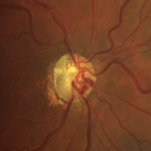
 Ciliary Artery Sparing Central Retinal Artery Occlusion (CRAO)
Oct 27 2024 by César Adrián Gómez Valdivia, MD
Ciliary artery sparing central retinal artery occlusion (CRAO) is a condition where the central retinal artery is blocked, but the cilioretinal artery is patent, allowing for the preservation of central vision.
Photographer: @eyemissu2
Imaging device: California ICG OPTOS
Condition/keywords: Ciliary artery sparing central retinal artery occlusion (CRAO), ciliary artery sparring, oclussion
-
 New Iris Melanoma
Oct 10 2024 by Virginia Gebhart
56 year old male with new amelanotic melanoma emanating from the ciliary body through the posterior iris epithelium. CT scan showed no evidence of metastatic disease. Pt scheduled for radioactive plaque and tumor biopsy
Photographer: Virginia Gebhart, Retina Consultants of Carolina
Imaging device: Samsung Galaxy
Condition/keywords: amelanotic melanoma, iris melanoma
-
 Uveal Effusion Syndrome
Sep 19 2024 by Virginia Gebhart
61 year old female with idiopathic uveal effusion syndrome. 360 degrees of choroidal thickening, especially anterior with exudative fluid inferior. Mild vitritis present. Unable to gain venous access for FA, ultrasound and UBM performed which confirm choroidal and ciliary body thickening. Pt sent for inflammatory work up including MRI of brain and orbits. Treatment pending results.
Photographer: Virginia Gebhart, Retina Consultants of Carolina
Imaging device: Optos California
Condition/keywords: idiopathic uveal effusion syndrome, uveal effusion
-
 Retinal Colomoba
Jul 21 2024 by César Adrián Gómez Valdivia, MD
Retinal Coloboma found in a female 41 year old patient. Iris, Lens, Ciliary Body, Zonules, Choroid and Retina were involved.
Photographer: Erika Paulina Ornelas Cazares
Imaging device: TOPCON TRC-50DX
Condition/keywords: coloboma
-
 New Choroidal Melanoma
Jan 4 2024 by Virginia Gebhart
77 year old male with a bilobed pigmented mass with exudative RD, and trace inflammation present in AV consistent with choroidal melanoma. Mass extends into ciliary body. Pt scheduled for MRI prior to plaque radiation to rule out metastasis.
Photographer: Virginia Gebhart
Imaging device: Optos California
Condition/keywords: ciliary body melanoma, exudative retinal detachment
-
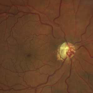
 Optociliary Shunts
Sep 12 2023 by Ben Serar
Fundus photograph showing Optociliary shunts with tortuous vessels.
Condition/keywords: Optociliary Shunts, tortuous vessels
-
 Optociliary Shunt
Aug 21 2023 by Harsh Vardhan Singh, MS
Optociliary shunt in a case of optic nerve glioma
Photographer: Harsh Vardhan Singh
Condition/keywords: Optociliary shunt
-
 Optociliary Shunt
Aug 21 2023 by Harsh Vardhan Singh, MS
Optociliary shunt in a case of optic nerve glioma
Photographer: Harsh Vardhan Singh
Condition/keywords: Optociliary shunt
-
 Optociliary Shunt
Aug 21 2023 by Harsh Vardhan Singh, MS
Optociliary shunt in a case of optic nerve glioma
Photographer: Harsh Vardhan Singh
Condition/keywords: Optociliary shunt
-
 Optociliary Shunt
Aug 21 2023 by Harsh Vardhan Singh, MS
Optociliary shunt in a case of optic nerve glioma
Photographer: Harsh Vardhan Singh
Condition/keywords: Optociliary shunt
-
 Amalric triangular sign (posterior ciliary artery occlusion)
Feb 8 2023 by Bruno DECAY, MD
Fundus photograph of a 61-year-old female (routine examination)
Photographer: Amélie DULAC , Centre Ophtalmologique Vic-Montaner, Vic en Bigorre, France
Imaging device: Centervue Eidon
Condition/keywords: Diabetes, Hypertension
-
 Iris Vascular Tuft
Jul 5 2022 by Olivia Rainey
Anterior segment imaging of a 66-year-old male with Vascular Disorders of Iris and Ciliary Body affecting his right eye. The physician stated that the findings are most consistent with a benign vascular tuft at the pupillary margin. The patient presented at the office with 20/20 vision in both eyes and had no ocular complaints at the time of his appointment.
Photographer: Olivia Rainey, OCT-C, COA
Imaging device: Heidelberg Spectralis, Slit Lamp with Samsung Galaxy 7
Condition/keywords: anterior segment, fluorescein angiogram (FA), heidelberg spectralis, infrared image, near infrared autofluorescence (NIRAF), slit lamp photo, vascular anomaly, vascular disorders of iris and ciliary body, vascular tuft
-
 Prominent Long Ciliary Nerve
Jan 25 2022 by Kachelle Brown
Ultra-wide field photograph of a 48-year-old female with a prominent long ciliary nerve. Patient presented asymptomatic, and was referred for a macula on retinal detachment. Patient was diagnosed with high myopia and a posterior vitreous detachment, and the physician discussed increased risk of floaters, myopic degeneration and retinal detachment associated with high myopia. -24.00 prior to cataract surgery OU per patient.
Photographer: Kachelle Brown
Imaging device: Optos California
Condition/keywords: fundus photograph, high myopia, long ciliary nerve, optos, right eye, ultra-widefield image
-
 Iridociliary Cyst
Oct 12 2021 by Jesus Lozano, MD
82 year old man with a brown mass beneath the iris, inside the pupil at 6-9 o’clock.
Photographer: Dr. Jesús Lozano Gutiérrez. Jerusalem. Israel.
Imaging device: iPhone 12
Condition/keywords: ciliary body cyst, cystic lesion, iris
-
 Ciliary Body Melanoma
Jul 4 2021 by Gerardo Rivera Arroyo
Clinical image taken in a slit lamp with a gonioscopy of a 39-year-old female patient with ciliary body melanoma before enucleation and pathological study.
Condition/keywords: ciliary body melanoma, gonioscopy
-
 Ciliary body melanoma
Jul 4 2021 by Gerardo Rivera Arroyo
Clinical image taken in a slit lamp with a gonioscopy of a 39-year-old female patient with ciliary body melanoma before enucleation and pathological study.
Condition/keywords: ciliary body melanoma, gonioscopy
-
 Suprachoroidal Hemorrhage
Sep 2 2020 by Rinal Pandit
Fundus photograph of left eye of a 56-year-old female with primary angle closure glaucoma showing massive hemorrhagic choroidal detachment that developed following trabeculectomy surgery. Suprachoroidal hemorrhage is defined as the accumulation of blood within the potential space between the choroid and sclera, with the source of the blood being the long or short posterior ciliary artery. Delayed suprachoroidal hemorrhage (DSHC) remains one of the most dreaded and sight threatening complications of glaucoma filtration surgery. The risk factors include old age, hypertension, high myopia, arteriosclerosis, chronically elevated IOP, sudden hypotony, trauma, aphakia/pseudophakia, prior vitrectomy, history of 5 FU injections and anti-platelet agents. The incidence of postoperative SCH after trabeculectomy varies between 0.6%- 1.4%. DSCH after surgery varies considerably in severity but is generally characterized by the sudden onset of severe pain, decreased vision, and a shallow anterior chamber usually associated with raised intraocular pressure. B-scan ultrasonography can help to distinguish serous from hemorrhagic choroidals.Suprachoroidal hemorrhages appear as dome-shaped elevations of the retina with increased echo densities that are often heterogeneous and within the suprachoroidal space. Choroidal effusions appear as dome-shaped elevations with hypoechoic suprachoroidal space. The first step in the management is the timely diagnosis. Medical management includes oral and topical antiglaucoma drugs to lower IOP, oral and topical steroids to control inflammation and topical cycloplegics and oral analgesics to tackle pain. Serial ultrasound B scans of the affected eye should be performed in order to monitor progression of the SCH and help determine apposition, height, and liquefaction of the SCH. Indications of surgical drainage include non resolution with medical management,concurrent retinal detachment, central retinal apposition (kissing choroidals) and incarceration of vitreous in the wound site. The ideal time of drainage is between 7-14 days depending upon clot lysis. The prognosis of both intraoperative and postoperative SCH is poor. An overwhelming majority of patients do not achieve pre-hemorrhage visual acuity and most do not recover to a visual acuity of 20/200 or better. The major determinants of good or bad visual outcomes of SCH’s are preoperative visual acuity and retinal detachment at the time of hemorrhage, respectively.
Imaging device: OPTOS,Ultra wide field retinal imaging system
Condition/keywords: suprachoroidal hemorrhage, trabeculectomy, ultra-wide field imaging
 Loading…