Search results (134 results)
-
 New Iris Melanoma
Oct 10 2024 by Virginia Gebhart
56 year old male with new amelanotic melanoma emanating from the ciliary body through the posterior iris epithelium. CT scan showed no evidence of metastatic disease. Pt scheduled for radioactive plaque and tumor biopsy
Photographer: Virginia Gebhart, Retina Consultants of Carolina
Imaging device: Samsung Galaxy
Condition/keywords: amelanotic melanoma, iris melanoma
-
 Persistent Fetal Vasculature
Oct 10 2012 by Audina M. Berrocal, MD FASRS
Elongated ciliary body processes and zonules
Photographer: Ditte Hess CRA, BPEI
Imaging device: Ret Cam
Condition/keywords: persistent fetal vasculature (PFV)
-
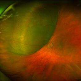
 Ciliary Body Metastasis
Mar 26 2025 by Virginia Gebhart
54 year old female referred for iris mass. UBM shows large solid mass originating in the ciliary body and eroding into the anterior chamber under the iris epithelium. Recent CT scans revealed multiple bilateral pulmonary and hepatic nodules. Pt has been scheduled for PET scan and liver biopsy by radiation oncologist.
Photographer: Virginia Gebhart, Retina Consultants of Carolina
Imaging device: Samsung Galaxy
Condition/keywords: choroidal metastasis, ciliary body mass, metastatic cancer
-
 Persistent Fetal Vasculature
Oct 10 2012 by Audina M. Berrocal, MD FASRS
Elongated ciliary body processes Posterior capsule opacification with posterior tunica vasculosa
Photographer: Ditte Hess CRA, BPEI
Condition/keywords: persistent fetal vasculature (PFV)
-
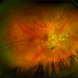 Prominent Long Ciliary Nerve
Jan 25 2022 by Kachelle Brown
Ultra-wide field photograph of a 48-year-old female with a prominent long ciliary nerve. Patient presented asymptomatic, and was referred for a macula on retinal detachment. Patient was diagnosed with high myopia and a posterior vitreous detachment, and the physician discussed increased risk of floaters, myopic degeneration and retinal detachment associated with high myopia. -24.00 prior to cataract surgery OU per patient.
Photographer: Kachelle Brown
Imaging device: Optos California
Condition/keywords: fundus photograph, high myopia, long ciliary nerve, optos, right eye, ultra-widefield image
-
 Central Retinal Artery Occlusion
May 26 2025 by yao zhang
Fundus photograph of an 84-year-old man with CRAO
Photographer: Yao Zhang,TongUniversity, Shanghai East Hospital, Department of ophthalmology
Condition/keywords: Ciliary artery sparing central retinal artery occlusion (CRAO)
-
 Ciliary Body Melanoma
Nov 2 2024 by Virginia Gebhart
53 year old male with a large mass behind the lens as well as prominent scleral vessels. Clinical exam and ultrasound findings consistent with melanoma. Pt will be scheduled for enucleation pending CT scan results. Edit: Sadly patient has canceled all appointments and has requested no further contact
Photographer: Virginia Gebhart, Retina Consultants of Carolina
Imaging device: Optos California
Condition/keywords: ciliary body mass, ciliary body melanoma, ciliary body tumor
-
 Ciliary Body Melanoma
Feb 12 2025 by Virginia Gebhart
91 year old female with large collar button tumor emanating from the ciliary body with resolving vitreous hemorrhage. Melanoma cells in the AV as well as studded on the entire retina surface. Pt scheduled for enucleation. CT scans of chest and abdomen showed no evidence of metastatic disease.
Photographer: Virginia Gebhart, Retina Consultants of Carolina
Imaging device: Optos California
Condition/keywords: asteroid hyalosis, ciliary body mass, ciliary body melanoma, vitreous hemorrhage
-
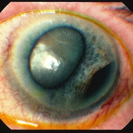
 Ciliary Body Melanoma
Jul 4 2021 by Gerardo Rivera Arroyo
Clinical image taken in a slit lamp with a gonioscopy of a 39-year-old female patient with ciliary body melanoma before enucleation and pathological study.
Condition/keywords: ciliary body melanoma, gonioscopy
-
 Ciliary body melanoma
Jan 11 2013 by Alex P. Hunyor, MD
Left inferotemporal ciliary body melanoma with displacement of pupil, cataract, and large dilated episcleral vessels.
-
 Disseminated Chorioretinitis With Unknown Etiology
Apr 5 2018 by Kim Barrett
Ultra-wide field fluorescein angiogram of a 31-year-old female with intermittent pain in her left eye. Her condition has been managed in Liberia until recently when she moved to the United States. She suffers from multiple modalities including central retinal artery occlusion, posterior synechiae of the iris, interstitial keratitis, disseminated chorioretinitis, as well as HIV. An infectious cause is high on the differential in light of her HIV status. DDx: hypertensive crisis, an embolism (? IV drug use), coagulopathy, trauma, infectious. Blood work was normal. Her current vision is 20/30 right eye and 20/400 left eye.
Photographer: Kim Barrett, COA
Imaging device: Optos
Condition/keywords: central retinal artery occlusion (CRAO), chorioretinal scar, ciliary artery sparring, disseminated chorioretinitis, HIV, left eye, optic atrophy, staining
-
 Enucleated Eye Showing Choroidal Melanoma
May 18 2020 by McGill University Health Centre
This enucleation specimen shows an aphakic eye with a large, solid choroidal tumor. The tumor is heavily pigmented; it shows different shades in some areas. The tumor reaches the ciliary body.
Condition/keywords: aphakic eye, choroidal tumor, enucleation
-
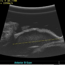 Ciliary mass ultrasound
Nov 7 2012 by Rajiv Anand, MD, FRCS, FASRS
Immersion ultrasound of mass shows cystic lesion 11mm in diameter
Photographer: Janae Sierocki
Condition/keywords: ciliary body mass, cystic lesion, immersion ultrasound
-
 Large, Dome-Shaped Peripheral Choroidal Melanoma - Widefield Color
Feb 13 2020 by Michael Seider, MD
Large, dome-shaped peripheral choroidal melanoma of the left eye with inferior exudative retinal detachment. Note the lack of obvious orange pigment over the tumor and apparent drusen anteriorly. A lack of ophthalmoscopically obvious lipofuscin is not uncommon among larger choroidal melanomas. B-Scan ultrasonography (transverse, 10 o’clock) confirms a low-moderate internally reflective dome-shaped choroidal lesion with a small adjacent retinal detachment. Ultrasound biomicroscopy (radial, 10 o’clock) confirms no ciliary body involvement of the tumor.
-
 Multiple Ciliary Body Cysts
Aug 7 2020 by Rinal Pandit
Ultrasound biomicroscopy image (Transverse scan) of a 30-year-old female with occludable angle. For all young patients presenting with angle closure ,UBM should preferably be performed. It helps establish the cause of narrowing , such as a pseudoplateau configuration in this case produced by multiple CB cysts.
Photographer: Dr Rinal Pandit, Raghudeep eye hospital, Ahmedabad
Imaging device: UBM, ABsolu, Quantel Medical
Condition/keywords: ciliary body cyst
-
 Aniridic Fibrosis Syndrome - #3 of 7
Jan 24 2013 by Christopher D. Riemann, MD
6-year-old pseudophakic girl with aniridic fibrosis syndrome. Nasal view with HD endoscope. Note: increasing fibrosis fibrosis clearly extending onto the ciliary body and enveloping ciliary processes.
Photographer: Christopher Riemann MD, Cincinnati Eye Institute, University of Cincinnati
Imaging device: Endoscope
Condition/keywords: aniridia, epiciliary membrane
-
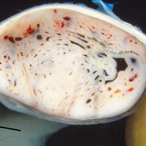
 Ciliary Body Melanoma
May 18 2020 by McGill University Health Centre
Uveal melanoma is the most common primary eye malignancy in adulthood, occurring mainly after age 60. The uveal tract — composed of the iris, ciliary body, and choroid — can be affected by uveal melanoma. Despite advances in treatment of the primary tumor, metastatic disease occurs in almost half of patients, generally affecting the liver and lungs via hematogenous dissemination of the primary tumor. Tumors have different levels of pigmentation, and some are amelanocytic (nonpigmented). The differential diagnosis for amelanotic choroidal melanoma is metastatic disease. Large tumors displace the lens. Of the 3 locations in the uveal tract, tumors of the ciliary body have the worst prognosis. The enucleation specimen in (A) shows a firm, dome-shaped, deeply pigmented tumor arising from the ciliary body (arrow). The lens has been removed, and a diffuse retinal detachment artifact is present.
Condition/keywords: enucleation, melanoma
-
 Iridociliary Cyst
Oct 12 2021 by Jesus Lozano, MD
82 year old man with a brown mass beneath the iris, inside the pupil at 6-9 o’clock.
Photographer: Dr. Jesús Lozano Gutiérrez. Jerusalem. Israel.
Imaging device: iPhone 12
Condition/keywords: ciliary body cyst, cystic lesion, iris
-
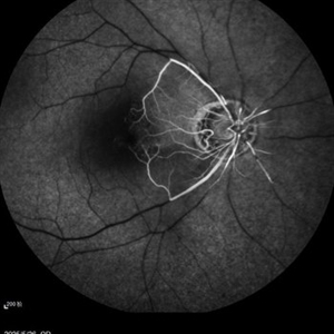
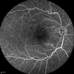 Optociliary Shunt Vessels in Old CRVO
Sep 8 2012 by Hamid Ahmadieh, MD
FA image of a 60-year-old woman with the history of central retinal vein occlusion.
Photographer: Hamid Ahmadieh, MD, Ophthalmic Research Center, Labbafinejad Medical Center, Shahid Beheshti University of Medical Sciences
Imaging device: Heidelberg Spectralis
Condition/keywords: central retinal vein occlusion (CRVO), shunts vessels
-
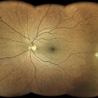 Amalric triangular sign (posterior ciliary artery occlusion)
Feb 8 2023 by Bruno DECAY, MD
Fundus photograph of a 61-year-old female (routine examination)
Photographer: Amélie DULAC , Centre Ophtalmologique Vic-Montaner, Vic en Bigorre, France
Imaging device: Centervue Eidon
Condition/keywords: Diabetes, Hypertension
-
 Amelanotic Choroidal Tumor
May 18 2020 by McGill University Health Centre
Choroidal melanoma is often asymptomatic and diagnosis is incidental. The tumors may grow beneath the retina, or may break through the Bruch membrane and disrupt the retina. Tumors breaking through the Bruch membrane and disrupting the retina have a characteristic “mushroom” shape. This enucleation specimen shows an amelanotic, dome-shaped choroidal tumor with several dilated blood vessels. The tumor has not infiltrated the sclera, ciliary body, or optic nerve. Note the retinal detachment next to the tumor (arrow).
Condition/keywords: amelanotic, choroidal tumor
-
 Aniridic Fibrosis Syndrome #2 of 7
Jan 24 2013 by Christopher D. Riemann, MD
6-year-old pseudophakic girl with aniridic fibrosis syndrome. Superonasal view with HD endoscope. Note: 20 gauge sclerotomy and very mild fibrosis barely extending onto the ciliary body.
Photographer: Christopher Riemann MD, Cincinnati Eye Institute, University of Cincinnati
Imaging device: Endoscope
Condition/keywords: aniridia, epiciliary membrane
-
 Aniridic Fibrosis Syndrome #4 of 7
Jan 24 2013 by Christopher D. Riemann, MD
6-year-old pseudophakic girl with aniridic fibrosis syndrome. Close up nasal view with HD endoscope. Note: increasing fibrosis fibrosis clearly extending onto the ciliary body and enveloping ciliary processes.
Photographer: Christopher Riemann MD, Cincinnati Eye Institute, University of Cincinnati
Imaging device: Endoscope
Condition/keywords: aniridia, epiciliary membrane
-
 Aniridic Fibrosis Syndrome #6 of 7
Jan 24 2013 by Christopher D. Riemann, MD
6-year-old girl with Aniridic Fibrosis Syndrome. Note: endoscopic view of 20 gauge vitreous cutter engaging epiciliary membrane.
Photographer: Christopher Riemann MD, Cincinnati Eye Institute, University of Cincinnati
Imaging device: Endoscope
Condition/keywords: aniridia, epiciliary membrane
-
 Aniridic Fibrosis Syndrome - #1 of 7
Jan 24 2013 by Christopher D. Riemann, MD
6-year-old pseudophakic girl with aniridic fibrosis syndrome. Superior view with HD endoscope. Note: complete absence of fibrosis, a normal ciliary body, normal pars plana and normal anterior retina.
Photographer: Christopher Riemann MD, Cincinnati Eye Institute, University of Cincinnati
Imaging device: Endoscope
Condition/keywords: aniridia, epiciliary membrane
 Loading…