Search results (96 results)
-
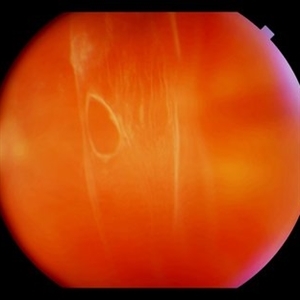
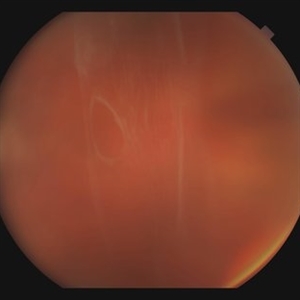
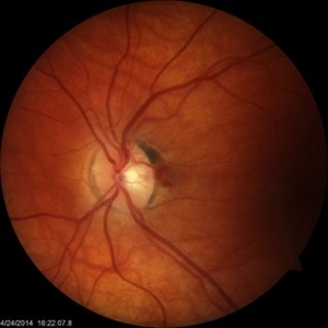
 ERM that Spontaneously Peeled
Oct 8 2012 by David R. Chow, MD, FRCS(C)
An ERM that through follow-up sponateously separated with the development of PVD.
Condition/keywords: epiretinal membrane (ERM), posterior vitreous detachment
-
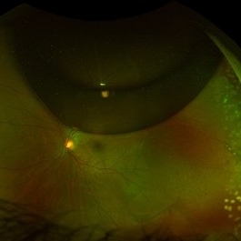
 Weiss Ring
Jan 9 2019 by John S. King, MD
77-year-old white male with ERM and PVD OD; sheet of vitreous with weiss ring in the nasal mid-vitreous cavity.
Photographer: Macey Highfill, RN
Imaging device: Topcon 50
Condition/keywords: posterior vitreous detachment, Weiss ring
-
 Lattice Degeneration
Nov 9 2012 by Norman Byer
Lattice degeneration in a 42-year-old man which has produced four atrophic holes in a linear arrangement surrounded by a subclinical retinal detachment of unknown duration. By age 63, 21 years later, a posterior vitreous detachment was diagnosed in this eye, which was not present four years earlier. Nevertheless, the appearance seen here has remained exactly the same for 30 years, more than eight years with a concurrent PVD.
Condition/keywords: atrophic retinal hole, lattice degeneration, posterior vitreous detachment
-
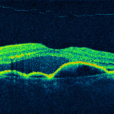 OCT Cirrus 5 Line Scan ARMD SRF RPED Stage 3 PVD
Mar 6 2013 by James B. Soque, CRA, OCT-C, COA, FOPS
Zeiss Cirrus OCT 4000, 5 Line Scan , 91-year-old white female with peripapillary SRN with subretinal heme, serous fluid, and a stage 3 PVD, still attached at the optic nerve.
Photographer: James Soque, CRA, COA, Island-Retina
Imaging device: Zeiss Cirrus 4000 SD OCT with 6.0.2.81 Software
Condition/keywords: optical coherence tomography (OCT)
-
 24 Hours Post Scleral Wound Closure+ Scleral Buckle+25 g Vitrectomy+Silicon Oil
Jan 23 2015 by Carlos Quezada-Ruiz, MD, FASRS
24 hours post op fundus photograph of a 43-year-old man who had perforating injury to the right eye with a small piece of plastic while he was hammering. OD LP, subconjunctival hemorrhage, clear cornea, hyphema, irido and ciclodyalisis as well as a luxated lens with traumatic cataract and a dense vitreous hemorrhage. B-US showed rhegmatogenous retinal detachment with a tear and a big inferior hemorrhagic choroidal detachment. 360 peritomy revealed 2-entry scleral wounds were found in zone II (M V and M VI) and closure was performed. 25 G PPV was performed with the infusion canal placed in the AC through the limbus. Lensectomy and removal of a dense recent vitreous hemorrhage revealed a white detached retina with an exit wound through the temporal inferior segment of the optic nerve with a nasal GRT and sub retinal hemorrhage as well as temporal inferior choroidal, PVD was induced and PFOs helped stabilizing the retina while vitrectomy and sub-retinal hemorrhage was removed through the GRT. Fluid air exchange was made and 360 endolaser over the buckle indentation was done and silicon oil was used as endotamponade. This picture was taken 24 hrs after the surgery.
Photographer: Lilibeth Rodriguez, Instituto de la Visión. Torreon, Mexico.
Condition/keywords: central retinal artery occlusion (CRAO), giant retinal tear, trauma
-
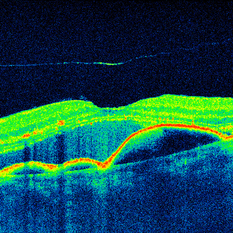 OCT Cirrus 5 Line HD Scan EDI ARMD SRF RPED Stage 3 PVD
Mar 6 2013 by James B. Soque, CRA, OCT-C, COA, FOPS
Zeiss Cirrus OCT 4000, EDI Aquired Using Enhanced Depth Mode, 91-year-old white female with peripapillary SRN with subretinal heme, serous fluid, and a stage 3 PVD, still attached at the optic nerve.
Photographer: James Soque, CRA, COA, Island-Retina
Imaging device: Zeiss Cirrus 4000 SD OCT with 6.0.2.81 Software
Condition/keywords: optical coherence tomography (OCT)
-
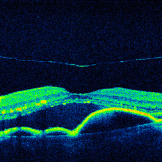 OCT Cirrus HD 5 Line Scan ARMD SRF RPED Stage 3 PVD
Mar 6 2013 by James B. Soque, CRA, OCT-C, COA, FOPS
Zeiss Cirrus OCT 4000, Hi Definition 5 Line Scan, 91-year-old white female with peripapillary SRN with subretinal heme, serous fluid, and a stage 3 PVD, still attached at the optic nerve.
Photographer: James Soque, CRA, COA, Island-Retina
Imaging device: Zeiss Cirrus 4000 SD OCT with 6.0.2.81 Software
Condition/keywords: optical coherence tomography (OCT)
-
 TA Stained Posterior Hyaloid Face
Apr 11 2014 by Subhendu Kumar Boral, MBBS, MD(AIIMS), DNB, FASRS (USA)
Intraoperative step of posterior hyaloid face staining by triamcinolone acetonide particles during PVD induction in a case of diabetic epiretinal membrane left eye in a 68-year-old gentleman.
Photographer: Subhendu Kumar Boral
Condition/keywords: hyaloid
-
 Color Fundus Photograph of Myope With PVD and Staphyloma
Jun 11 2016 by Philip J. Polkinghorne, MD
Color photograph of patient with PVD and staphyloma.
Imaging device: Optos
Condition/keywords: degenerative myopia, peripheral vascular disease (PVD), staphyloma
-
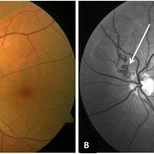
 Complete PVD
Dec 10 2012 by Yale L. Fisher, MD
Dr. Yale Fisher presents a sagittal view of a complete posterior detachment demonstrated by the thin preretinal reflection (yellow arrow). Scleral depression (green arrow) at the ora serrata demonstrates the ability to register anatomical position on ultrasound using a scleral depressor.
Condition/keywords: video
-
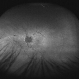
 Stage 1 Macular Hole Pre PVD
May 26 2014 by John T. Thompson, MD
OCT of late stage 1 macular hole with vitreomacular traction on fovea.
Imaging device: Heidelberg Spectralis
Condition/keywords: late stage 1 macular hole
-
 Peri-papillary Vascular Loop
Jun 2 2020 by Dhaivat Shah
Peri-papillary vascular loops (PVL) are rare congenital vascular malformations, which are usually detected as accidental finding during routine fundus examination. They can often be confused with tributary vein occlusion or racemose hemangioma. Although benign and asymptomatic, they can be rarely associated with vitreous hemorrhage and arterial occlusion. We herein present a case of a 60-year-old hypertensive male, who was diagnosed elsewhere to have a tributary vein occlusion and was referred to us. FFA was advised to rule out neovascularization, surrounding capillary non perfusion and mass lesion (hemangioma). On FFA, the arterial loop showed a slightly delayed filling (3-5 seconds) as compared to the other arterial vessels and the original vessel appeared to be a branch arising from central retinal artery. The choroidal filling was delayed in the area supplied by the loop. A cilioretinal artery was also noted. The patient was diagnosed to have a Peri-papillary vascular arterial loop (PVL), likely to be congenital in origin. The patient was reassured and was advised yearly follow up. These loops are usually accidental findings discovered during routine fundus examination. Since these vessels are looped and tortuous, they exhibit a slower and laminar blood flow, which make them more prone for arterial occlusions. The vitreous in this area tends to be adherently attached, so during PVD induction, it is likely to cause a tear and hemorrhage leading to vitreous hemorrhage. Until and unless there is a break, this hemorrhage tends to resolve on its own and does not warrant treatment. If there is an evident break, it can be dealt with laser barrage.
Photographer: Choithram Netralaya
Condition/keywords: congenital prepapillary vascular loop
-
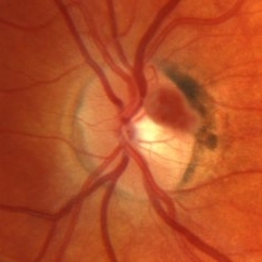
 Vitreous Traction on Disc / PVD
Feb 24 2015 by David Callanan, MD
43-year-old female, vitreous traction on disc / PVD.
Condition/keywords: posterior vitreous detachment, vitreous traction
-
 Triamcinolone Acetonide stained PVD induction by cutter
Apr 11 2014 by Subhendu Kumar Boral, MBBS, MD(AIIMS), DNB, FASRS (USA)
Intra operative step of PVD induction in a case of diabetic epiretinal membrane in Left Eye in a 68 years old gentleman
Photographer: Subhendu Kumar Boral
Condition/keywords: PVD induction
-
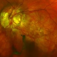
 Detached NVE During PVD induction
Apr 27 2018 by Michael J. Koss, MD, PhD, MBA
A 73-year-old woman with macular pucker underwent a pars plana vitrectomy with membrane peeling. Additionally the patient suffers from diabetic retinopathy after being diagnosed with type 2 diabetes mellitus sixteen years ago. Prior to the procedure she was treated with a series of intravitreal Bevacizumab-injections due to diabetic macular edema. There was no history of a proliferative DRP. During the vitrectomy a branch of an obliterated NVE spontaneously detached and floated freely in the vitreous. The 3D shot was captured via Alcon’s NGENUITY® 3D Visualization System in form of photograph and video providing an outstandingly detailed image of the branched NVE.
Photographer: Michael Koss, Augenzentrum Nymphenburger Hoefe
Imaging device: Alcon’s NGENUITY® 3D Visualization System
Condition/keywords: diabetes, diabetic retinopathy, neovascularization elsewhere (NVE), pars plana vitrectomy (PPV), PVD induction
-
 Weiss Ring
Jan 9 2019 by John S. King, MD
77-year-old white male with ERM and PVD OD; sheet of vitreous with weiss ring in the nasal mid-vitreous cavity.
Photographer: Macey Highfill, RN
Imaging device: Topcon 50
Condition/keywords: posterior vitreous detachment, Weiss ring
-
 Vitreous Traction on Disc / PVD
Feb 24 2015 by David Callanan, MD
43-year-old female, vitreous traction on disc / PVD.
Condition/keywords: posterior vitreous detachment, vitreous traction
-
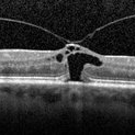
 Partial Vitreous Separation in a High Myope With a Posterior Staphyloma
Dec 10 2012 by Yale L. Fisher, MD
This B-scan demonstrates a partial PVD. A posterior vitreous detachment (PVD) may occur in a normal aging eye or may be associated with pathology such as vitreous hemorrhage or inflammation. In a normal eye, as in this example, the PVD appears as a thin and smooth line (arrow) on B-scan. When the globe is moved voluntarily by the patient, real time echography demonstrates a quick jerky motion of the sheet-like echo with movements continuing after the globe movement has ceased. This is helpful in differentiating a PVD from a retinal detachment, which typically has a slower undulating pattern of motion. If there was presence of blood or inflammatory debris associated with the PVD, the echogenic line might appear thicker, especially in the most gravity dependent portions of the globe (i.e., posterior and inferior).
Condition/keywords: video
-
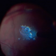
 VHL "Free Floating" Juxtapapillary Hemangioblastoma
Jul 1 2014 by John S. King, MD
30-year-old female with fhx VHL and CNS hemangioblastomas and visceral lesions. P/C with a floater (no PVD or VH) after episodes of vomiting. - this photo and images following taken a few months after initial presentation (images before this one)
Photographer: Wayne A Ladlee Jr
Condition/keywords: retinal hemangioblastoma, Von Hippel-Lindau
-
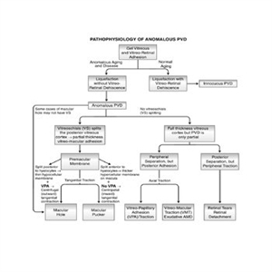 Pathophysiology of Anomalous PVD
Sep 1 2020 by J. Sebag, MD, FACS, FRCOphth, FARVO
This unifying concept of vitreo-retinopathies hypothesizes that the pathogenesis of several vitreoretinal diseases that were previously considered very disparate, are actually all manifestations of the same underlying pathophysiology – anomalous PVD. Note that vitreo-papillary adhesion (VPA) and traction can cause primary optic neuropathy, but might also play a role in facilitating/promoting cell migration and proliferation during pathologic neovascularization of the optic disc. Further, VPA seems to alter the vector of tangential forces exerted by a membrane, in some cases full-thickness posterior vitreous cortex and in some cases the outer layer of the posterior vitreous cortex left attached to the macula after vitreoschisis. While not all cases of macular holes have vitreoschisis, they feature vitreomacular adhesion and traction almost always with VPA. [From Sebag J: Anomalous PVD – a unifying concept in vitreo-retinal diseases. Graefe’s Arch Clin Exp Ophthalmol 2004;242:690-8 and Sebag J, Niemeyer M, Koss M: Anomalous PVD and vitreoschisis. In: Vitreous – in Health & Disease (J. Sebag, ed.) Springer, New York, 2014, pg. 252; image © Springer Nature, reprinted with permission]
Condition/keywords: pathology, peripheral vascular disease (PVD)
-
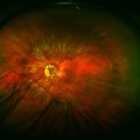
 Morning-Glory-Syndrome
Dec 22 2017 by James B. Soque, CRA, OCT-C, COA, FOPS
68-year-old WM with Morning Glory Syndrome with PVD OS with Staphyloma surrounding optic nerve and extending into the macula. Also, esotropia OS from V1 nerve paresis from birth, with amblyopia.
Photographer: James B Soque, CRA OCT-C COA FOPS
Imaging device: Optos Daytona
Condition/keywords: color photo, esotropia, fundus photograph, Optomap, Optos, peripheral vascular disease (PVD), posterior vitreous detachment, staphyloma, ultra-wide field imaging, wide angle imaging
-
 VHL "Free Floating" Juxtapapillary Hemangioblastoma
Jul 1 2014 by John S. King, MD
30-year-old female with fhx VHL and CNS hemangioblastomas and visceral lesions. P/C with a floater (no PVD or VH) after episodes of vomiting.
Photographer: Wayne A Ladlee Jr
Condition/keywords: retinal hemangioblastoma, Von Hippel-Lindau
-
 Chronic Retinal Detachment after Pneumatic Retinopexy
Jan 8 2022 by Parnian Arjmand, MD, MSc, FRCSC, DABO
This is a fundus photo in the eye of a young phakic patent who presented with a 6 month history of "difficulty seeing at night" and subjective nasal "blurriness" in the left eye. There was a chronic temporal RD, OS, extending to the arcades (Mac on). This photo is week 1 s/p Pneumatic retinopexy with SF6 gas and laser retinopexy to temporal breaks (6 holes, lattice); no PVD. As you can see, there is a "bleb" of viscous schlieren given the chronic nature of this RD that persist posterior to the breaks and temporal to the macula. This type of sub retinal fluid may take months to years to resorb.
Condition/keywords: chronic retinal detachment, pneumatic retinopexy
-
 Myope With Staphyloma and Vitreous Detachment
Jun 11 2016 by Philip J. Polkinghorne, MD
Fundus autofluorescence of a myope with PVD and staphyloma.
Imaging device: Optos FAF
Condition/keywords: degenerative myopia, myopia, staphyloma
-
 Vitreous Traction on Disc / PVD
Feb 24 2015 by David Callanan, MD
43-year-old female, vitreous traction on disc / PVD.
Condition/keywords: posterior vitreous detachment, vitreous traction
 Loading…