Search results (96 results)
-
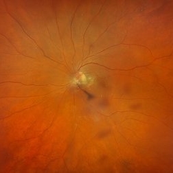
 Central Retinal Vein Occlusion with Macular Edema
Jan 29 2025 by Kimberly Wakester
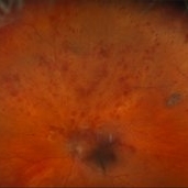
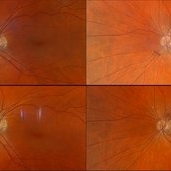
Fundus photograph of a 62-year-old man with central retinal vein occlusion with macular edema and a new PVD with an operculated retinal tear in the left eye. Laser to retinal tear was completed. Patient will return in 2-3 weeks for follow up exam with possible intravitreal injection for the CRVO with edema and to follow up on the operculated retinal tear s/p retinal tear laser.
Photographer: Kimberly Wakester, COA
Imaging device: Optos California
Condition/keywords: central retinal vein occlusion (CRVO), operculated tear, PVD
-
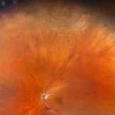
 Evolving Weiss Ring
Sep 11 2022 by Michael B Green, MD, MBA
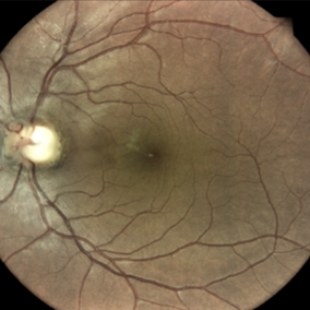
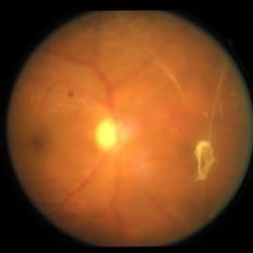
Fundus photograph of a 62-year-old female with an evolving Weiss-ring in the process of separating from the optic disc.
Condition/keywords: posterior vitreous detachment, PVD, Weiss ring
-
 Folds in Detached Posterior Vitreous Cortex
May 31 2022 by Joshua Friedman
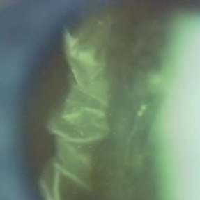
Slit lamp (video) image showing folds in the posterior vitreous cortex in an eye with PVD.
Photographer: Martin Snead, MD, Cambridge, England
Condition/keywords: folds, posterior vitreous cortex, PVD, vision degrading myodesopsia, vitreous
-
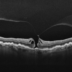
 Myopic Traction Maculopathy
Mar 17 2025 by Drew Mitchell
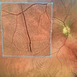
HD 1 line 100x 9 mm scan of a right eye with MTM at stage 3c. Macular Schisis Detachment.
Photographer: Drew Mitchell OCT-C
Imaging device: Zeiss Cirrus 5000
Condition/keywords: full thickness macular hole, Macular hole, myopic foveoschisis, myopic macular schisis, myopic traction maculopathy, PVD
-
 Posterior Vitreous Detachment
Nov 1 2023 by ANKIT JAIN
USG B SCAN image showing membranous echoes with low to moderate spikes with free after movements with no attachment to disc suggestive of posterior vitreous detachment.
Photographer: DR ANKIT JAIN
Condition/keywords: B scan ultrasound, posterior vitreous detachment, PVD, ultrasound
-
 Posterior Vitreous Detachment
Sep 28 2025 by Sanauddin Samejo , Diploma (Ophthalmic Technician Training Course)
Posterior Vitreous Detachment (PVD)
Photographer: Sanauddin Samejo
Imaging device: Optos Silver Stone
Condition/keywords: posterior vitreous detachment, PVD
-
 Posterior Vitreous Detachment
Sep 28 2025 by Sanauddin Samejo , Diploma (Ophthalmic Technician Training Course)
Posterior Vitreous Detachment (PVD)
Photographer: Sanauddin Samejo
Imaging device: Optos Silver Stone
Condition/keywords: posterior vitreous detachment, PVD
-
PVD induction in a retinal detachment
Oct 24 2022 by Manish Nagpal, MD, FRCS (UK), FASRS
This video highlights the PVD induction technique in a case of retinal detachment with mobile retina, triamcinolone staining allows ease of visualizing the pvd attachment which is gradually removed from the retinal attachment using suction.
Photographer: Manish Nagpal
Condition/keywords: posterior hyaloid, PVD, triamcinolone, video, vitrectomy
-
 Retinal Detachment with Single Break
Feb 5 2025 by Virginia Gebhart
61 year old male with mac-off retinal detachment with single horseshoe tear. Macula has been off for several days and has developed associated cystic edema. Visual prognosis guarded. Pt schedule for PPV/Laser/GFE
Photographer: Virginia Gebhart, Retina Consultants of Carolina
Imaging device: Optos California
Condition/keywords: horseshoe tear, PVD, retinal detachment
-
 Retinal Tear
Sep 4 2025 by Kimberly Wakester
Optomap RBG of a 55-year-old woman with a retinal tear at 12 with bridging vessel and some fluid. Treatment with prophylaxis laser was recommended. Patient is to continue follow up care post operatively.
Photographer: Kimberly Wakester, COA, OCT-C
Imaging device: Optos California
Condition/keywords: left eye, PVD, Retinal tear
-
 Stage 2 Macular Hole From VMT
Mar 21 2025 by Drew Mitchell
HD 1 line 100x OCT showcasing a full thickness macular hole caused by vitreomacular traction on fovea. Choroidal folds can also be seen on scan.
Photographer: Drew Mitchell OCT-C
Imaging device: Zeiss Cirrus 6000
Condition/keywords: Choroidal Folds, FTMH, macular hole, OCT, PVD
-
 Staining-the-vitreous
Jan 10 2022 by Parnian Arjmand, MD, MSc, FRCSC, DABO
This is an example of staining the posterior hyaloid in an eye with no PVD with triamcinolone actinide to assist with inducing a PVD prior to repairing the macular hole.
Condition/keywords: macular hole, PVD, triamcinolone, triescence
-
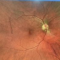
 Weiss Ring
Jan 21 2025 by Kimberly Wakester
Fundus photographs of a 70-year-old woman with a PVD with Weiss ring present in the left eye. Doing sweeps of the left eye shows how changing the patient's gaze can reposition the Weiss ring in the patient's eye.
Photographer: Kimberly Wakester, COA
Imaging device: Optos California
Condition/keywords: PVD, Weiss ring
-
 Bidding Adieu to Attachments: Weiss Ring
Jan 7 2022 by Gayathri Mohan
Colour fundus photograph showing a Weiss ring following PVD.
Photographer: Dr. GAYATHRI MOHAN
Imaging device: Canon
Condition/keywords: PVD induction, Weiss ring
-
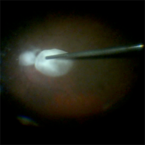
 Posterior Vitreous Detachment
Jan 31 2025 by Thirumalesh Mochi Basavaraj, MD
Intraoperative view of Triamcinolone-assisted posterior vitreous detachment.
Photographer: Thirumalesh Mochi Basavaraj
Condition/keywords: PVD induction, triamcinolone
-
 PVD Induction
Feb 2 2022 by Manish Nagpal, MD, FRCS (UK), FASRS
Intraoperative photo of PVD induction being carried out. Hyaloid has been stained using triamcinolone dye and PVD induction is carried out using high suction on the cutter and engaging the stained hyaloid.
Photographer: Manish Nagpal, Retina Foundation, Ahmedabad, India
Imaging device: Sony PMW -10 MD surgical camera
Condition/keywords: Hyaloid staining, PVD induction, triamcinolone
-
 PVD Induction in Progress
Jan 10 2022 by Manish Nagpal, MD, FRCS (UK), FASRS
Intraoperative image of PVD induction with triamcinolone staining clearly showing the Weiss ring, which is being lifted with suction from the cutter.
Photographer: Manish Nagpal, Retina Foundation, Ahmedabad, india
Imaging device: Sony PMW -10 MD surgical camera
Condition/keywords: PVD induction, triamcinolone, Weiss ring
-
 PVD induction with IVTA staining
Nov 1 2022 by Shobhit Chawla, M.S.
This is an intraoperative photograph of Pvd induction showing both the macular and disc attatchments stained with triamcinolone.
Photographer: Shobhit Chawla
Condition/keywords: PVD induction, triamcinolone
-
 PVD With Vitreous Attachment to Retinal Tear
Dec 10 2012 by Yale L. Fisher, MD
There is a posterior vitreous face separation with remaining attachment to a retinal flap tear. Movement of the tear is visible during voluntary motion of the patient's eye. There is strong reflectivity from the flap tear (yellow arrow) and moderate reflectivity from the vitreous face (green arrow). The peripheral retinal tear is seen in this sagittal nasal cut near the medial rectus muscle insertion, which localizes the tear to the ora serrata around the 3 o'clock position.
Condition/keywords: video
-
 Triamcinolone Acetonide stained PVD induction by cutter
Apr 11 2014 by Subhendu Kumar Boral, MBBS, MD(AIIMS), DNB, FASRS (USA)
Intra operative step of PVD induction in a case of diabetic epiretinal membrane in Left Eye in a 68 years old gentleman
Photographer: Subhendu Kumar Boral
Condition/keywords: PVD induction
-
 24 Hours Post Scleral Wound Closure+ Scleral Buckle+25 g Vitrectomy+Silicon Oil
Jan 23 2015 by Carlos Quezada-Ruiz, MD, FASRS
24 hours post op fundus photograph of a 43-year-old man who had perforating injury to the right eye with a small piece of plastic while he was hammering. OD LP, subconjunctival hemorrhage, clear cornea, hyphema, irido and ciclodyalisis as well as a luxated lens with traumatic cataract and a dense vitreous hemorrhage. B-US showed rhegmatogenous retinal detachment with a tear and a big inferior hemorrhagic choroidal detachment. 360 peritomy revealed 2-entry scleral wounds were found in zone II (M V and M VI) and closure was performed. 25 G PPV was performed with the infusion canal placed in the AC through the limbus. Lensectomy and removal of a dense recent vitreous hemorrhage revealed a white detached retina with an exit wound through the temporal inferior segment of the optic nerve with a nasal GRT and sub retinal hemorrhage as well as temporal inferior choroidal, PVD was induced and PFOs helped stabilizing the retina while vitrectomy and sub-retinal hemorrhage was removed through the GRT. Fluid air exchange was made and 360 endolaser over the buckle indentation was done and silicon oil was used as endotamponade. This picture was taken 24 hrs after the surgery.
Photographer: Lilibeth Rodriguez, Instituto de la Visión. Torreon, Mexico.
Condition/keywords: central retinal artery occlusion (CRAO), giant retinal tear, trauma
-
 Age-Related Differences in the Structure of the Human Vitreous Body
Sep 1 2020 by J. Sebag, MD, FACS, FRCOphth, FARVO
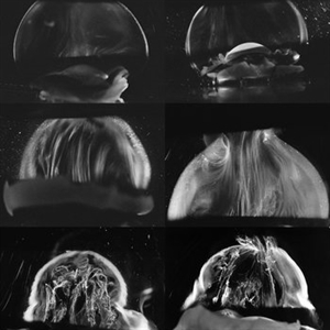
Dark-field slit microscopy was performed on fresh, unfixed, post-mortem human eyes that had undergone dissection to peel off the sclera, choroid, and retina. The vitreous body remains attached to the anterior segment which is seen below, while the posterior pole is above in these images. The top panel demonstrates the absence of internal vitreous structures that scatter light in youth (left image from an 11 year-old girl, right image from a 14 year-old boy. The middle panel demonstrates light scattering from linear, fibrous structures that have an antero-posterior orientation with insertions into the vitreous base peripherally and the posterior vitreous cortex, typical in middle age (left image from a 56 year-old and right image from a 59 year-old). The bottom panel illustrates advance fibrous liquefaction in old age (88-year-old subject). [From Sebag J, Niemeyer M, Koss M: Anomalous PVD and vitreoschisis. In: Vitreous – in Health & Disease (J. Sebag, ed.) Springer, New York, 2014, pg. 245; image © Springer Nature, reprinted with permission]
Condition/keywords: vitreous
-
 Asteroid Hyalosis
Apr 12 2019 by Gary R. Cook, MD, FACS
B-Scan image of visually-significant asteroid hyalosis in the right eye of a 79-year-old white male showing hyperechoic echoes from the asteroid of high internal reflectivity on simultaneous A-scan, and a PVD in the eye; VA= 20/80
Imaging device: Ophthscan
Condition/keywords: asteroid hyalosis, B scan ultrasound
-
 Chronic Retinal Detachment after Pneumatic Retinopexy
Jan 8 2022 by Parnian Arjmand, MD, MSc, FRCSC, DABO
This is a fundus photo in the eye of a young phakic patent who presented with a 6 month history of "difficulty seeing at night" and subjective nasal "blurriness" in the left eye. There was a chronic temporal RD, OS, extending to the arcades (Mac on). This photo is week 1 s/p Pneumatic retinopexy with SF6 gas and laser retinopexy to temporal breaks (6 holes, lattice); no PVD. As you can see, there is a "bleb" of viscous schlieren given the chronic nature of this RD that persist posterior to the breaks and temporal to the macula. This type of sub retinal fluid may take months to years to resorb.
Condition/keywords: chronic retinal detachment, pneumatic retinopexy
-
 Cloquet Canal
Sep 20 2024 by Jordyn Beckman
79 year old male with wet AMD, no PVD, presents with stable cloquet canal.
Photographer: Jordyn Beckman
Imaging device: Optos California
Condition/keywords: Cloquet Canal, hyaloid membrane, punctum caecum, stilling's canal
 Loading…