Search results (96 results)
-
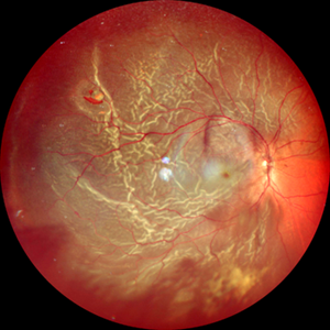
 Posterior Vitreous Detachment
Sep 28 2025 by Sanauddin Samejo , Diploma (Ophthalmic Technician Training Course)
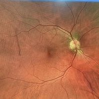
Posterior Vitreous Detachment (PVD)
Photographer: Sanauddin Samejo
Imaging device: Optos Silver Stone
Condition/keywords: posterior vitreous detachment, PVD
-
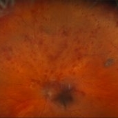
 Posterior Vitreous Detachment
Sep 28 2025 by Sanauddin Samejo , Diploma (Ophthalmic Technician Training Course)
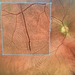
Posterior Vitreous Detachment (PVD)
Photographer: Sanauddin Samejo
Imaging device: Optos Silver Stone
Condition/keywords: posterior vitreous detachment, PVD
-
 Retinal Tear
Sep 4 2025 by Kimberly Wakester
Optomap RBG of a 55-year-old woman with a retinal tear at 12 with bridging vessel and some fluid. Treatment with prophylaxis laser was recommended. Patient is to continue follow up care post operatively.
Photographer: Kimberly Wakester, COA, OCT-C
Imaging device: Optos California
Condition/keywords: left eye, PVD, Retinal tear
-
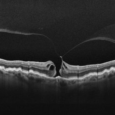 Stage 2 Macular Hole From VMT
Mar 21 2025 by Drew Mitchell
HD 1 line 100x OCT showcasing a full thickness macular hole caused by vitreomacular traction on fovea. Choroidal folds can also be seen on scan.
Photographer: Drew Mitchell OCT-C
Imaging device: Zeiss Cirrus 6000
Condition/keywords: Choroidal Folds, FTMH, macular hole, OCT, PVD
-
 Myopic Traction Maculopathy
Mar 17 2025 by Drew Mitchell
HD 1 line 100x 9 mm scan of a right eye with MTM at stage 3c. Macular Schisis Detachment.
Photographer: Drew Mitchell OCT-C
Imaging device: Zeiss Cirrus 5000
Condition/keywords: full thickness macular hole, Macular hole, myopic foveoschisis, myopic macular schisis, myopic traction maculopathy, PVD
-
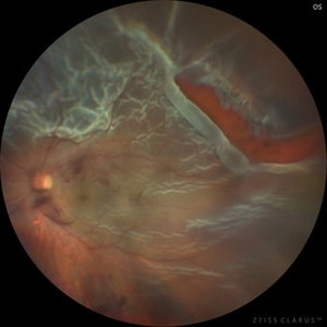
 Retinal Detachment Secondary to Anomalous PVD
Mar 13 2025 by Fabricio Dolores
This color wide-field clinical image depicts the right eye of a female patient who experienced a sudden loss of vision one month earlier. She was initially diagnosed with a vitreous hemorrhage and managed with conservative treatment. Upon presentation to our institute one month later, a superior rhegmatogenous retinal detachment was identified, extending across the 12 o’clock meridian. This was accompanied by an inferior vitreous hemorrhage and a solitary superior retinal lesion located at M11 in the superior triangle of the ora serrata, in alignment with Lincoff's second law.
Photographer: Fabricio Dolores-Villanueva, MD
Imaging device: Nidek Mirante
Condition/keywords: Retinal Detachment
-
 Retinal Detachment with Single Break
Feb 5 2025 by Virginia Gebhart
61 year old male with mac-off retinal detachment with single horseshoe tear. Macula has been off for several days and has developed associated cystic edema. Visual prognosis guarded. Pt schedule for PPV/Laser/GFE
Photographer: Virginia Gebhart, Retina Consultants of Carolina
Imaging device: Optos California
Condition/keywords: horseshoe tear, PVD, retinal detachment
-
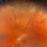
 Eales Disease
Jan 31 2025 by Thirumalesh Mochi Basavaraj, MD
Ultra wide field image of a 24 year-old young healthy adult male with a visible sea fan neovascularization with partial PVD secondary to Scatter LASER photocoagulation with Vitreous and subhyaloid hemorrhage.
Photographer: Puttaswamy N K
Condition/keywords: Eales disease, Neovascularisation elsewhere (NVE), sea fan
-
 Eales Disease
Jan 31 2025 by Thirumalesh Mochi Basavaraj, MD
Ultra-wide field image of a 24 year old young healthy adult male with a visible sea fan neovascularization with partial PVD with vitreous and subhyaloid hemorrhage.
Photographer: Puttaswamy
Condition/keywords: Eales disease, sea fan, Ultra-wide field retinal imaging
-
 Posterior Vitreous Detachment
Jan 31 2025 by Thirumalesh Mochi Basavaraj, MD
Intraoperative view of Triamcinolone-assisted posterior vitreous detachment.
Photographer: Thirumalesh Mochi Basavaraj
Condition/keywords: PVD induction, triamcinolone
-
Macular Hole Surgery: Inverse Flaps
Jan 31 2025 by Thirumalesh Mochi Basavaraj, MD
This video demonstrates, PVD induction , followed by ILM peeling in multiple flower petal flap technique.
Condition/keywords: ILM flaps, ILM peel, induction
-
 Central Retinal Vein Occlusion with Macular Edema
Jan 29 2025 by Kimberly Wakester
Fundus photograph of a 62-year-old man with central retinal vein occlusion with macular edema and a new PVD with an operculated retinal tear in the left eye. Laser to retinal tear was completed. Patient will return in 2-3 weeks for follow up exam with possible intravitreal injection for the CRVO with edema and to follow up on the operculated retinal tear s/p retinal tear laser.
Photographer: Kimberly Wakester, COA
Imaging device: Optos California
Condition/keywords: central retinal vein occlusion (CRVO), operculated tear, PVD
-
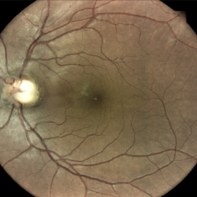
 Weiss Ring
Jan 21 2025 by Kimberly Wakester
Fundus photographs of a 70-year-old woman with a PVD with Weiss ring present in the left eye. Doing sweeps of the left eye shows how changing the patient's gaze can reposition the Weiss ring in the patient's eye.
Photographer: Kimberly Wakester, COA
Imaging device: Optos California
Condition/keywords: PVD, Weiss ring
-
 Cloquet Canal
Sep 20 2024 by Jordyn Beckman
79 year old male with wet AMD, no PVD, presents with stable cloquet canal.
Photographer: Jordyn Beckman
Imaging device: Optos California
Condition/keywords: Cloquet Canal, hyaloid membrane, punctum caecum, stilling's canal
-
 New Retinal Detachment 6w s/p RD repair
Nov 16 2023 by Virginia Gebhart
13 year old male presented with new blind spot 6 weeks s/p RD repair with cryo/scleral buckle/prophylaxis laser with gas bubble. New RD involving the macula, posterior to scleral buckle, secondary to PVD. Small gas bubble remaining. Pt was brought back to OR for repeat PPV and silicone oil repair
Photographer: Virginia Gebhart
Imaging device: Optos
Condition/keywords: gas bubble, Retinal Detachment, retinal detachment of the macula, scleral buckle
-
 Posterior Vitreous Detachment
Nov 1 2023 by ANKIT JAIN
USG B SCAN image showing membranous echoes with low to moderate spikes with free after movements with no attachment to disc suggestive of posterior vitreous detachment.
Photographer: DR ANKIT JAIN
Condition/keywords: B scan ultrasound, posterior vitreous detachment, PVD, ultrasound
-
 Total Rhegmatogenous retinal detachment with lattice degeneration & Vitreous haemorrhage
Jul 31 2023 by Harsh Vardhan Singh, MS
72-year male presented PVD induced total retinal detachment with vitreous hemorrhage
Photographer: Dr Harsh Vardhan Singh, AIIMS, Guwahati
Imaging device: Zeiss Clarus 700
Condition/keywords: chronic retinal detachment, hemorrhage, rrd
-
Vitrectomy for Macular Hole
Jan 13 2023 by Manish Nagpal, MD, FRCS (UK), FASRS
This is a case of Macular hole for which vitrectomy is being done. After doing core vitrectomy triamcinolone dye is injected to stain the hyaloid. High aspiration is used on cutter to engage the hyaloid and gradually pull it anteriorly. PVD induction is carried out. After this brilliant blue dye is injected to stain the internal limiting membrane. ILM is peeled using a 25 gauge forceps in a tangential manner. After this i use a instrument called the massager which we have developed to gently and atraumatically massage concentrically the edges of the hole. This releases the subtle contaction on the edges of the hole and relaxes the margins. After this air fluid exchange is carried out followed by low vacuum aspiration over the hole. The hole approximates itself gradually as the aspiration dries up the edges.
Condition/keywords: forceps, hyaloid, ILM, macular hole, peeling, staining, video, vitrectomy
-
Vitrectomy for epiretinal membrane removal
Nov 30 2022 by Manish Nagpal, MD, FRCS (UK), FASRS
Vitrectomy is carried out for epiretinal membrane removal. After doing core vitrectomy and pvd induction a 25 gauge forceps is used to pinch and peel the macular pucker. After finding a edge the forceps is gradually moved tangentially over the retinal surface to remove the membrane.
Photographer: Manish Nagpal
Condition/keywords: epiretinal membrane, forceps, macular pucker, peeling, video, vitrectomy
-
 PVD induction with IVTA staining
Nov 1 2022 by Shobhit Chawla, M.S.
This is an intraoperative photograph of Pvd induction showing both the macular and disc attatchments stained with triamcinolone.
Photographer: Shobhit Chawla
Condition/keywords: PVD induction, triamcinolone
-
PVD induction in a retinal detachment
Oct 24 2022 by Manish Nagpal, MD, FRCS (UK), FASRS
This video highlights the PVD induction technique in a case of retinal detachment with mobile retina, triamcinolone staining allows ease of visualizing the pvd attachment which is gradually removed from the retinal attachment using suction.
Photographer: Manish Nagpal
Condition/keywords: posterior hyaloid, PVD, triamcinolone, video, vitrectomy
-
 Evolving Weiss Ring
Sep 11 2022 by Michael B Green, MD, MBA
Fundus photograph of a 62-year-old female with an evolving Weiss-ring in the process of separating from the optic disc.
Condition/keywords: posterior vitreous detachment, PVD, Weiss ring
-
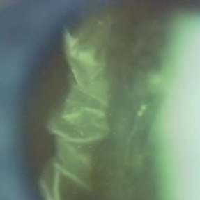 Folds in Detached Posterior Vitreous Cortex
May 31 2022 by Joshua Friedman
Slit lamp (video) image showing folds in the posterior vitreous cortex in an eye with PVD.
Photographer: Martin Snead, MD, Cambridge, England
Condition/keywords: folds, posterior vitreous cortex, PVD, vision degrading myodesopsia, vitreous
-
 Epiretinal Membrane
Feb 2 2022 by Manish Nagpal, MD, FRCS (UK), FASRS
Intraoperative photo of a epiretinal membrane, glistening reflex noted. Prior to this capture, PVD induction has been done, which has left a small splinter hemorrhage around the disc attachment of hyaloid.
Photographer: Manish Nagpal, Retina Foundation, Ahmedabad, India
Imaging device: Sony PMW -10 MD surgical camera
Condition/keywords: epiretinal membrane formation, ERM, ILM flap, PVD induction
-
 PVD Induction
Feb 2 2022 by Manish Nagpal, MD, FRCS (UK), FASRS
Intraoperative photo of PVD induction being carried out. Hyaloid has been stained using triamcinolone dye and PVD induction is carried out using high suction on the cutter and engaging the stained hyaloid.
Photographer: Manish Nagpal, Retina Foundation, Ahmedabad, India
Imaging device: Sony PMW -10 MD surgical camera
Condition/keywords: Hyaloid staining, PVD induction, triamcinolone
 Loading…