Search results (43 results)
-
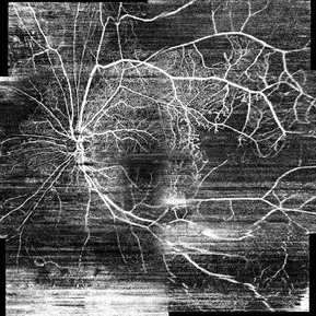
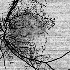
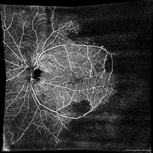
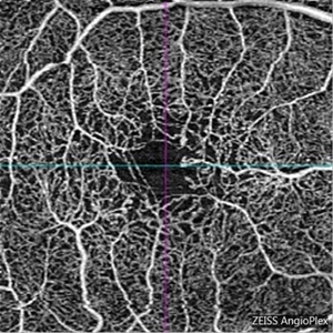 Macular Telangiectasia Type 2
Mar 8 2018 by Daniel R Agarwal, MD
OCT Angiography image in a 51-year-old male with fogging of vision and leaking on fluorescein angiography.
Photographer: Jen Welsh
Imaging device: Zeiss Angioplex OCTA
Condition/keywords: macular telangiectasia, macular telangiectasia type 2
-
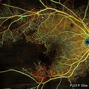
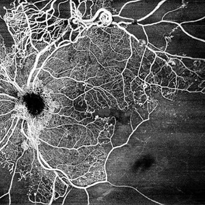 Central Retinal Vein Occlusion by OCT Angiography
Jun 13 2022 by JORGE SOBERANES
A 63 year old man with a central retinal vein oclussion. In the OCT angiogram we could observe retinal isquemia, neovascularization and arteriovenous shunts.
Photographer: Jorge I. Soberanes MD
Imaging device: PLEX Elite 9000, Zeiss
Condition/keywords: Central vein oclussion, neovascularization, OCT angiography, retina, Shunts
-
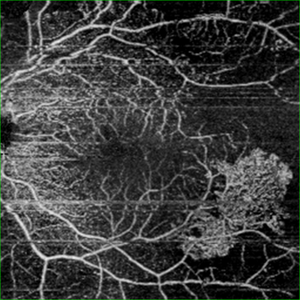 OCT Angiography- PDR
May 11 2020 by Gayathri Mohan
OCT angiography image of the superficial plexus showing neovascularisation infero-temporal to macula. Avascular areas are seen temporally.
Photographer: Gayathri Mohan, Retina Foundation
Imaging device: Mirante, Nidek
Condition/keywords: neovascularization (NV), optical coherence tomography (OCT), proliferative diabetic retinopathy (PDR)
-
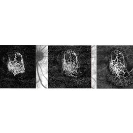 OCT Angiography
Jul 1 2018 by Mark H. Nelson, MD, MBA
82-year-old male, s/p 14 x ranibizumab injections, with persistent exudation and neovascularization on IVFA/ICG/OCTA. The three images reveal the progression of OCTA imaged neovascularization during the course of the anti-VEGF monotherapy.
Photographer: B.J. Graham, CRA
Condition/keywords: exudative age-related macular degeneration
-
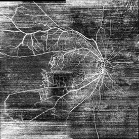
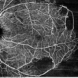
 Proliferative Diabetic Retinopathy
Oct 16 2021 by Timur Shaimov
32 y.o. female with Type 1 Diabetes with no glucose compensation for several years. A manual montage of several 8x8 mm OCT angiograms were obtained for this Widefield OCTA image.
Photographer: Timur Shaimov
Imaging device: RTVue xR Avanti
Condition/keywords: OCT Angiography, proliferative diabetic retinopathy (PDR)
-
 Proliferative Diabetic Retinopathy
Mar 1 2021 by Avris Romario Diparaja Siahaan
Swept source OCT angiography (12.0 mm X 12.0 mm) of a 57-year-old woman with proliferative diabetic retinopathy in her both eyes.
Photographer: Nanda Lessi Hafni Eka Putri, MD (Ophthalmologist) & Ryan Mishbahuddin (Nurse), Ciawi General Hospital (Rumah Sakit Umum Daerah Ciawi)
Imaging device: DRI OCT Triton Plus
Condition/keywords: fundus photograph, montage, optical coherence tomography (OCT), swept source, wide angle imaging
-
 Proliferative Diabetic Retinopathy
Mar 1 2021 by Avris Romario Diparaja Siahaan
Swept Source OCT angiography (montage photography) of a 57-year-old woman with proliferative diabetic retinopathy in her both eyes.
Photographer: Nanda Lessi Hafni Eka Putri, MD (Ophthalmologist) & Ryan Mishbahuddin (Nurse), Ciawi General Hospital (Rumah Sakit Umum Daerah Ciawi)
Imaging device: DRI OCT Triton Plus
Condition/keywords: fundus photograph, montage, optical coherence tomography (OCT), swept source, wide angle imaging
-
 Retinal Arterial Macroaneurysm
Apr 8 2023 by Yousef A Fouad, MD, FRCS (Glasg.)
Multimodal imaging of a retinal arterial macroaneurysm in the right eye of a 73-year-old male patient with uncontrolled hypertension. Fundus photography shows hemorrhage surrounding an arterial branch of the upper temporal arcade. Optical coherence tomography (OCT) through the lesion shows inner retinal hyperreflectivity with back shadowing, and adjacent cystoid macular edema in the outer retina. En face OCT centered on the lesion delineates the fusiform dilatation of the affected vessel, and OCT angiography confirms the presence of blood flow within the aneurysmal dilatation.
Photographer: Yousef Fouad, Ain Shams University, Egypt
Condition/keywords: arteriolar macroaneurysm, enface imaging, macroaneurysm, macroarterial aneurysm, OCT Angiography, OCTA
-
 Proliferative Diabetic Retinopathy
Mar 1 2021 by Avris Romario Diparaja Siahaan
Swept source OCT angiography (montage photography) of a 57-year-old woman with proliferative diabetic retinopathy in her both eyes.
Photographer: Nanda Lessi Hafni Eka Putri, MD (Ophthalmologist) & Ryan Mishbahuddin (Nurse), Ciawi General Hospital (Rumah Sakit Umum Daerah Ciawi)
Imaging device: DRI OCT Triton Plus
Condition/keywords: fundus photograph, montage, optical coherence tomography (OCT), swept source, wide angle imaging
-
 Proliferative Diabetic Retinopathy with Macular Isquemia by OCT Angiography
Oct 14 2022 by JORGE SOBERANES
Depth-encoded OCT angiography of a patient with proliferative diabetic retinopathy showing vascular changes and extensive ischemia including macular area.
Photographer: Jorge I. Soberanes, Asociación para Evitar la Ceguera en México.
Imaging device: PLEX Elite 9000, Zeiss
Condition/keywords: diabetic retinopathy, OCT Angiography
-
 Retinal Hemorrhage
Sep 2 2021 by Avris Romario Diparaja Siahaan
Swept source OCT angiography of a 58-year-old man with hemorrhage in his left eye.
Photographer: Nanda Lessi Hafni Eka Putri, MD (Ophthalmologist) & Ryan Mishbahuddin (Nurse), Ciawi General Hospital (Rumah Sakit Umum Daerah Ciawi)
Imaging device: DRI OCT Triton Plus (Topcon)
Condition/keywords: fundus photograph, optical coherence tomography (OCT)
-
 Acute Posterior Multifocal Placoid Pigment Epitheliopathy
Feb 20 2024 by Soobien Lee
12x12mm OCT Angiography of a 20-year-old caucasian female with viral prodrome and vision loss OS>OD secondary to Acute Posterior Multifocal Placoid Pigment Epitheliopathy (APPME). Imaging shows multifocal flow voids.
Photographer: Kim Seay, Elman Retina Group
Imaging device: 12x12mm OCT-Angiography
Condition/keywords: acute posterior multifocal placoid pigment epitheliopathy (APMPPE), bacillary layer detachment, OCT, OCT Angiography, Uveitis, white dot syndrome
-
 Reverse Polarity OCT Angiography of Proliferative Diabetic Retinopathy
Aug 31 2021 by RUSHIK PATEL
Reverse polarity OCTA image of left eye of 50 year-old diabetic male with proliferative diabetic retinopathy.
Photographer: Rushik Patel, Netralaya Super Speciality Eye Hospital
Condition/keywords: OCT Angiography, proliferative diabetic retinopathy (PDR)
-
 Proliferative Diabetic Retinopathy
Mar 1 2021 by Avris Romario Diparaja Siahaan
Swept Source OCT angiography (9.0 mm X 9.0 mm) of a 62-year-old woman with proliferative diabetic retinopathy in her both eyes.
Photographer: Nanda Lessi Hafni Eka Putri, MD (Ophthalmologist) & Ryan Mishbahuddin (Nurse), Ciawi General Hospital (Rumah Sakit Umum Daerah Ciawi)
Imaging device: DRI OCT Triton Plus
Condition/keywords: fundus photograph, montage, optical coherence tomography (OCT), swept source, wide angle imaging
-
 Proliferative Diabetic Retinopathy
Mar 1 2021 by Avris Romario Diparaja Siahaan
Swept source OCT angiography (montage photography) of a 62-year-old woman with proliferative diabetic retinopathy in her both eyes.
Photographer: Nanda Lessi Hafni Eka Putri, MD (Ophthalmologist) & Ryan Mishbahuddin (Nurse), Ciawi General Hospital (Rumah Sakit Umum Daerah Ciawi)
Imaging device: DRI OCT Triton Plus
Condition/keywords: fundus photograph, montage, optical coherence tomography (OCT), swept source, wide angle imaging
-
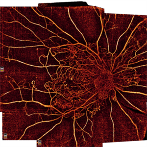
 Retinal neovascularization
Feb 28 2023 by Nassim Alejandro Abreu Arbaje, MD
OCT and OCTa of a diabetic patient with severe PDR, showing the anatomical location and blood flow of neovessels
Photographer: Nassim Abreu, Centro de Oftalmologia y Glaucoma
Imaging device: Topcon Triton Plus
Condition/keywords: neovascularization (NV), OCT, OCT Angiography, PDR
-
 Ischemic CRVO
Jun 23 2021 by Eduardo Torres-Porras, MD
Montage 8x8 mm OCT angiography of the left eye of a 36-year-old male who had an ischemic CRVO following a hypertensive emergency secondary to consumption of high doses of cocaine. Areas of non perfusion can be seen within the posterior pole which extends temporally to the mid-periphery.
Photographer: Eduardo Torres-Porras, Provissia, Laser y Ultrasonido Ocular
Imaging device: Cirrus 600, Carl Zeiss
Condition/keywords: central retinal vein occlusion (CRVO), ischemic CRVO, optical coherence tomography (OCT), ultra-wide field imaging
-
 Proliferative Diabetic Retinopathy
Mar 1 2021 by Avris Romario Diparaja Siahaan
Swept source OCT angiography (montage photography) of a 62-year-old woman with proliferative diabetic retinopathy in her both eyes.
Photographer: Nanda Lessi Hafni Eka Putri, MD (Ophthalmologist) & Ryan Mishbahuddin (Nurse), Ciawi General Hospital (Rumah Sakit Umum Daerah Ciawi)
Imaging device: DRI OCT Triton Plus
Condition/keywords: fundus photograph, montage, optical coherence tomography (OCT), swept source, wide angle imaging
-
 Diabetic Retinopathy
Sep 2 2021 by Avris Romario Diparaja Siahaan
Swept source OCT angiography (montage photography) and fundus photography of a 61-year-old woman with proliferative diabetic retinopathy in her right eye.
Photographer: Nanda Lessi Hafni Eka Putri, MD (Ophthalmologist) & Ryan Mishbahuddin (Nurse), Ciawi General Hospital (Rumah Sakit Umum Daerah Ciawi)
Imaging device: DRI OCT Triton Plus (Topcon)
Condition/keywords: diabetic retinopathy, fundus photograph, optical coherence tomography (OCT)
-
 Proliferative Diabetic Retinopathy
Mar 1 2021 by Avris Romario Diparaja Siahaan
Swept source OCT angiography (12.0 mm X 12.0 mm) of a 57-year-old woman with proliferative diabetic retinopathy in her both eyes.
Photographer: Nanda Lessi Hafni Eka Putri, MD (Ophthalmologist) & Ryan Mishbahuddin (Nurse), Ciawi General Hospital (Rumah Sakit Umum Daerah Ciawi)
Imaging device: DRI OCT Triton Plus
Condition/keywords: fundus photograph, montage, optical coherence tomography (OCT), swept source, wide angle imaging
-
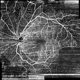
 From Ora to Ora
Aug 26 2024 by Nassim Alejandro Abreu Arbaje, MD
Ultra-wide field OCT angiography of a 39 year-old healthy male. The photo attempts to explore retinal vasculature up to the ora serrata.
Photographer: Johel Arrieta, TowardPi
Imaging device: TowardPi BMizar 400khz
Condition/keywords: OCT Angiography, OCTA, ultra-wide field imaging
-
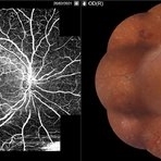
 OCT Angiography of a 360 retinotomy for closed funnel combined retinal detachment
Jan 1 2023 by Malek Yassine, MD
this is an OCTA image of 12X12 MM, showing all the 3 vascular plexi of the residual posterior retinal, with a good perfusion in the superior and central area, a ratification in the intermediate plexus in the inferior area, a non perfused temporal area, and some macular cysts. There's almost none macular translocation
Imaging device: Topcon Triton DRI-OCT
Condition/keywords: combined retinal detachment, OCT Angiography, retinotomy
-
 Proliferative Diabetic Retinopathy
Mar 1 2021 by Avris Romario Diparaja Siahaan
Swept Source OCT angiography (9.0 mm X 9.0 mm) of a 62-year-old woman with proliferative diabetic retinopathy in her both eyes.
Photographer: Nanda Lessi Hafni Eka Putri, MD (Ophthalmologist) & Ryan Mishbahuddin (Nurse), Ciawi General Hospital (Rumah Sakit Umum Daerah Ciawi)
Imaging device: DRI OCT Triton Plus
Condition/keywords: fundus photograph, montage, optical coherence tomography (OCT), swept source, wide angle imaging
-
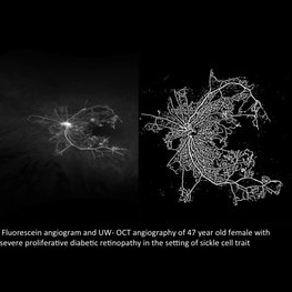 Severe Proliferative Diabetic Retinopathy
Jan 9 2018 by Kasra Rezaei, M.D.
Fluorescein angiogram and UW- OCT angiography of 47-year-old female with severe proliferative diabetic retinopathy in the setting of sickle cell trait.
Photographer: Kasra A Rezaei, University of Washington
Imaging device: Zeiss OCTA and Optos
Condition/keywords: proliferative diabetic retinopathy (PDR), ultra-wide field imaging
-
 Ischemic CRVO
Jun 23 2021 by Eduardo Torres-Porras, MD
HD 8x8 mm OCT angiography of a 36-year-old male who had an ischemic CRVO following a hypertensive emergency secondary to consumption of high doses of cocaine. The posterior pole has areas of non perfusion.
Photographer: Eduardo Torres-Porras, Provissia, Laser y Ultrasonido Ocular
Imaging device: Cirrus 600, Carl Zeiss
Condition/keywords: central retinal vein occlusion (CRVO), ischemic CRVO, optical coherence tomography (OCT), ultra-wide field imaging
 Loading…