 Initializing download.
-
 By Malek Yassine, MD
Centre d’Ophtalmologie Dr Malek Yassine - Uploaded on Jan 1, 2023.
- Last modified by Joshua Friedman on Jan 2, 2023.
- Rating
- Appears in
- 360 Retinotomy in a closed Funnel combined Tractional and rhegmatogenous retinal detachment
- Condition/keywords
- OCT Angiography, combined retinal detachment, retinotomy
- Imaging device
-
Optical coherence tomography system
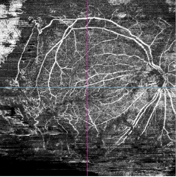
Topcon Triton DRI-OCT - Description
- this is an OCTA image of 12X12 MM, showing all the 3 vascular plexi of the residual posterior retinal, with a good perfusion in the superior and central area, a ratification in the intermediate plexus in the inferior area, a non perfused temporal area, and some macular cysts. There's almost none macular translocation


