Search results (431 results)
-
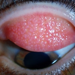 Giant Papillary Conjunctivitis, Left Upper Eyelid
Jul 22 2013 by Jason S. Calhoun
Contact lens wearer in for exam. Has rough feeling underneath both eyelids. Patient thought it was through SCL wear. Patient VA was 20/20. right eye, 20/30, left eye. Underneath the left upper eyelid, you can see papillary inflammation and redness.
Photographer: Jason S. Calhoun, Department of Ophthalmology, Mayo Clinic Jacksonville, Florida
Imaging device: TOPCON D-90 SL NIKON CAMERA
Condition/keywords: giant papillary conjunctivitis
-
 Auto-Enucleation with Tire Iron
Oct 19 2012 by Larry Halperin, MD
Auto-enucleation with tire iron
Condition/keywords: enucleation, self-inflicted, trauma
-
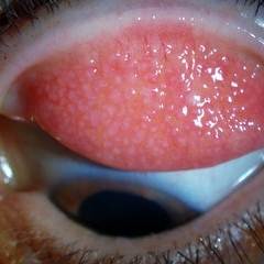 Giant Papillary Conjunctivitis, Left Upper Eyelid
Jul 22 2013 by Jason S. Calhoun
Contact lens wearer, in for exam. Has rough feeling underneath both eyelids. Patient thought it was through SCL wear. Patient VA was 20/20. right eye, 20/30, left eye. Underneath the left upper eyelid, you can see papillary inflammation and redness.
Photographer: Jason S. Calhoun, Department of Ophthalmology, Mayo Clinic Jacksonville, Florida
Imaging device: TOPCON D-90 SL NIKON CAMERA
Condition/keywords: giant papillary conjunctivitis
-
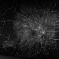 "Starry Sky" Fundus in Vogt-Koyanaki-Harada Syndrome
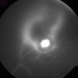
Jan 10 2018 by Peter H. Tang, MD, PhD
Fluorescein angiography imaging of a 27-year-old male with acute inflammation as part of Vogt-Koyanagi-Harada Syndrome.
Imaging device: Optos California
Condition/keywords: chorioretinal inflammations, retina, uveitis, Vogt-Koyanagi-Harada
-
---thumb.jpg/image-square;max$300,300.ImageHandler) Myelinated Nerve Fiber Layer
Myelinated Nerve Fiber Layer
-
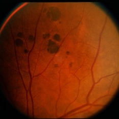 Bear Tracks / CHRPE / Myelinated NFL
Jul 12 2014 by David Callanan, MD
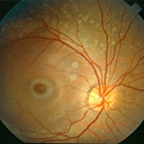
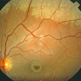
58-year-old female, bear tracks / CHRPE / myelinated NFL.
Condition/keywords: bear tracks, congenital hypertrophy of the retinal pigment epithelium (CHRPE), myelinated nerve fibers
-
---thumb.jpg/image-square;max$300,300.ImageHandler) Progressive Outer Retinal Necrosis
Progressive Outer Retinal Necrosis
Feb 15 2013 by From the Collections of Thomas M. Aaberg, MD and Thomas M. Aaberg Jr., MD
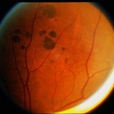
Color fundus photograph showing extensive confluent retinal whitening, retinal exudation, intraretinal hemorrhage, and sheathing of retinal vessels consistent with infectious retinitis such as progressive outer retinal necrosis (PORN).
Condition/keywords: occlusive retinitis, retinal necrosis
-
---thumb.JPG/image-square;max$300,300.ImageHandler) Vitreous Snow Balls
Vitreous Snow Balls
Nov 18 2013 by Mallika Goyal, MD
Clumps of inflammatory cells in the inferior vitreous in a patient with bilateral vitreitis.
Photographer: Mallika Goyal, MD, Apollo Health City, Hyderabad
Condition/keywords: vitreous snowballs
-
---thumb.jpg/image-square;max$300,300.ImageHandler) Birdshot Choroidopathy
Birdshot Choroidopathy
Oct 9 2013 by Maurice F. Rabb
Forty two year old white female first noted flashing lights in her left eye at the age of 30. Although she had many previous eye examinations for low grade myopia, she had never had a dilated fundus examination. The evaluation twelve years ago disclosed 20/20 acuity in each eye with a myopic correction, an afferent pupillary defect on the left, no evidence of anterior segment inflammation in either eye, a full field on the right and markedly constricted field on the left, fundus pigmentary abnormalities extending beyond the equator in each eye, and narrow vessels with pigment migration into the retina in the left eye only.
Condition/keywords: birdshot choroidopathy
-
 OCT in Patient With IIH Showing Thickened RNFL
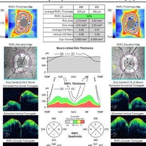
Jan 16 2019 by John S. King, MD
18-year-old African American female with increased BMI with a history of headaches, nausea, transient diplopia and vision loss that she notices when getting up from her bed (and goes away after standing upright) for the last two weeks. Went to PCP and was treated for the flu, and after no improvement and visual symptoms known, was sent to ED. MRI did not show any masses and showed empty sella turcia. Vision 20/30 OD and 20/20 OS; no RAPD; IOP 15OU; no anterior segment or vitreous inflammation; discs are elevated with obscuration of the disc margins and some of the smaller vessels; there are no SVPs; there are mild Patton's lines temporally (see Initial Photos). The optic disc cube shows 360 degrees of RNFL thickening (see OCT). Was referred to near-ophthalmologist, Dr. Doyle. She obtained additional work-up, and LP opening pressure was high, and MRV showed bilateral transverse sinus stenosis. Patient showed steady improvement with medical therapy, that included weight loss and oral diamox. On her last visit with Dr. Doyle, vision has remained stable at 20/20-20/25 without an enlarged blindspot; there are SVPs and optic disc edema has resolved (see Post Treatment Photos); she is currently on 1000 mg of diamox and has lost 15 pounds, and no stinting procedure needed.
Imaging device: Cirrus
Condition/keywords: benign idiopatic intracranial hypertension, optic disc edema, papilledema
-
---thumb.jpg/image-square;max$300,300.ImageHandler) Ocular Toxoplasmosis
Ocular Toxoplasmosis
Feb 15 2013 by From the Collections of Thomas M. Aaberg, MD and Thomas M. Aaberg Jr., MD
Diffuse slit-lamp photograph of the right eye of a patient with ocular toxoplasmosis showing strands of vitreous inflammation.
Condition/keywords: ocular toxoplasmosis, vitritis
-
 Dry Age-Related Macular Degeneration
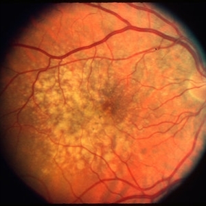
Mar 29 2013 by Henry J. Kaplan, MD
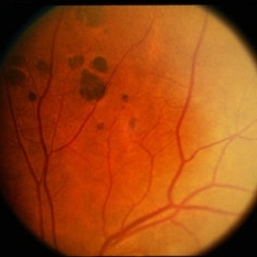
Dry AMD with multiple confluent soft drusens
Condition/keywords: age-related macular degeneration (AMD), dry age-related macular degeneration (dry AMD)
-
 Choroidal Granuloma Secondary to Tuberculosis
Mar 14 2013 by Eduardo Torres-Porras, MD
OCT scan through the granuloma shows attachment of the retinal pigment epithelial-choriocapillaris layer and the neurosensory retina over the granuloma (“contact” sign), inflammatory retinal infiltrate in the deeper retinal layers and subretinal fluid.
Photographer: Eduardo Torres Porras, Laser y ultrasonido ocular de Puebla
Imaging device: Cirrus
Condition/keywords: optical coherence tomography (OCT), tubercular choroidal granuloma
-
 Toxoplasmosis Slide 1
Oct 22 2012 by Ronald C. Gentile, MD
Focal, white area of chorioretinitis with overlying vitreous inflammation adjacent to an old chorioretinal scar in a patient complaining of photophobia, floaters and a decrease in vision of the right eye. The focal area of chorioretinitis is involving the inferior nasal macula and adjacent optic nerve with surrounding retinal and peri-papillary edema.
Photographer: The New York Eye & Ear Infirmary Department of Medical Imaging
Condition/keywords: posterior uveitis, toxoplasmosis
-
 Toxoplasma Gondii Chorioretinitis, Fluorescein Angiogram
Aug 23 2012 by Gerardo Garcia-Aguirre, MD
Fluorescein angiogram of the superior periphery showing a highly hyperfluorescent lesion adjacent to a hypofluorescent lesion, surrounded by a hyperfluorescent halo.
Photographer: Noemí Hernández, Asociación para Evitar la Ceguera en México
Imaging device: Zeiss FF4
Condition/keywords: chorioretinal inflammations, toxoplasmosis
-
 Toxoplasmosis Slide 2
Oct 22 2012 by Ronald C. Gentile, MD
One month following treatment with Bactrim, Clindamycin, and oral prednisone the focal area chorioretinitis has coalesced with a decrease in overlying vitreous inflammation. Kyrieleis plaques can be seen along the inferior retinal arteriole.
Photographer: The New York Eye & Ear Infirmary Department of Medical Imaging
Condition/keywords: posterior uveitis, toxoplasmosis
-
 Niemann Pick Disease Type B
Aug 6 2013 by Hamid Ahmadieh, MD
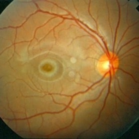
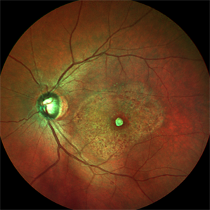
Color fundus photograph of the right eye of a patient with Niemann Pick Type B with typical macular halo and confluent deposits in mid-periphery.
Photographer: Ali Mohammad-Rabie, Ophthalmic Research Center, Labbafinejad Medical Center, Tehran
Condition/keywords: fleck retinopathy, macular halo
-
 Choroidal Granuloma Secondary to Tuberculosis
Mar 14 2013 by Eduardo Torres-Porras, MD
OCT scan through the granuloma shows attachment of the retinal pigment epithelial-choriocapillaris layer and the neurosensory retina over the granuloma (“contact” sign), inflammatory retinal infiltrate in the deeper retinal layers and subretinal fluid.
Photographer: Eduardo Torres Porras
Imaging device: Cirrus
Condition/keywords: optical coherence tomography (OCT), tubercular choroidal granuloma
-
 Bear Tracks / CHRPE / Myelinated NFL
Jul 12 2014 by David Callanan, MD
58-year-old female, bear tracks / CHRPE / myelinated NFL.
Condition/keywords: bear tracks, congenital hypertrophy of the retinal pigment epithelium (CHRPE), myelinated nerve fibers
-
 Niemann Pick Disease Type B
Aug 6 2013 by Hamid Ahmadieh, MD
Color fundus photograph of the right eye of a patient with Niemann Pick Type B with typical macular halo and confluent deposits in mid-periphery.
Photographer: Ali Mohammad-Rabie, Ophthalmic Research Center, Labbafinejad Medical Center, Tehran
Condition/keywords: macular halo
-
 Bear Tracks / CHRPE / Myelinated NFL
Jul 12 2014 by David Callanan, MD
58-year-old female, bear tracks / CHRPE / myelinated NFL.
Condition/keywords: bear tracks, congenital hypertrophy of the retinal pigment epithelium (CHRPE), myelinated nerve fibers
-
 Syphilis Neuroretinopathy
Apr 2 2018 by JEFFERSON R SOUSA, Tecg.º (Biomedical Systems Technology)
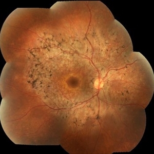
Female patient, 21-years-old, with complaint of low vision in the right eye for 3 years. According to information from the patient's history, at the time she noticed the low vision, it also coincided with a picture of a strong urinary infection as well as episodes of constant tonsillitis. Yes, the patient did not seek medical attention and self-medicated with antibiotics. In ophthalmologic evaluation, as well as examinations of color retinography and ocular fundus autofluorescence, important pigmentary alterations were observed following vascular arches with pigment mobilization in osteoclasts (aspect of a unilateral pigmentary retinitis secondary to the inflammatory process). Which suggested inflammatory process sequelae. Through the laboratory tests, he had positive (+) confirmation for SYPHILIS NEURORETINOPATHY .
Photographer: JEFFERSON R SOUSA - Study Center and Ophthalmological Research Dr. Andre M V Gomes, Institute Dr. Suel Abujamra São Paulo-Brazil
Imaging device: Fundus camera Topcon TRC-50 DX, Imaginet 5.0, angle de 50 graus. Flash 36 / Mosaic with 10 images.
Condition/keywords: neurosyphilitic optic atrophy, retinitis pigmentosa, syphilis, syphilis neuroretinopathy
-
 Mild Patton's Lines in IIH - Initial Photos
Jan 16 2019 by John S. King, MD
18-year-old African American female with increased BMI with a history of headaches, nausea, transient diplopia and vision loss that she notices when getting up from her bed (and goes away after standing upright) for the last two weeks. Went to PCP and was treated for the flu, and after no improvement and visual symptoms known, was sent to ED. MRI did not show any masses and showed empty sella turcia. Vision 20/30 OD and 20/20 OS; no RAPD; IOP 15OU; no anterior segment or vitreous inflammation; discs are elevated with obscuration of the disc margins and some of the smaller vessels; there are no SVPs; there are mild Patton's lines temporally (see Initial Photos). The optic disc cube shows 360 degrees of RNFL thickening (see OCT). Was referred to near-ophthalmologist, Dr. Doyle. She obtained additional work-up, and LP opening pressure was high, and MRV showed bilateral transverse sinus stenosis. Patient showed steady improvement with medical therapy, that included weight loss and oral diamox. On her last visit with Dr. Doyle, vision has remained stable at 20/20-20/25 without an enlarged blindspot; there are SVPs and optic disc edema has resolved (see Post Treatment Photos); she is currently on 1000 mg of diamox and has lost 15 pounds, and no stinting procedure needed.
Photographer: Gretchen Harper
Imaging device: Topcon 50
Condition/keywords: idiopathic intracranial hypertension, optic disc edema, papilledema, Patton's Lines
-
 Niemann Pick Disease Type B
Aug 6 2013 by Hamid Ahmadieh, MD
Color fundus photograph of the left eye of a patient with Niemann Pick Type B with typical macular halo and confluent deposits in mid-periphery.
Photographer: Ali Mohammad-Rabie, Ophthalmic Research Center, Labbafinejad Medical Center, Tehran
Condition/keywords: fleck retinopathy, macular halo
-
 Macular Scar Due to Cysticercosis Fundus Photo
Aug 8 2017 by Manuel A Paez-Escamilla, MD, FICO
Fundus photograph of a 69-year-old patient with a long history of eye inflammation and progressive decrease in vision. Multiple trips to Asia and eating undercooked pork.
Photographer: Mark Erickson CRA, COT. The Macula Center. Clearwater, Florida
Condition/keywords: cysticercosis, uveitis
 Loading…